Vol. 41, No. 1, March 2015, 9-20 http://dx.doi.org/10.15230/SCSK.2015.41.1.9
각질세포에서 자외선B가 유도한 세포 사멸, 산화적 스트레스 및 matrix metalloproteinase 1 발현에 대한 죽여추출물의 영향
석 진 경⋅곽 준 엽⋅서 형 호*⋅서 화 진**⋅부 용 출†
경북대학교 의학전문대학원 분자의학교실, 세포기질연구소, *(주)루비크라운, **(재)경북천연염색산업연구원 (2014년 10월 20일 접수, 2014년 11월 24일 수정, 2015년 1월 9일 채택)
Effects of Bambusae Caulis in Taeniam Extract on the UVB-induced Cell Death, Oxidative Stress and Matrix Metalloproteinase 1 Expression in Keratinocytes
Jin Kyung Seok, Jun Yup Kwak, Hyeong Ho Seo*, Hwa Jin Suh**, and Yong Chool Boo†
Department of Molecular Medicine, Cell and Matrix Research Institute, BK21 Plus KNU Biomedical Convergence Program, Kyungpook National University School of Medicine, Daegu, 700-422, Republic of Korea,
*Ruby Crown Co., Ltd., Daegu, Republic of Korea
**Gyeongbuk Natural Color Industry Institute, Gyeongbuk, Republic of Korea (Received October 20, 2014; Revised November 24, 2014; Accepted January 9, 2015)
요 약: 자외선은 피부 광노화의 주요 요인이며, 효과적인 자외선 차단제가 피부의 건강과 아름다움을 위해 필요
하다. 본 연구는 세포 실험을 통하여 자외선에 의해 유도된 세포 사멸, 산화적 스트레스, matrix metal- loproteinase 1 발현에 미치는 죽여추출물의 영향을 알아보고자 수행하였다. HaCaT 인간 각질세포를 여러 농 도의 죽여추출물 유무 조건에서 자외선을 조사하고 세포의 생존율과 생화학적 과정들의 변화를 분석하였다. 죽 여추출물은 자외선을 조사한 세포의 생존율을 증진시켰고, procaspase 3가 활성화 형태로 절단되고, Bax/Bcl-2 비율이 증가하는 세포 자살 과정을 완화시켰다. 죽여추출물은 자외선에 노출된 세포에서 활성산소 의 발생과 지질 과산화도 감소시켰다. 또한 죽여추출물은 자외선에 의해 자극된 matrix metalloproteinase 1의 발현과 c-Jun N-terminal kinase의 인산화를 억제하였다. 본 연구는 죽여추출물이 자외선에 의한 세포 사멸, 산화적 스트레스, 그리고 matrix metalloproteinase 1 발현을 억제함을 보여 주었으며, 이 추출물이 피부 광노 화의 일부 현상을 억제하는 화장품 원료로 유용함을 시사하였다.
Abstract: Ultraviolet radiation (UV) is a major cause of skin photoaging, and effective UV protecting agents are needed for the skin health and beauty. This study was undertaken to examine the effects of Bambusae caulis in Taeniam extract (BCTE) on UVB-induced cell death, oxidative stress and matrix metalloproteinase 1 (MMP1) expression in cell-based assays. HaCaT human keratinocytes were exposed to UVB in the presence of BCTE at different concentrations and re- sulting changes in cell viability and biochemical events were determined. The results showed that BCTE enhanced the viabilities of UVB-exposed cells, and attenuated apoptotic events such as cleavage of procaspase 3 to its active form, and the increase of Bax to Bcl-2 ratios. BCTE also attenuated the reactive oxygen generation and lipid peroxidation in cells exposed to UVB. Additionally, it attenuated the expression of matrix metalloproteinase 1 and the phosphor- ylation of c-Jun N-terminal kinase stimulated by UVB. Conclusively, the present study demonstrated that BCTE pro
1)
† 주 저자 (e-mail: [email protected]) call: 053)420-4946
tected skin cells from the UVB-induced cell death, oxidative stress and MMP1 expression, suggesting its potential use as a cosmetic ingredient mitigating some features of the skin photoaging.
Keywords: UVB, Bambusae Caulis in Taeniam, oxidative stress, apoptosis, matrix metalloproteinase 1, keratinocytes
1. Introduction
Intrinsic aging of human skin is dependent on time and genetics, whereas extrinsic skin aging is affected by environmental factors such as solar radiation[1]. Intrinsic aging, also called natural or chronological aging, is in- evitable, but extrinsic photoaging can be avoided with good habits minimizing exposure to ultraviolet radiation (UV)[2]. The sun is a primary source of UV inducing skin photoaging which alters the normal phenotypes of the skin such as texture, wrinkles, tone, and color[3]. Of course, UV is a major cause of photo-carcinogenesis[4].
UV-induced apoptotic cell death is involved in skin photoaging[5]. Apoptosis usually involves the changes of pro-apoptotic (Bax, Bak and Bid) and anti-apoptotic (Bcl-2 and Bcl-x) members of the Bcl-2 protein fam- ily[6]. Caspases are known to mediate UV-induced apop- tosis in keratinocytes[7]. UV induces oxidative stress in cells by stimulating production of reactive oxygen spe- cies (ROS) and depleting antioxidants[8,9]. As the re- sults, oxidative damages of biomolecules such as lipids, nucleic acids, and proteins increase in cells exposed to UV[10,11]. Skin photoaging due to UV exposure in- volves the activation of the matrix metalloproteinases (MMPs), a group of zinc endopeptidases, which regulate the turnover of extracellular matrix macromolecules in- cluding type I collagen[12,13]. These MMPs can regulate connective tissue remodeling leading to the formation of wrinkles, and other phenotypes of photoaged skin[14, 15]. MMP1 secreted not only from dermal fibroblasts and but from epidermal keratinocytes is known to partic- ipate in collagen destruction in the skin[16,17].
Sunscreen products have become essential products to those who pursue skin health and beauty[18]. Although the weight of current evidence is that sunscreen agents of either inorganic or organic forms remain on the surface
of the skin, there also is a concern regarding the pene- tration of sunscreen agents into the skin and their harm- ful effects[19,20]. There is considerable interest in the development of safer, more effective cosmetic ingredients that mitigate the harmful effects of UV. In this regard, plant extracts with UV-shielding and anti-oxidative prop- erties are an attractive prospect. We have previously ob- served the protective effects of plant extracts derived from Gardenia jasminoides and Sasa quelpaertensis against the UV-induced injury in cells and animal mod- els[21,22].
Bambusae caulis in Taeniam has been used as a health food additive and a traditional medicine for the treat- ments of atherosclerosis, hyperlipidemia, hypertension, fatigue and so on[23-25]. Its activity is thought be related partly to potent antioxidant properties[16,26]. In the pres- ent study, we examined the effects of BCTE on UVB-in- duced cell death, oxidative stress and MMP1 expression in HaCaT keratinocytes. This plant material was chosen because it attenuated the UVB-induced HaCaT cell death effectively and showed the lowest cytotoxicity among the numerous plant extracts tested in a preliminary experiment. The present study demonstrated the benefi- cial properties of BCTE protecting skin cells from the UVB-induced damages, suggesting its potential use in the photoprotection of the skin.
2. Materials and Methods
2.1. Preparation of BCTE
Bambusae caulis in Taeniam (stem) was purchased
from a local market (http://www.jchanbang.com/),and ex-
tracted with 70% aqueous ethanol at room temperature
for 7 days, followed by evaporation under a reduced
pressure to obtain BCTE at a yield of 4.5%.
2.2. UV spectrophotometry
BCTE was dissolved in phosphate buffered saline (PBS) at 10, 30, and 100 µg/mL, and its absorption spec- tra were recorded in the 200 ~ 400 nm range using a Shimadzu UV ‐1650PC spectrophotometer (Shimadzu Corporation, Kyoto, Japan).
2.3. HPLC analysis
HPLC analysis of BCTE was done using a Gilson HPLC system (Gilson, Inc. Middleton, WI, USA) equip- ped with a 321 pump and UV/VIS 151 detector.
Aqueous solution of BCTE (1.0 mg/mL) was injected at 20 µL. Separation was done on a 5 mm Hector-M C
18column (4.6 mm × 250 mm)(RStechco, Daejeon, Korea).
The mobile phase consisted of 0.5% formic acid (A) and acetonitrile (B). The gradient was programmed as fol- lows: 0 ~ 6 min, 100% A; 6 ~ 10 min, a linear gradient from 0 to 12% B; 10 ~ 35 min, 12 to 21% B; 35 ~ 60 min, 21 to 41%; 60 ~ 70 min, 41 to 100% B; 70 ~ 80 min, 100% B. The flow rate was 0.8 mL/min. The de- tector was set at 280 nm. p-Coumaric acid (p-CA) stand- ard was purchased from Sigma-Aldrich (St. Louis, MO, USA).
2.4. Free radical scavenging effects
The free radical scavenging activities were determined against the radical cation of 2,2’-azinobis-(3-ethylbenzothiazo- line-6-sulfonic acid) (ABTS)[27], and 2,2-diphenyl-1- picrylhydrazyl radial (DPPH)[28]. The ABTS radical was generated by mixing 7.4 mM ABTS (Sigma-Aldrich) and 2.6 mM potassium persulfate (Sigma-Aldrich) in equal quantities, and allowing them to react for 24 h at room temperature in the dark. Each serial dilution of a plant extract in methanol (100 µL) was reacted with 100 µL of 0.2 mM ABTS radical in methanol at room temper- ature for 3 min, followed by measurement of the absorb- ance at 734 nm with a BioRad Model 680 microplate reader (Bio-Rad Laboratories, Inc., Hercules, CA, USA).
For DPPH assay, the reaction mixture containing 100 µL of a serially diluted plant extract in methanol and 100 µL of 0.2 mM DPPH (Sigma-Aldrich) in methanol was kept
at room temperature for 3 min, and the absorbance was taken at 517 nm using a microplate reader. Data are pre- sented as SC
50(µg/mL) which is the concentration of a sample showing 50% radical scavenging activity.
2.5. HaCaT cells culture
HaCaT cells (a human immortalized keratinocyte cell-line) were grown in DMEM/F ‐12 medium (GIBCO‐
BRL, Grand Island, NY, USA) supplemented with a 10%
fetal bovine serum, 100 U/mL penicillin, 0.1 mg/mL streptomycin, 0.25 µg/mL amphotericin B, and 10 µg/mL hydrocortisone. Cells were cultured at 37 ℃ in a humidi- fied atmosphere containing 5% CO
2and 95% air.
2.6. UVB-exposure of HaCaT cells
HaCaT cells were seeded on a six ‐well plate at a den- sity of 3 × 10
5cells per well and grown in culture me- dium for 24 h. Cells were then exposed to UVB in PBS containing the test material at the specified concentrations. UVB irradiation was conducted using a UVB-18 lamp (ULTRA*LUM. Inc., Claremont, CA, USA) that emitted radiation in the wavelength range 280 to 340 nm with maximum intensity at 300 nm. UV was administered to cells in culture plates at an intensity of 80 µW/cm
2for different durations to provide different doses of UVB (5, 10, or 15 mJ/cm
2). Following irradi- ation, cells were fed with a growth medium and in- cubated for the specified time.
2.7. Cell viability test
Cell viability was determined using 3 ‐[4,5‐dimethylth- iazol ‐2‐yl]‐2,5‐ diphenyltetrazolium bromide (MTT).
Briefly, cells were washed with PBS and incubated in 1
mL of culture media supplemented with 1 mg/mL MTT
(AMRESCO, Solon, OH, USA) for 3 h. The medium
was aspirated off and cells were extracted with iso-
propanol to dissolve the formed formazan. The solutions
were moved to a 96-well plate and absorbance was de-
termined at 595 nm using a microplate reader.
2.8. Detection of ROS
The production of ROS was determined with the oxi- dant-sensitive probe dihydrorhodamine 123 (DHR123) (Sigma-Aldrich). Cells were treated with 1.0 µM DHR123 prior to UVB irradiation. Images of cells fluo- rescing due to the oxidation of DHR123 to rhodamine 123 were obtained with a Nikon Eclipse TE2000-U mi- croscope (Nikon, Melville, NY, USA). The formed rhod- amine123 was extracted from cells using ice-cold 70%
ethanol/0.1 N HCl, followed by spinning down at 13,000 rpm for 15 min. The supernatants were neutralized with 1 M NaHCO
3and spinned down again to obtain clear su- pernatants, of which fluorescence intensity was measured at an excitation wavelength at 485 nm and an emission wavelength of 590 nm, using the Gemini EM fluo- rescence microplate reader (Molecular devices, Sunnyvale, CA, USA).
2.9. Analysis of lipid peroxidation
As a marker of lipid peroxidation, 2-thiobarbituric acid-reactive substances (TBARS) was quantified[29].
Cell lysates were prepared using a lysis buffer (20 mM Tris-Cl, 2.5 mM EDTA, 1.0% SDS, pH 7.5). Cell lysates (300 mg protein in 100 mL) was mixed with 0.9 mL of 1.0% phosphoric acid and 1.0 mL of 0.9% 2-thio- barbituric acid (Sigma-Aldrich), and then heated on a boiling water bath for 45 min. Standard solutions of 1,1,3,3-tetramethoxypropane (Sigma-Aldrich), a precursor of malondialdehyde, were treated in the same way as cell lysates. After cooling, 1.5 mL of 1-butanol was added and the mixture was centrifuged at 13,000 rpm for 15 min to separate into two layers. The fluorescence in- tensity of the 1-butanol layer was measured at an emis- sion wavelength of 590 nm (excitation at 540 nm) using a fluorescence microplate reader.
2.10. Western blotting
Whole cell lysates were prepared using a lysis buffer (10 mM Tris-Cl, pH 7.4, 120 mM NaCl, 25 mM KCl, 2 mM EGTA, 1 mM EDTA, 0.5% Triton X-100, and protease inhibitor cocktail). Aliquots of whole cell lysates
were resolved by the electrophoresis on a sodium do- decyl sulfate polyacrylamide under a denaturing condition. Proteins were transferred from the gel to a polyvinylidene fluoride membrane (Millipore, Billerica, MA, USA), which were incubated with a primary anti- body overnight at 4 ℃ and then with a secondary anti- body conjugated to horseradish peroxidase for 1 h at room temperature. Immunoreactive bands were detected using SuperSignal West Femto Western Reagent kit (Thermo Scientific, Waltham, MA, USA) and subjected to densitometric analysis. Rabbit polyclonal antibodies for caspase-3 (#9662), Bax (#2774), Bcl-2 (#2876), c-Jun N-terminal kinase (JNK) (#9252) and phospho-JNK (Thr
183/Tyr
182) (#4668) were purchased from Cell Signaling (Danvers, MA, USA). The rabbit polyclonal MMP1 antibody (ab38929) was purchased from Abcam (Life Sciences-Biotech, Cambridge, UK). The mouse monoclonal β-actin antibody (#A5441) was purchased from Sigma-Aldrich.
2.11. Quantitative reverse transcription polymerase chain reaction (qRT-PCR)
Cellular RNA was extracted with the RNeasy Kit
(Qiagen, Valencia, CA, USA) and its 1 mg aliquot was
reverse transcribed using the High Capacity cDNA
Archive Kit (Applied Biosystems, Foster City, CA, USA)
to prepare cDNA. PCR amplification was conducted us-
ing the StepOnePlus
TMReal-Time PCR System in re-
action mixtures (20 µL) containing SYBR® Green PCR
Master Mix, 60 ng cDNA, and 2 pmol gene-specific pri-
mer sets (Macrogen, Seoul, Korea). The sequences of
primers are as follows: MMP1 (GenBank accession num-
bers NM_001145938.1) 5 ′-CAT ATA TGG ACG TTC
CCA AAA TCC-3 ′(forward) and 5′-GTG CGC ATG
TAG AAT CTG TCT TTA A-3 ′(reverse). Reactions
were performed under the following conditions: 50 ℃ for
2 min, 95 ℃ for 10 min, 40 amplification cycles (95 ℃
for 15 s and 60 ℃ for 1 min), followed by a dissociation
step. Melting curve analysis showed single peaks, which
supported the homogeneity of amplicons. The mRNA ex-
pression level relative to the internal control GAPDH
(A) (B)
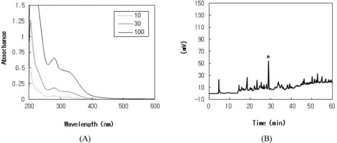
Figure 1. Absorption spectra and fingerprint HPLC of BCTE. In A, the UV absorption spectra of BCTE in PBS were measured at the indicated concentrations (µg/mL). In B, a typical HPLC chromatogram of BCTE monitored at 280 nm is shown. *The peak of p-CA was assigned by cochromatography with the authentic standard.
(A) (B)
Figure 2. Effects of BCTE on the viability of HaCaT keratinocytes in the absence and presence of UVB irradiation. In A, Cells were treated with BCTE at the indicated concentrations for 24 h and cell viabilities were determined. In B, cells were irradiated with 10 mJ/cm2 UVB in PBS in the absence and presence of BCTE (10, 30, or 100 µg/mL) or p-CA (10 µg/mL). Irradiated cells were incubated for 24 h and cell viabilities were determined. Data are presented as percentages of the unirradiated control (means ± SEs; n = 3). #p < 0.05 versus the unirradiated control. *p < 0.05 versus the irradiated vehicle control.
was calculated using the comparative threshold cycle method.
2.12. Statistical analysis
Data are presented as the means ± SEs of three or more independent experiments. The significances of dif- ferences between groups were determined using the Student’s t-test, at p values < 0.05.
3. Results
The UV absorption spectra of BCTE at different con-
centrations are shown in Fig. 1A. Typical HPLC pattern
of BCTE is shown in Fig. 1B. Because p-CA has been
reported as a major phenolic compound contained in
many bamboo species such as Phyllostachys pubescens
(Pradelle) Mazel ex J. Houz [30] and Sasa quelpaertensis
Nakai[31], we analyzed the content of p-CA in BCTE
using HPLC and it was determined to be 1.7%.
(A) (B)
Figure. 3. Effects of BCTE on the apoptotic pathway in HaCaT cells. HaCaT cells were irradiated with UVB (10 mJ/cm2) in the absence and presence of BCTE (10, 30, or 100 µg/mL) or p-CA (10 µg/mL), followed by incubation for 24 h. The whole cell lysates were subjected to western blot analysis for procaspase 3 and its cleaved form (A), and Bax and Bcl-2(B). Data are expressed as fold changes (means± SEs; n = 3). #p < 0.05 versus the unirradiated control. *p < 0.05 versus the irradiated vehicle control.
The cytotoxicity of BCTE was determined in cultured HaCaT human keratinocytes. As shown in Fig. 2A, BCTE was nontoxic up to 100 µg/mL whereas it showed significant cytotoxicity at 300 and 1000 µg/mL. The pro- tective effects of BCTE against UVB-induced cell death were examined at non-toxic concentrations. As shown in Fig. 2B, BCTE enhanced the viabilities of UVB-exposed cells at 100 µg/mL. p-CA was used as a positive control and it exhibited a protective effect at 10 µg/mL.
Occurrence of apoptosis was monitored by the cleav- age of procaspase 3 and the increase of pro-apoptotic Bax relative to anti-apoptotic Bcl-2 protein. As shown in Fig. 3A. UVB increased the cleavage of procaspase 3 to its active form, and this change was attenuated by BCTE as well as by p-CA. The relative ratios of Bax to Bcl-2 increased in UVB-irradiated cells and the change was at- tenuated by either BCTE or p-CA (Fig. 3B).
The effects of BCTE on the UVB-induced ROS gen- eration and lipid peroxidation were determined in cells exposed to UVB. As shown in Fig. 4A, UVB exposure increased ROS generation, and this increase was attenu- ated by BCTE at 100 µg/mL. p-CA at 10 µg/mL also at-
tenuated the cellular ROS generation due to UVB exposure. We determined the levels of TBARS to mon- itor lipid peroxidation[29]. The results showed that lipid peroxidation increased in HaCaT cells exposed to UVB and this change was attenuated by BCTE and by p-CA (Fig. 4B).
The free radical scavenging activities of BCTE were determined in vitro against ATBS and DPPH radicals.
Ascorbic acid and p-CA were used as positive controls.
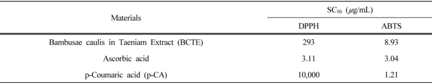
As shown in Table 1, ascorbic acid showed strongest scavenging activity against DPPH radial, while p-CA was the least active. Crude BCTE appeared to scavenge DPPH radical more effectively than p-CA. In the case of ABTS radical, p-CA showed the strongest free radical scavenging activity, followed by ascorbic acid and BCTE, in the order.
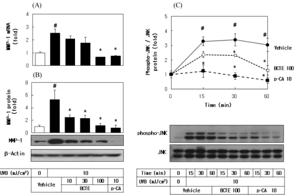
Skin photoaging involves changes in dermal ex-
tracellular matrix composition and collagen loss due to
overexpression of MMPs via mitogen-activated protein
(MAP) kinase-dependent pathways in response to UVB
exposure[12-15]. As shown in Fig. 5A and B, UVB ex-
posure increased MMP1 expression at the mRNA and
(A) (B)
Figure 4. Effects of BCTE on UVB-induced ROS generation and lipid peroxidation in HaCaT cells. HaCaT cells were treated with 1.0 µM DHR123 and irradiated with UVB (10 mJ/cm2)in the absence and presence of BCTE (10, 30, or 100 µg/mL) or p-CA (10µg/mL). After 24h, fluorescence and optical images of cells were captured and then cells were harvested to extract oxidized rhodamine 123. In B, cells were irradiated with UVB (10 mJ/cm2) in the absence and presence of BCTE (10, 30, or 100 µg/mL) or p-CA (10 µg/mL), followed by incubation for 24 h. Lipid peroxidation was determined by quantifying TBARS.
Data are expressed as fold changes versus the vehicle control (means ± SEs; n = 3). #p < 0.05 versus the unirradiated control.
*p < 0.05 versus the irradiated vehicle control.
Materials
SC50 (µg/mL)
DPPH ABTS
Bambusae caulis in Taeniam Extract (BCTE) 293 8.93
Ascorbic acid 3.11 3.04
p-Coumaric acid (p-CA) 10,000 1.21
Data are presented as SC50 which is the concentration of a sample showing 50% free radical scavenging activity.
Table 1. Free Radical Scavenging Effects of BCTE, p-CA and Ascorbic acid against ABTS and DPPH Free Radicals
protein levels in cells and these changes were attenuated by BCTE and by p-CA. As also shown in Fig. 5C, UVB stimulated phosphorylation of JNK in a time-dependent manner and this change were attenuated by BCTE and p-CA.
4. Discussion
Plant extracts enriched with secondary metabolites which can provide UVB-screening and anti-oxidative ef- fects are an attractive prospect as cosmeceuticals. The
present study demonstrated that BCTE attenuated the UVB-induced cytotoxicity and oxidative stress in cul- tured keratinocytes (Fig. 2 and 4), suggesting its potential use as a cosmeceutical for the photoprotection of the skin.
BCTE was shown to attenuate the UVB-induced apop- totic events such as the cleavage of procaspase 3 to its active form, and the increase of Bax relative to Bcl-2 (Fig. 3). BCTE might have suppressed the apoptotic pathway mainly because of its ability absorbing UV (Fig.
1). Additionally, the extract might have scavenged ROS
(A)
(B)
(C)
Figure 5. Effects of BCTE on UVB-induced MMP1 expression and JNK phosphorylation in HaCaT cells. (A), HaCaT cells were irradiated with UVB (10 mJ/cm2)in the absence and presence of BCTE (10, 30, or 100 µg/mL) or p-CA (10 µg/mL), followed by incubation for 24 h. The MMP1 mRNA levels were determined by qRT-PCR analysis using GAPDH as a control, and MMP1 protein levels were determined by Western blot analysis of whole cell lysates, using β-actin as a control. (B), HaCaT cells were irradiated with 10 mJ/cm2 UVB in the absence and presence of BCTE (100 µg/mL) or p-CA (10 µg/mL), and then incubated for the specified time. Total and phosphorylated forms of JNK were analyzed by western blot analysis. Data are expressed as fold changes (means ± SEs; n = 3). #p < 0.05 versus the unirradiated control. *p < 0.05 versus the irradiated vehicle control.
and free radicals implicated in apoptotic signal trans- duction (Table 1, Fig. 4).
Like the cases of other bamboo species[30,31], p-CA was assumed to contribute to bioactivities of BCTE. The protective effects of p-CA against the UV-induced in- flammatory responses or injury has been previously ob- served in cells and in vivo models[32-34]. We suppose that p-CA is one of the active constituents of BCTE based on the observations in the current study, but other studies reported that BCTE contained various other con- stituents with antioxidant properties[16,17]. Thus, the ac- tivity of BCTE can be attributed to the multiple con- stituents rather than any single compound. Of interest, BCTE scavenged DPPH free radicals more effectively than p-CA (Table 1), indicating that other constituents al-
so could contribute to the antioxidative effects of BCTE.
Both BCTE and p-CA were more reactive to ABTS radi- cal than to DPPH radical whereas ascorbic acid showed similar reactivity to both free radicals (Table 1). Further studies are needed to understand this matter.
MMPs regulate the turnover of extracellular matrix
such as type I collagen[12,13]. UV exposure elevates the
expression of MMPs in skin cells and triggers skin tissue
remodeling, resulting in the formation of wrinkles and
other phenotypes of skin aging[14,15,35]. Thus, the sup-
pression of UV-induced MMPs expression is pursued as
a strategy to reduce the photoaging process. In the cur-
rent study, UVB exposure increased MMP1 mRNA and
protein levels in cells and these changes were attenuated
by BCTE and by p-CA (Fig. 5), suggesting the useful-
Figure 6. A summary; BCTE attenuates the UVB-induced biochemical events associated with skin photoaging.