Protective Effects of Portulaca oleracea L. Extract against Matrix
Metalloproteinase Production and Reactive Oxygen Species Generation Induced by Ultraviolet B Radiation in Human Keratinocytes
Jung Hwan Oh
1, Fatih Karadeniz
2, Jung Im Lee
3, So Young Park
1, Youngwan Seo
3,4and Chang-Suk Kong
1,2*
1Department of Food and Nutrition, College of Medical and Life Sciences, Silla University, Busan 46958, Korea
2Marine Biotechnology Center for Pharmaceuticals and Foods, College of Medical and Life Sciences, Silla University, Busan 46958, Korea
3Division of Marine Bioscience, Korea Maritime and Ocean University, Busan 49112, Korea
4Ocean Science and Technology School, Korea Maritime and Ocean University, Busan 49112, Korea Received April 13, 2018 /Revised May 15, 2018 /Accepted May 17, 2018
Portulaca oleracea L. is an edible plant widely consumed in daily diet throughout Europe, Asia and America. In this study, protective effects of P. oleracea L. extracts against oxidative stress and matrix metalloproteinase (MMP) activity induced by ultraviolet B (UVB) radiation were investigated using HaCaT immortal human keratinocytes. In this context, the mRNA and protein productions of MMPs (MMP-1, -2, and -9) and type I procollagen, which are major markers of photoaging induced by UVB radiation in HaCaT keratinocytes, were evaluated. Furthermore, UVB-induced reactive oxygen species (ROS) generation and mRNA and protein expression levels of superoxide dismutase-1 (SOD-1), oxy- genase-1 (OH-1), and nuclear factor-erythroid 2-related factor-2 (Nrf-2), all of which are associated with the antioxidant balance, were investigated. As shown by the results, UVB radiation induced ROS formation and led to increased production of MMPs and decreased collagen production in human ker- atinocytes, which resulted in skin photoaging or photodamage. The treatment with P. oleracea L. ex- tracts downregulated MMP (MMP-1, -2, and -9) production and upregulated type I procollagen ex- pression in UVB-induced HaCaT cells. Furthermore, treatment with the extracts decreased UVB-in- duced ROS generation and increased the expression of antioxidant enzymes, such as SOD-1 and OH-1, through the Nrf-2 pathway. Taken together, these results suggest that P. oleracea L. extracts could be a potential cosmeceutical agent for the prevention of skin photoaging or photodamage.
Key words : Antioxidant activity, human keratinocytes, matrix metalloproteinase, Portulaca oleracea L.
extracts, ultraviolet B (UVB)
*Corresponding author
*Tel : +82-51-999-5429, Fax : +82-51-999-5457
*E-mail : [email protected]
This is an Open-Access article distributed under the terms of the Creative Commons Attribution Non-Commercial License (http://creativecommons.org/licenses/by-nc/3.0) which permits unrestricted non-commercial use, distribution, and reproduction in any medium, provided the original work is properly cited.
Journal of Life Science 2018 Vol. 28. No. 8. 892~899 DOI : https://doi.org/10.5352/JLS.2018.28.8.892
Introduction
Skin aging is classified into two types, intrinsic and ex- trinsic aging. Intrinsic or chronological aging is caused by the passage of time. Extrinsic aging result from ultraviolet irradiation, which is frequently referred to as photoaging.
Photoaging is characterized by sagging, thickness, roughness and deep coarse wrinkles, which is a result of decomposed extracellular matrix (ECM). ECM proteins, including colla- gen, elastin proteoglycans and fibronectin, act as conferring strength and resiliency of the human skin [30].
The solar ultraviolet (UV) radiation can be divided into three segments based on the wavelengths: long wave (UVA;
320-400 nm), mid-wave (UVB; 280-320 nm) and short-wave (UVC; 200-280 nm). Especially UVB radiation can penetrate into the epidermis of skin and induce acute and chronic pho- todamages [18, 21].
Matrix metalloproteinases (MMPs) are an important fam- ily of zinc-dependent endoproteinases that have the capacity to cleave the ECM proteins. MMPs are overexpressed by UV irradiation and oxidative stress, resulting in a decrease of collagen in human skin. Therefore, inhibition of MMP ex- pression is important to prevent photoaging [26].
Reactive oxygen species (ROS) are a main factor in photo-
damaged skin by UV-irradiation. The higher concentrations
of ROS produced through UV exposure can cause an im-
balance in antioxidant defense mechanism, leading to oxida-
tive stress [18]. ROS play a important role in collagen metab-
olism and increased ROS generation leads to the enhance
of MMPs expression, resulting in a decrease of collagen in photoaging skin [30]. Thus, ROS detoxifying enzymes and antioxidants are important factors to prevent photogaing or photodamage in ROS defense mechanism.
P. oleracea L. belongs to the family of Portulacacea and is widely distributed in temperate and tropical regions [25].
P. oleracea L. is rich nutrition with contents of ω-3 and ω-6 fatty acids, linolenic acid, tocopherol, ascorbic acid [22] and dietary minerals such as K, Mg, Ca, and Fe [29]. Also, it has been used as herbal medicine because of its many health functions, such as antioxidant [4, 15], anti-inflammatory [14]
and anti-bacterial [13] activities. Nevertheless, the effect of P. oleracea L. extracts on photoaging or photodamage in rela- tion to MMP expression has not been well established.
Therefore, the purpose of this study is to evaluate the capa- bility of P. oleracea L. extracts to prevent human keratino- cytes against UVB-induced photodamage via suppressing the formation of ROS and the expression of MMPs.
Materials and Methods
Plant Materials
P. oleracea L. was purchased from Hyosung Food, Inc.
(Hongcheon, Korea). Whole plant of P. oleracea L. was dried in the shade and ground to fine powder. The dried and ground samples (100 g) were extracted for 48 hr with CH
2Cl
2and CH
3OH at room temperature, combined and con- centrated to dryness in a rotary evaporator, which yield crude extracts (57.3 g).
Cell culture
A human keratinocytes cell line (HaCaT, ATCC) was grown in Dulbecco's modified Eagle medium (DMEM) con- taining 10% fetal bovine serum, 100 units/ml of penicillin/
streptomycin in a humidified atmosphere with 5% CO
2at 37℃. For sub-culturing HaCaT cells, the medium was re- moved, and cells were washed with phosphate-buffered sal- ine (PBS) and detached with trypsin-EDTA. The cells were fed fresh medium every 2-3 days.
UVB Irradiation
HaCaT cells were grown in 12-well plates for 24 hr. After 24 hr, the cells were washed with PBS and exposed to 15 mJ/cm
2UVB (315 nm UVB light source, Bio-Sun lamp, Vilber Lourmat, Marine, France). After UVB irradiation, the cells were incubated with or without P. oleracea L. extracts for 24 hr.
Cell viability
Cytotoxic levels of crude extracts from P. oleracea L. on cultured cells were measured using a MTT (3-(4,5-dime- thylthiazol-2-yl)-2,5-diphenyltetrazolium bromide) assay.
The cells were grown in 96-well plates at a density of 5×10
3cells/well. After 24 hr, the cells were treated with crude ex- tracts from P. oleracea L. at the concentrations of 10, 50 and 100 μg/ml. After incubation for 24 hr, MTT (1 mg/ml) re- agent was added to each well and then the cells were in- cubated for additional 4 hr. Finally, the media were removed and the formed formazan crystals were dissolved in 100 μl of dimethyl sulfoxide. Absorbance values were measured at 540 nm using a microplate reader (Teacan Group Ltd., Mannedorf, Swiss).
Quantitation of Intracellular ROS
Generation of intracellular ROS in the human keratino- cytes was detected by the 2',7'-dichlorofluorescein diacetate (DCFH-DA) assay. The cells were seeded at 5×10
3cells in black 96-well plates. After 24 hr, the cells were loaded with 20 μM DCFH-DA in PBS and incubated for 20 min in the dark. The cells were treated with from P. oleracea L. extracts (10, 50, 100 μg/ml) or retinoic acid (1 μM) as positive control for 1 hr. After rinsing the cells with PBS, 15 mJ/cm
2UVB was applied to produce ROS. The fluorescence of 2',7'-di- chlorofluorescein (DCF) was detected at the excitation wave- length of 495 nm and the emission wavelength of 620 nm every 30 min using an Infinite F200 Pro fluorescence micro- plate reader (Teacan Group Ltd., Mannedorf, Swiss).
RNA isolation and RT-PCR (reverse-transcription polymerase chain reaction)
Total RNA was isolated from HaCaT cells treated with or without P. oleracea L. extracts using Trizol reagent (Invi- trogen, CA, USA). The RNA concentration and purity were examined by measuring the absorbance at 260 and 280 nm.
The absorbance 260/280 values ranged from 1.8 to 2.0 for all extracted RNA indicated little protein contamination. For the synthesis of cDNA, the reaction of reverse-transcription was carried out using an oligo (dT) and 2 μg total RNA.
RT-PCR was performed using an automatic T100 Thermo
Cycler (Bio-rad, CA, USA). The reaction steps followed: de-
naturation for 2 min at 94℃ followed by 40 cycles consisting
of denaturation for 2 min at 95℃, annealing for 2 min at
60℃ and extension for 2 min at 72℃. The PCR products
were separated by electrophoresis (Mupid CO., Ltd., Tokyo,
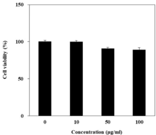
Fig. 1. Effect of P. oleracea L. extracts with different concen- trations on viability of HaCaT cells.
Japan) on 1.5% agarose gel for 10 min at 100 V. Gels vi- sualized with UV light using Davinch-Chemi imager
TM(Davinch-K, Seoul, Korea). The sequences of specific oligo- nucleotide primers are as followings: sense 5'-AGGGCATCA TCAATTTCGAG-3' and antisense 5'-TGCCTCTCTTCATCC TTTGG-3' for SOD-1; sense 5'-ACATCTATGTGGCCCTGG AG-3' and antisense 5'-CGCTTCACATAGTGCTGCAT-3' for HO-1; sense 5'-GCGACGGAAAGAGTATGAGC-3' and anti- sense 5'-GTTGGCAGATCCACTGGTTT-3' for Nrf-2; sense 5'-GATGTGGAGTGCCTGATGTG-3' and antisense 5'-TGCT TGACCCTCAGAGACCT-3' for MMP-1; sense 5'-ATGGCA AGTACGGCTTCTGT-3' and antisense 5'-ATACTTCTTGTC GCGGTCGT-3' for MMP-2; sense 5'-CTCGAACTTTGACA GCGACA-3' and antisense 5'-GCCATTCACGTCGTCCTTAT- 3' for MMP-9; sense 5'-CTCGAGGTGGACACCACCCT-3' and antisense 5'-CAGCTGGATGGCCACATCGG-3' for Type
Ⅰ procollagen; sense 5'-AGCCATGTACGTAGCCATCC-3' and antisense 5'-TCCCTCTCAGCTGTGGTGGT-3' for β-actin.
Western blot
Protein expression levels were analyzed by Western blot analysis. Whole cells were lysed with RIPA (Sigma-Aldrich, MO, USA) buffer. After centrifugation, total protein quantifi- cation was estimated using the BCA protein assay kit (Thermo scientific, MA, USA). An aliquot of supernatant containing equal amounts of proteins was separated by 10%
SDS-PAGE and transferred onto the polyvinylidene fluoride membrane (Amersham Pharmacia Biotech, England, UK).
The membrane was blocked with 5% skim milk for 1 hr and incubated overnight with primary antibodies such as SOD-1, HO-1, Nrf-2, MMP-1, MMP-2, MMP-9, Type Ⅰ procollagen (Cell Signaling Technology Inc., MA, USA) and β-actin (Santa Cruz Biotechnology Inc., TX, USA). After washing with TBS plus Tween 20 (TBS-T) buffer, horseradish perox- idase-conjugated secondary antibody was added and in- cubated at room temperature for 1 hr. The protein band was visualized on a Davinch-Chemi imager
TM(Davinch-K, Seoul, Korea) and activated by chemiluminescence using ECL kit (GE healthcare, Little Chalfont, UK).
Statistical analysis
Results were presented as mean ± standard division of triplicate experiments. Statistical differences between in- dividual groups were analyzed by one-way analysis of var- iance (ANOVA) using SPSS version 12.0 (SPSS Inc., IL, USA).
The significance of differences was determined at the p<0.05
level.
Results and Discussion
Cell viability of extracts
The cytotoxic effect of P. oleracea L. extracts in HaCaT cells was evaluated at various concentrations for 24 hr by MTT assay. As shown in Fig. 1, P. olerace L. extracts did not show any cytotoxicity up to 100 μg/ml (Fig. 1). Therefore, all fol- lowed experiments were conducted within a concentration range up to 100 μg/ml.
Effect on UVB-induced MMP expression
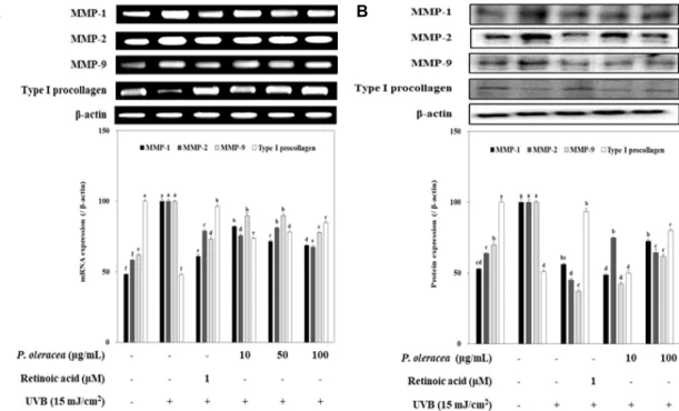
Exposure of skin to UV irradiation causes human skin photoaging, which is related to increased matrix metal- loproteinases (MMPs). There are more than 25 types of MMPs divided into collagenases, gelatinases, stromelysins, membrane type MMP and other non-classified MMP accord- ing to their substrate and function [7]. MMPs are initiates cleavage of collagen, which is the critical structural compo- nent of the ECM and released from keratinocytes and dermal fibroblasts by photoaging stress. Among them, MMP-1 (collagenase), MMP-2 (gelatinase A) and MMP-9 (gelatinase B) are mainly responsible for collagen degradation in in pho- toaging skin [19, 26]. Therefore, the effect of P. oleracea L.
extracts on MMPs expression induced by UV radiation in HaCaT cells was investigated. A significantly increased ex- pression of MMPs was observed in the cells radiated with the UVB compare to the non-UVB irradiated blank cells.
RT-PCR analysis showed that presence of P. oleracea L. ex-
A B
A B
Fig. 2. Effect of P. oleracea L. extracts on MMPs expression in HaCaT cells exposed to 15 mJ/cm2 of UVB. Cells were exposed to 15 mJ/cm2 of UVB irradiation and treated with different concentrations of P. oleracea L. extracts for 24 hr. The expression levels of these MMPs were detected using RT-PCR (A) and western blot (B) analysis. β-actin was used as an internal standard.
a-fMeans with different letters are significantly different (p<0.05) by Duncan's multiple range test. Values are means ± SD (n=3).
tracts inhibits UV-induced elevation of MMP-1, MMP-2 and MMP-9 mRNA levels, whereas treatment of P. oleracea L. ex- tracts promoted type Ⅰ procollagen mRNA levels in UVB- induced cells (Fig. 2A). In parallel with the mRNA ex- pression, P. oleracea L. extracts down-regulated the induction of MMP-1, MMP-2 and MMP-9 protein and up-regulated type Ⅰ procollagen protein in UVB-induced HaCaT cells (Fig. 2B). In these results predicted that the higher potential of P. oleracea L. extracts are due to presence of flavonoid and polyphenolic compounds [4, 31], which demonstrated by a previous study showing the key role of its hydroxyl groups in MMPs down-regulation and type Ⅰ procollagen up-regulation [2, 23]. In performed assay, P. oleracea L. ex- tracts confirmed to be a potentially effective extracts against photoaging, and thus processed for investigation of intra- cellular ROS levels and the antioxidant expression levels in relation to its photo-protective potential.
Effect on UVB-induced ROS generation
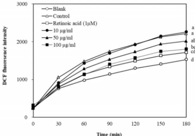
Excessive exposure to UV light generates ROS in the skin [24]. Uncontrolled generation of ROS leads to increased for- mation of MMPs and decreased formation of collagen in hu- man keratinocytes and fibroblasts. To determine the
ROS-scavenging activity of various concentrations of P. oler- acea L. extracts on cytosolic ROS production in UVB-induced HaCaT cells, the level of ROS generation was detected by the fluorescence dye DCFH-DA. As shown in Fig. 3, UVB exposure elevated the intracellular ROS levels in HaCaT cells compared to without UVB, whereas cells treated with P. oler- acea L. extracts indicated significant suppression of ROS for- mation in a concentration-dependent manner. The results are in the correlation with MMPs levels, which is linked to ROS formation. Therefore P. oleracea L. extracts have a pro- tective effect on cellular damage attributed to UVB-induced oxidative stress. These results speculated that the reduction of ROS production in the cells treated with P. oleracea L. ex- tracts can be due to the presence of antioxidant compounds such as flavonoids, polyphenols and alkaloids, which scav- enge the free radical [4, 14, 15, 31]. In support of our results, a study proved that P. oleracea L. extracts have radical scav- enging effects and inhibits lipid peroxidation [12].
Crude extracts of plant materials abundant in phenolic
content are of interest from the cosmeceutical industry be-
cause of their antiphotoaging and antioxidation activities by
blocking the activation of MMPs. Several studies have re-
ported that flavonoid-riched crude extracts of Agastache ru-
Fig. 3. Effect of P. oleracea L. extracts on intracellular ROS gen- eration induced by UVB irradiation. Cells were treated with DCFH-DA and exposed 15 mJ/cm2 of UVB irradi- ation. ROS levels were measured by fluorescence in- tensity of DCF-DA after extracts treatment with various concentrations in a time-dependent manner. a-dMeans with different letters are significantly different (p<0.05) by Duncan's multiple range test. Values are means ± SD (n=3).
gose leaf [21] and Louicerae japonicae flos [28] have capabilities to prevent human skin cells against UVB-induced photo- aging by suppressing the formation of ROS. Additionally, the polyphenolic compound, epicatechin-3-gallate (ECG), in green tea protects keratinocytes against UVB-induced photo skindamage [8].
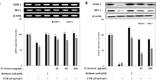
Effect on antioxidant enzyme expression
An enzymatic antioxidant defense system is important for the protection of the skin from UV exposed oxidative stress [3, 9]. To elucidate the intracellular radical scavenging effect of P. oleracea L. extracts in detail, the expression levels of antioxidant enzymes were measured by RT-PCR and immunoblotting. Presence of P. oleracea L. extracts remark- ably increased not only the mRNA expression of SOD-1 and HO-1 but also the protein production of SOD-1 and HO-1 compared to that of the UVB-induced control group of HaCaT cells (Fig. 4A, Fig. 4B). Taken together, P. oleracea L. extracts are able to increase the reduction of SOD-1 and HO-1 by UVB exposure, implying that the SOD-1 and HO-1- enhancing effect of P. oleracea L. extracts might contribute to these reducing activities on the UVB-induced ROS for- mation and MMPs expression.
UV exposure to the human skin results in the generation of ROS. ROS are constantly produced in UV irradiated kera- tinocytes and fibroblast, and are rapidly eliminated by non-
enzymatic and enzymatic antioxidant substances. Non-enzy- matic antioxidants such as vitamin C, plant polyphenols and flavonoids play a critical role in interrupting free radical chain reactions. Enzymatic antioxidant enzymes play a role in breaking down and eliminating free radical. The anti- oxidant enzymes such as SOD, catalase and glutathione, con- vert the superoxide anion to oxygen and hydrogen peroxide and then to water [20]. Among these enzymes, SOD-1 is a key endogenous enzyme that catalyzes the dismuation of superoxide radicals into hydrogen peroxide [6]. HO-1 is a key antioxidant defense enzyme that catalyzes the degrada- tion of the heme to biliverdin, carbon monoxide and free iron, which act as potentially biologically active molecules and relate to antioxidant defenses [10, 17].
Effect on Nrf-2 expression
Transcription factor Nrf-2 has shown to play an important role in the cellular ROS defense mechanism by inducing ex- pression of a variety of ROS neutralizing enzymes and anti- oxidants [27]. Therefore, the Nrf-2 level in P. oleracea L. ex- tracts mediated cytoprotection effects against photo-oxida- tive stress was investigated. The mRNA and protein ex- pressions of Nrf-2 were significantly down-regulated in UVB-induced HaCaT cells. However, presence of P. oleracea L. extracts remarkably up-regulated Nrf-2 expression com- pared to that of the only UVB irradiated control group (Fig.
5A, Fig. 5B). These results implicated that P. oleracea L. ex- tracts enhanced SOD-1 and HO-1 expression by stimulating of the nuclear translocation of Nrf-2.
Nrf-2 plays a role as a sensor for electrophilic and oxida- tive stress [1]. In normal physiological condition, Nrf-2 is in the cytosol before being ubiquitinated and cleavaged by Kelch like ECH-associated protein (keap 1). In oxidative stress condition, the electrophiles and ROS bind to residues of keap1. As a result, Nrf-2 released from keap1 and trans- located to the nucleus, where it heterodimerizes with MAF proteins, to conjugate the antioxidant responsive element (ARE) in the promoter region of antioxidant genes such as SOD-1 and HO-1 [16]. Some research has shown that the photo-protective effect of active compounds, sargachrome- nol [5] and youngiasides [11] by activation of Nrf-2 and anti- oxidant enzyme namely SOD-1 and HO-1.
In conclusion, the present study confirmed that protective
effect of P.oleracea L. extracts against UVB damage in kerati-
nocytes via down-regulating MMP-1, -2, -9 expression and
up-regulating type Ⅰ procollagen. Furthermore, P.oleracea L.
A B
Fig. 4. Effect of P. oleracea L. extracts on antioxidant enzyme expression in HaCaT cells exposed to 15 mJ/cm2 of UVB. Cells were exposed to 15 mJ/cm2 of UVB irradiation and treated with different concentrations of P. oleracea L. extracts for 24 hr. The expression levels of these antioxidant enzymes were detected using RT-PCR (A) and Western blot (B) analysis. β-actin was used as an internal standard. a-eMeans with different letters are significantly different (p<0.05) by Duncan's multiple range test. Values are means ± SD (n=3).
A B
Fig. 5. Effect of P. oleracea L. extracts on Nrf-2 expression in HaCaT cells exposed to 15 mJ/cm2 of UVB. Cells were exposed to 15 mJ/cm2 of UVB irradiation and treated with different concentrations of P. oleracea L. extracts for 24 hr. The expression levels of Nrf-2 were detected using RT-PCR (A) and Western blot (B) analysis. β-actin was used as an internal standard.
a-dMeans with different letters are significantly different (p<0.05) by Duncan's multiple range test. Values are means ± SD (n=3).
extracts suppressed the generation of ROS and elevated lev- els of the antioxidant enzymes such as SOD-1 and OH-1 through the Nrf-2 pathway. Therefore, our results suggested that P. oleracea L. extracts possess photoaging inhibiting com- ponents, which may be used as a potential source for cosme- ceutical agents. More work should be encouraged to isolate
the active compounds from P. oleracea L. extracts and its fractions.
Acknowledgement
This research was supported by Basic Science Research
Program through the National Research Foundation of Korea (NRF) funded by the Ministry of Education (2017009588).
Also, this work was supported by the BB21+ project in 2018.
References
1. Cesar, L. C., Elena, A. C., Mavil, L. C., Carlos, P. P., Elizebeth, A. S. and Laurence, A. M. 2012. Protein kinase and tran- scripition factors activation in response to UV-radiation of skin: Implications for carcinogenesis. Int. J. Mol. Med. Sci.
13, 142-172.
2. Cho, Y. H., Bahuguna, A., Kim, H. H., Kim, D. I., Kim, H.
J., Yu, J. M., Jung, H. G., Jang, J. Y., Kwak, J. H., Park, G.
H., Kown, O. J., Cho, Y. J., An, J. Y., Jo C. R., Kang, S. C.
and An, B. J. 2017. Potential effect of compounds isolated from Coffea arabica against UV-B induced skin damage by protecting fibroblast cells. J. Photochem. Photobiol. B. 174, 323- 332.
3. Diego, S. C., Natalia, P., Carla, Z., Pagono, E., Karina, B.
and Gustavo, Y. 2017. Heme oxygenase up-regulation under ultraviolet-B radiation in not epigenetically restricted and involves specific stress-related transcriptions factors. Redox Biol. 12, 549-557.
4. Erkan, N. 2012. Antioxidant activity and phenolic com- pounds of fractions from Portulaca oleracea L. Food Chem. 133, 775-781.
5. Fernando, P. M. D. J., Piao, M. J., Hewage, S. R. K. M., Kang, H. K., Yoo, E. S., Koh, Y. S., Ko, M. H., Ko, C. S., Byeon, S. H., Mun, S. R., Lee, N. H. and Hyun, J. M. 2016.
Photo-protective effect of sargachromenol against UVB radi- ation-induced damage through modulating cellular anti- oxidant systems and apoptosis in human kerinocyte.
Environ. Toxicol. Pharmacol. 43, 112-119.
6. Fridovich, I. 1978. The biology of oxygen radicals. Science 201, 875-880.
7. Herouy, Y. 2004. The role of matrix metalloproteinases (MMPs) and their inhibitor in venous leg ulcer healing.
Phlebolymphology 44, 231-243.
8. Huang, C. C., Wu, W. B., Fang, J. Y., Chiang, H. S., Chen, S. K., Chen, B. H., Chen, Y. T. and Hung, C. F. 2007.
(-)-Epicatechin-3-gallate, a green tea polyphenol is a potent agent against UVB-induced damage in HaCaT keratinocytes.
Molecules 12, 1845-1858.
9. Jang, J. Y., Ye, B. R., Heo, S. J., Oh, C. H., Kang, D. H., Kim, J. H., Affan, A., Yoon, K. T., Choi, Y. U., Park, S. C., Han, S. H., Qian, Z. J., Jung, W. K. and Choi, I. W. 2012.
Photo-oxidative stress by ultraviolet-B radiation and anti- oxidative defense of eckstolonol in human keratinocytes.
Environ. Toxicol. Pharmacol. 34, 926-934.
10. Kikuchi, G., Yoshida, T. and Noguchi, M. 2005. Heme oxy- genase and heme degradation. Biochem. Biophys. Res. Com- mun. 338, 558-567.
11. Kim, M. S., Park, Y. G., Lee, H. J., Lim, S. J. and Nho, C.
W. 2015. Youngiasides A and c isolated from Younia dentic- ulatum inhibit UVB-induced MMP expression and promote
type Ⅰ procollagen production via repression of MAPK/
AP-1/NF-κB and activation of AMPK/Nrf2 in HaCaT cells and human dermal fibroblast. J. Agric. Food Chem. 63, 5428- 5438.
12. Lee, H. J., Lee, J. B., Lee, D. S. and Seo, Y. W. 2003. DPPH radical scavenging effect and in vitro lipid peroxidation in- hibition by Portulaca oleracea. Kor. J. Biotechnol. Bioeng. 18, 165-169.
13. Lei, X., Li,. J. Liu, B., Zhang, N. and Liu, H. 2015. Separation and identification of four new compounds with antibacterial activity from Portulaca oleracea L. Molecules 20, 16375-16387.
14. Li, C. Y., Meng, Y. H., Ying, Z. M., Xu, N., Hao, D., Gao, M. Z., Zhang, W. J., Xu, L., Gao, Y. C. and Ying, X. X. 2016.
Three novel alkaloids from Portulaca oleracea L. and their anti-inflammatory effects. J. Agric. Food Chem. 64, 5837-5844.
15. Lim, Y. Y. and Quah, E. P. L. 2007. Antioxidant properties of different cultivars of Portulaca oleracea. Food Chem. 103, 737-740.
16. Liu, C., Vojnovic, D., Kochevar, I. E. and Jurkunas, U. V.
2016. UV-A irradiation activates Nrf2-regulated antioxidant defense and induced p53/caspase3-dependent apoptosis in corneal endothelial cells. Invest. Ophthalmol. Vis. Sci. 57, 2319-2327.
17. Marines, M. D. 1997. The heme oxygenase system: A regu- lator of second messenger gases. Annu. Rev. Pharmacol. Toxi- col. 37, 517-554.
18. Matsumura, Y. and Ananthaswamy, H. N. 2004. Toxic ef- fects of ultraviolet radiation on the skin. Toxicol. Appl.
Pharmacol. 195, 298-308.
19. Nagase, H., Visse, R. and Murphy, G. 2006. Structure and function of matrix metalloproteinases and TIMPs. Cardiovasc.
Res. 69, 562-573.
20. Nimse, S. B. and Pal, D. 2015. Free radicals, natural anti- oxidants, and their reaction mechanisms. Mol. Omics 5, 27986-28006.
21. Oh, Y. R., Lim, H. W., Huang, Y. H., Kwon, H. S., Jin, C.
D., Kim, K. H. and Lim, C. J. 2016. Attenuating properties of Agastache rugose leaf extract against ultraviolet-B-induced photoaging via up-regulating glutathione and superoxide dismuatase in a human keratinocyte cell line. J. Photochem.
Photobiol. B. 163, 170-176.
22. Palaniswamy, U. R., McAvoy, R. J. and Bible, B. B. 2001.
Stage of harvest and polyunsaturated essential fatty acid concentrations in Purslane (Portulaca oleracea) leaves. J.
Agric. Food Chem. 49, 3490-3493.
23. Park, E. K., Ahn, S. R., Kim, D. H., Lee, E. W., Kwon, H.
J., Kim, B. W. and Kim, T. H. 2014. Effects of unripen apple polyphenols on the expression of matrix metaproteinase-1 and type-1 procollagen in ultraviolet irradiated human skin fibroblast. J. Kor. Soc. Appl. Biol. Chem. 57, 49-455.
24. Podhaisky, H. P., Riemschneider, S. and Wohlrab, W. A.
2002. UV light and oxidative damage of the skin. Pharmazie 57, 30-33.
25. Radhakrishnan, R., Zakaria, M. N. M., Islam, M. W., Chen, H. B., Kamil, M., Chan, K. and Al-Attas, A. 2001. Nurophar- macological actions of Portulaca oleracea L v.sativa (Hawk).
초록:쇠비름 추출물의 UVB 자외선 조사에 의한 인간각질형성세포 손상에 대한 보호 효과
오정환
1․파티 카라데니즈
2․이정임
3․박소영
1․서영완
3,4․공창숙
1,2*
(1신라대학교 의생명과학대학 식품영양학과, 2신라대학교 해양식의약소재융합기술연구소, 3한국해양대학교 해양
과학기술대학 해양환경·생명과학부, 4한국해양대학교 해양과학기술전문대학 해양과학기술융합학과)
쇠비름(Portulaca oleracea.L)은 쇠비름과에 속하는 한해살이풀로서 리놀렌산과 같은 불포화지방산, 페놀성 화합 물, 플라보노이드, 비타민 C, 미네랄 함량이 높은 것으로 보고되어 있다. 본 연구에서는 쇠비름 추출물을 이용하여 UVB를 조사한 인간각질형성세포에서 광노화 억제능을 확인하였다. Matrix metalloproteinases는 세포의 기질을 분해하는 효소로 MMP-1는 collagenase, MMP-2와 MMP-9는 gelatinases로 피부 진피층을 구성하는 type Ⅰ colla- gen을 분해시키는데 영향을 미친다. UVB를 조사한 인간각질형성세포에서 쇠비름 추출물을 처리했을 때 MMP-1, -2, -9의 발현이 감소하였으며, type Ⅰ procollagen의 발현은 증가하는 것으로 나타났다. 또한 쇠비름 추출물을 처리한 군에서 UV에 의한 ROS 생성이 감소하였는데 이는 Nrf-2의 활성화를 통한 항산화 인자 SOD-1과 OH-1의 발현 증가로 인해 세포내 ROS 생성이 감소한 것으로 사료된다. 따라서 본 연구 결과를 통해 쇠비름 추출물이 UVB를 조사한 인간각질형성세포에서 MMP 인자 및 항산화 인자의 발현 조절을 통해 광노화로부터의 세포 보호 능을 가지는 것을 확인하였으며 나아가 화장품 소재로서의 개발 가능성을 확인하였다.
J. Ethnopharmacol. 76, 171-176.
26. Rittie, L. and Fisher, G. J. 2002. UV-light-induced signal cas- cades and skin aging. Ageing Res. Rev. 1, 705-720.
27. Schafer, M., Dutsch, S., Keller, U. A. D. and Werner, S. 2010.
A central regulator of UV protection in the epidermis. Cell Cycle 9, 2917-2918.
28. Seo, S. H., Bae. G. S., Choi, S. B., Jo, I. J., Kim, D. G., Shin, J. Y., Song, H. J., Park, S. J. and Choi, M. O. 2014. The anti- oxidative and cytoprotective effect of Lonicerae japonicae flots water extracts on the ultraviolet (UV)B-induced human HaCaT keratinocytes. Kor. J. Herbology 29, 63-71.
29. Uddin, M. K., Juraimi, A. S., Ali, M. E. and Ismail, M. R.
2012. Evaluation of antioxidant properties and mineral com- position of Purslane (Portulaca oleracea L.) at different growth stage. Int. J. Mol. Med. Sci. 13, 10257-10267.
30. Wenk, J., Brenneisen, P., Meewes, C., Wlaschek, M., Peters, T., Blaudschun, R., Ma, W., Kuhr, L., Schneider, L. and Kohanek, S. 2001. UV-induced oxidative stress and photo- aging. Curr. Probl. Dermatol. 29, 83-94.
31. Xu, X., Yu, L. and Chen, G. 2006. Determination of fla- vonoids in Portulaca oleracea L. by capillary electrophoresis with electrochemical detection. J. Pharm. Biomed. Anal. 41, 493-499.