Official Journal of The Korean Medicine Society For The Herbal Formula Study Available at http://www.formulastudy.comHFS
Original Article / 원저
야교등의 항산화 및 간보호효과
전창권#, 정지윤#, 박정아, 지선영, 김상찬*
대구한의대학교 한의과대학
Effect of Polygoni Multiflori Ramulus extract against arachidonic acid and iron-induced oxidative stress in HepG2 cell and CCl
4-induced liver
injury in mice
Chang Kwon Jeon#, Ji Yun Jung#, Chung A Park, Seon Young Jee, Sang Chan Kim*
College of Korean Medicine, Daegu Haany University
ABSTRACT
Objectives : Polygoni Multiflori Ramulus has been widely used as a traditional medicinal herb for the treatment of insomnia, limb pain and itch. The extract of Polygoni Multiflori Ramulus (PMRE) is known to have a modulatory effect of many inflammatory responses. This study was performed to investigate the hepatoprotective effect of PMRE against arachidonic acid (AA) + iron-induced oxidative stress on HepG2 cell and carbon tetrachloride (CCl4)-induced liver injury on mice.
Methods : The effects of PMRE on cell viability was assessed by MTT assay. And flow cytometric analysis was performed to estimate the effects on mitochondrial function. To investigate its underlying mechanism, apoptosis- related proteins were analysed by using immunoblot analysis. In addition, ICR mouse were administrated (po) with the PMRE (30, 100 mg/kg) for 3 days and then, injected (ip) with CCl4 (0.5 ml/kg body weight) to induce acute liver damage. The level of pro-caspase-3 was measured.
Results : Treatment of PMRE increased relative cell viability, prevented a cleavage of poly (ADP ribose) polymerase and pro-caspase-3, and also reduced mitochondrial membrane permeability against AA + iron-induced oxidative stress. In addition, PMRE treatment decreased liver injury induced by CCl4, as evidenced by increases in pro-caspase-3 level.
Conclusions : These results demonstrate that PMRE has an ability to anti-oxidant and hepatoprotective effect against AA + iron-induced oxidative stress and CCl4-induced liver injury.
ⓒ 2017 The Korean Medicine Society For The Herbal Formula Study
This paper is available at http://www.formulastudy.com which permits unrestricted non-commercial use, distribution, and reproduction in any medium, provided the original work is properly cited.
Key words : Polygoni Multiflori Ramulus, Oxidative stress, Hepatoprotective effect
Ⅰ. 서론4)
간은 간실질세포, 쿠퍼 (Kupffer) 세포 및 간 성상 세포 (hepatic stellate cell)를 포함하는 다양한 세포 로 구성되며1), 산화적 스트레스 (oxidative stress)를 억제하여 해독에 관여하는 중요한 기관이다2,3). 산화 적 스트레스는 손상 및 세포사멸을 포함한 세포 기능 장애에서 흔히 발생하는 기전으로 손상 및 염증의 유 도를 통한 조직손상은 활성산소종 (reactive oxygen species, ROS) 생산과 세포의 항산화 활동 사이의 불균형과 관련되어 있다4,5). 과도한 ROS 생산은 막 유동성 및 단백질 구조 변형을 통해 인지질막을 변형 시키며 이를 통해 세포 사멸을 유도한다6). 따라서 산 화적 스트레스의 과도한 증가는 급만성 간질환을 포 함한 다양한 질환을 야기하는 주요 원인으로 보고되 어 왔다7,8).
염증 매개체 역할을 하는 오메가-6 불포화 지방산 인 아라키돈산 (arachidonic acid, AA)은 증가된 칼 슘을 통한 phospholipase A2의 활성화에 의해 유리
된다9,10). AA는 과도한 ROS 생산을 통해 일차 산화
생성물인 hydroperoxide를 생산하고, 염증, 세포 이 동, 세포사멸 및 혈소판 응집의 조절에 관여한다11). 이와 더불어 iron와 AA의 병용처치시 활성산소의 생 성 및 미토콘드리아 막전위 저하를 통한 세포 독성이 가속화됨이 보고되었다12).
夜交藤 (Polygoni Multiflori Ramulus)은 마디풀 (Polygonaceae)과 식물에 속하는 何首烏 (Polygonium multiflorum Thunb.)의 덩굴 혹은 잎이 달린 덩굴을 말하며, 夜交藤 추출물은 lipopolysaccharide로 자극 된 대식세포가 생성하는 염증성 매개물인 nitric oxide와 inducible nitric oxide synthase, cyclooxygenase (COX)-2, interleukin-6, tumor necrosis factor alpha 등을 유의적으로 감소시키며, 이는 nuclear factor kappa B의 핵 내 이동 저해에서 비롯된 것으
로 보고되었다13,14). 또한, 夜交藤 추출물은 심근 보호
15), 신경 보호16), 노화 방지17), 인지증진효과를 나타 내며18), 관절염의 통증 및 피부 감염19) 등에 효과가 있음이 보고되었다. 그러나 아직까지 AA와 iron의 처치에 의해 유도된 산화적 스트레스에 대한 夜交藤 의 항산화효과 및 간세포 보호 작용에 대해서는 연구 된 바가 없었다. 따라서, 본 연구에서는 夜交藤 추출 물 (PMRE; Polygoni Multiflori Ramulus water Extract)의 간보호 효능을 AA + iron의 처치를 통해 산화적 스트레스를 유발한 HepG2 cell 독성 모델과, 사염화탄소 (carbon tetrachloride, CCl4)의 처치를 통해 간독성을 유발한 동물모델을 활용하여 항산화 및 간보호효능을 평가하였다.
Ⅱ. 재료 및 방법
1. 夜交藤추출물의 제조
夜交藤은 대원약업사 (Daegu, Korea)에서 구입하 였으며, 200 g을 2 L의 물로 3 시간 전탕하였다. 전 탕액을 거즈로 1차 여과한 후, 0.2 μm 필터 (Nalgene, New York, NY, USA)로 여과하였다. 여과된 여액을 rotary evaporator (EYELA, Tokyo, Japan)로 70℃
에서 감압 농축한 후, ultra-low temperature freezer (Nihon freezer, Japan)로 동결하고, 동결된 물추출 물을 동결건조기 (Labconco, USA)로 동결 건조하여 21.8 g의 PMRE를 얻었다 (수율; 10.9 %). 동결된 PMRE는 실험기간 동안 –20℃에 보관하였으며, in vitro에서는 농도에 맞게 배지에 녹여 사용하였고, in vivo에서는 물에 녹여 동물에 투여하였다.
2. 시약
Fetal bovine serum (FBS)와 Dulbecco's modified Eagle's medium은 Life Technologies (Gaithersburg, MD, USA)와 BioWhittaker사 (Walkersville, MD,
# These authors contributed equally to this work.
* Corresponding author:Sang Chan Kim. College of Korean Medicine, Daegu Haany University, 1, Hanuidae-ro, Gyeongsan-si, Gyeongsangbuk-do, 38610, Republic of Korea.
Tel : +82-53-819-1862, Fax : +82-53-819-1860, E-mail : [email protected]
∙ Received : May 16, 2017 / Accepted : May 24, 2017
USA)로부터 구입하였으며, streptomycin과 penicillin은 Gibco/BRL (Eggenstein, Germany)로부터 구입하였 다. AA와 rhodamine 123 (Rh123)은 Calbiochem (Santacruz, CA, USA)로부터 구입하였다. Glutathione (GSH) determination kit는 Oxis International (South Tampa, FL, USA)에서 구입하였다. Pro-caspase-3, HRP-conjugated anti-rabbit IgG, HRP-conjugated anti-mouse IgG 항체는 Cell Signaling Technology (Beverely, MA, USA)로부터 구입하였으며, poly (ADP ribose) polymerase (PARP) 항체는 Santa Cruz Biotechnology (Santa Cruz, CA, USA)에서 구입하 였다. 2′7′-dichlorofluorescein diacetate (DCFH-DA)와 ferric nitrilotriacetate acid (Fe-NTA), 3-(4,5- dimethylthiazol-2-yl)-2,5-diphenyltetrazolium bromide (MTT)와 anti-β-actin 및 다른 시약들은 Sigma (St. Louis, MO, USA)에서 구입하였다.
3. 세포배양
Human hepatocyte-derived cell line인 HepG2 cell은 American Type Culture Collection (ATCC;
Rockville, MD, USA)로부터 구입하였으며, 10% FBS와 50 mg/ml streptomycin, 50 U/ml penicillin을 포함한 Dulbecco's modified Eagle's medium 배지를 사용 하여 5%의 CO2 및 37℃가 유지되는 배양기 (Sanyo, Japan)에서 배양하였다. 실험에 사용된 모든 세포는 confluence가 80-90% 이상으로 배양하여 실험에 사 용하였다. HepG2 cell에서 PMRE의 세포보호효과를 평가하기 위하여, 배양후 FBS가 제거된 배지에서 24 시간 배양한 후, PMRE를 농도별로 처치하고 1 시간 후, 10 μM의 AA를 12 시간 처치하고, 이어서 5 μM의 iron을 1 시간 처치하였다.
4. 세포생존율 측정
MTT assay로 세포생존율을 측정하였다. 세포생존 율 측정을 위해 24-well plate의 각 well당 HepG2 cell을 3×10⁵개로 0.5 ml씩 분주하여 배양한 후, FBS를 제거한 배지로 24 시간 더 배양하였다. 이후 PMRE를 최종농도 3, 10, 30, 100 ㎍/ml로 처치하고 1 시간 후, AA (12 시간), iron (1 시간)을 처치하여 세포생존율을 측정하였다. 처치가 완료된 세포의 각 well에 MTT (0.5 ㎍/ml) 용액을 가한 후, 4 시간 더 배양하고, 배지를 제거한 후, MTT에 의해 환원된
formazan crystal을 dimethyl sulfoxide를 가하여 용해시켰다. 이후 각 well의 흡광도를 570 nm에서 automatic microplate reader (Infinite 200 PRO, Tecan, Männedorf, Switzerland)를 이용하여 측정 하였다. 세포생존율은 control cell에 대한 백분율 (%)로 나타내었다.
Cell viability (%) = absorbance of PMRE
× 100 absorbance of control
5. Immunoblot analysis
세포자멸사 (apoptosis)에 관련된 단백질의 발현을 평가하기 위하여, 6-well plate에 각 well당 HepG2 cell을 5×10⁵개로 분주하고, PMRE 및 AA 및 iron 을 처치하였다. 처치가 완료된 세포에서 전추출액 (whole cell lysate)을 추출하기 위하여 20mM Tris·HCl (pH 7.5), 25 mM β-glycerophosphate, 1% Triton X-100, 1 mM phenyl methyl sulfonyl fluoride, 10% glycerol, 137 mM sodium chloride, 2 mM EDTA, 1 mM sodium orthovanadate, 2 mM sodium pyrophosphate, 1 mg/ml leupeptin을 포함하는 buffer를 사용하여 4℃에서 세포를 용해시켰다. 이 세포추출액을 15,000 ×g에서 30분간 원심분리하여 찌꺼기를 버리고, 세포의 전추출액인 상등액을 얻었 다. 전추출액을 BCA protein assay kit (Piece, Rockford, IL, USA)를 이용하여 각 실험군들의 단백 질을 정량하고, 동일한 농도의 추출액을 sodium dodecyl sulfate-polyacrylamide gel electrophoresis (SDS-PAGE) 로 분리시킨 후, acrylamide gel을 amido black으로 염색하여 최종 보정하고, 다시 SDS-PAGE 로 분리 한 후, acrylamide gel의 단백질을 전기영동하여 nitrocellulose (NC) membrane으로 전이시켰다. 단 백질이 전이된 NC membrane에 비특이적인 결합을 제거하기 위하여 blocking buffer (5% non-fat milk와 0.1% Tween 20을 함유한 phosphate buffer saline) 로 1 시간 blocking한 후, 각각의 1차 항체를 결합시 키고, 2차 항체를 결합시켜 ECL® chemiluminescence detection kit (Amersham Biosciences, Buckinghamshire, UK)로 각 protein의 band를 발색하였다. 발색 후 각 단백질의 발현량은 Image analyzing system (Imager 600, Amersham Biosciences, Buckinghamshire, UK) 으로 density를 측정 및 비교하였다.
6. 세포내 활성산소종 생성량 측정
PMRE의 항산화능을 평가하기 위하여 DCFH-DA를 이 용하였다. DCFH-DA는 cell-permeable non-fluorescent probe로 intracellular esterases 및 H2O2에 의하여 2′7′-dichlorofluorescein으로 변환되며, 이것의 형광강도를 평가하여 H2O2의 생성량을 상대적으 로 평가할 수 있다. 본 연구에서는 HepG2 cell을 48-well plate에 각 well 당 3×10⁵개로 분주하여 배 양하고, PMRE, AA, iron을 처치하였다. 이후 10 μM 의 DCFH-DA을 처치하고 37℃에서 1 시간 배양하여 2′7′-dichlorofluorescein의 형광강도를 측정하 였다. DCFH의 형광강도는 excitation 485 nm와 emission 530 nm로 automatic microplate reader (Tecan)를 사용하여 측정하였다.
7. GSH 함량측정
GSH 함량을 측정하기 위하여, 6-well plate에 각 well당 HepG2 cell을 5×10⁵개로 분주하고, PMRE, AA 및 iron을 순차적으로 처치하였다. 처치가 완료 된 세포에 well당 500 ㎕의 metaphosphoric acid를 처치하여 용해하고, 원심분리 (3,000 ×g, 4℃, 10 min)하여 단백질을 포함하는 상층액을 얻었다. 상층 액을 GSH detection kit (Oxis International, Portland, USA)를 사용하여 405 nm에서 automatic microplate reader (Tecan)를 이용하여 측정하였다.
8. Mitochondrial membrane potential (MMP)의 측정 MMP는 membrane-permeable cationic fluorescent 의 염색약인 Rh123을 이용하여 측정하였다. PMRE, AA, iron을 순차적으로 처치 및 배양한 HepG2 cell 에 0.05 ㎍/ml의 Rh123을 처치하고, 차광상태에서 1 시간 염색하였다. 이후 trypsin을 처치하여 cell을 회 수하고, 회수된 세포를 1% FBS를 함유하는 phosphate buffer saline으로 세척한 후, 이를 Partec GmbH FACS Calibur flow cytometer (Münster, Germany)로 분석하였다.
9. 실험동물의 사육
In vivo study를 위한 실험동물은 6주령 ICR mouse (male, 23-30 g)를 새론바이오㈜ (경기, 한국)로부터 구입하여 1주간 실험실환경에 적응시킨 후 실험에 사 용하였다. 사육실 환경은 온도 20-23℃, 습도 50%,
12 시간 간격의 명암주기를 유지하고, 사료 (Nestle Purina Petcare Korea, Seoul, Korea)와 물은 자유 롭게 섭취하도록 하였다. 동물실험은 대구한의대학교 동물실험윤리위원회 (DHU IACUC)의 규정에 따라 동 물실험윤리위원회의 승인 (DHU2016-098)을 획득한 후 시행하였다. 본 연구에서는 급성 간독성을 유발하기 위하여 CCl4를 0.5 ml/kg로 복강 투여하여 간독성을 유발하였다. CCl4로 간독성을 유발하기 전 3일 동안 (매일 1회) PMRE를 30, 100 mg/kg으로 투여하였다.
10. 간조직에서 세포자멸사 관련 단백질 발현 측정 채취한 간 조직 0.1 g에 total lysis buffer를 1 ml 넣고 균질화한 후 20분 동안 원심분리 (15,000 ×g, 4℃)하여 상층액을 얻고, 이 상층액을 bicinchoninic acid assay로 정량하였다. 단백질 20 µg을 10% SDS- PAGE로 전기영동한 후 NC membrane에 단백질을 전이시켰다. 단백질이 전이된 NC membrane에 pro- caspase-3 antibody와 2차 항체로 각각 반응시킨 후 ECL® chemiluminescence detection kit (Amersham Biosciences, Buckinghamshire, UK)로 pro-caspase-3 의 발현을 관찰하였다.
12. 통계적 검증
실험 결과는 mean ± S.D.로 나타내었으며, 처치 군간의 유의성은 one way analysis of variance (ANOVA) 로 검정한 후 Tukey test로 사후 검정하였다. 통계적 유의성은 p<0.05 또는 p<0.01로 하였다.
Ⅲ. 실험결과
1. PMRE의 AA와 iron에 의해 유도된 세포자멸사에 대한 보호 효과
HepG2 cell에서 PMRE의 처치가 세포생존율에 미 치는 영향을 평가하기 위해 PMRE를 3, 10, 30, 100
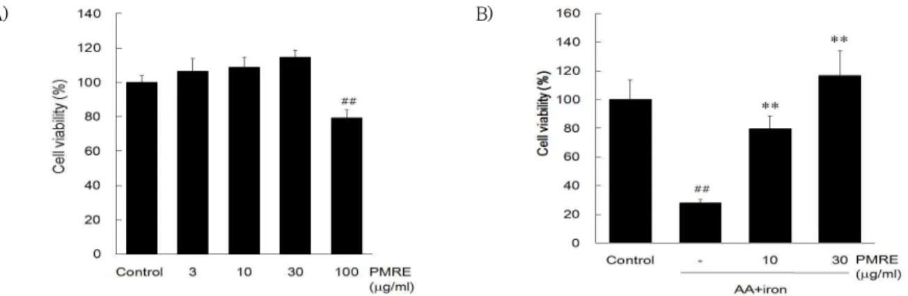
㎍/㎖의 농도로 처치한 결과, 대조군과 비교하였을 때 낮은 농도에서는 통계적으로 유의한 차이가 없으 나 100 ㎍/㎖ 농도에서는 통계적으로 유의하게 감소 하였다 (Fig. 1A). 이와 같은 결과로 PMRE가 세포독 성을 나타내지 않는 농도를 사용하여 이후 실험을 실 시하였다. 이후 PMRE가 AA + iron로 산화적 스트 레스를 가하였을 때 세포생존율에 미치는 영향을 MTT assay로 평가한 결과, AA + iron만을 처치한 실험군
의 경우, 대조군 (100%)과 비교하였을 때 평균 27.82
± 2.69 %의 세포생존율을 나타내어 통계적으로 유 의한 차이가 있었다. 이로 보아 AA + iron로 유도된 산화 스트레스는 세포독성을 유도하는 것을 확인할 수 있었으며, PMRE를 전처치한 실험군에서는 PMRE 가 농도 의존적으로 세포생존율을 증가시키는 것을 확인할 수 있었다 (Fig. 1B).
이후, 선행 연구결과를 통해 AA + iron으로 유도 된 산화적 스트레스가 apoptosis를 일으킨다는 보고
12)를 바탕으로 본 연구에서는 apoptosis와 관련된 지 표단백질들을 immunoblot analysis를 통해 그 발현 을 관찰하였다. 그 결과 AA + iron 처리군에서는 pro-caspase-3, PARP의 발현 정도가 감소한 것을 관찰할 수 있었으며, PMRE를 처리할 경우, pro-caspase-3 와 PARP의 발현이 회복되는 것을 확인하였다. PMRE의 단독 처치는 pro-caspase-3와 PARP에 대한 발현 변화는 관찰되지 않았다 (Fig. 2).
A) B)
Fig. 1. The effect of PMRE on AA + iron-induced cell death.
The effect of PMRE on cell viability was assessed using MTT assay. (A) HepG2 cells were incubated in 3, 10, 30, 100 ㎍/ml of PMRE alone for 24 h. (B) HepG2 cells were incubated in 10, 30 ㎍/ml of PMRE for 1 h and were continuously incubated with 10 μM AA for 12 h, followed by treatment of 5 μM iron for 1 h. Data represent the mean ± S.D. of four replicates (significant as compared with vehicle-treated control, ##p<0.01; significant as compared with AA + iron alone, **p<0.01).
A) B) C)
Fig. 2. Protective effect of PMRE on AA+iron-induced apoptosis.
HepG2 cells were treated with AA (10 μM) for 12 h, followed by iron (5 μM) for 1 h. PMRE was pretreated 1 h earlier than AA treatment. (A) Immunoblottings of apoptosis-related proteins.
Immunoblot analyses were performed on the lysates of HepG2 cells that had been incubated with 10 and 30 ㎍/ml PMRE for 1 h, continuously treated with 10 μM AA for 12 h, and then exposed to 5 μM iron for 1 h. Immunoblotting of β-actin was used as a loading control. The relative density levels of pro-caspase-3 and PARP protein bands were measured by scanning densitometry (B, C). Data represent the mean ± S.D. of four replicates (significant as compared with vehicle-treated control,
##p<0.01; significant as compared with AA+iron alone, *p<0.05, **p<0.01).
2. PMRE가 AA와 iron에 의해 유도된 mitochondrial dysfunction에 미치는 영향
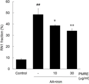
AA+iron에 의한 산화적 스트레스로 유도되는 apoptosis는 MMP의 증가와 관련되어 있다. PMRE가 AA+iron에 의한 미토콘드리아 기능이상을 억제할 수 있는지 알아보기 위해 Rh123 염색을 통해 flow cytometric analysis를 수행하였다. RN1 fraction은 Rh123의 염색양이 적은 분획으로 미토콘드리아의 막
전위가 파괴된 정도로 AA+iron만을 처치한 실험군의 경우 전체 세포 중 RN1 fraction의 비율이 48.40%
로 대조군 (8.35%)과 비교해 큰 차이를 보였으며, 반면 PMRE와 AA+iron을 함께 처리한 실험군 (33.84%)의 경우, 유의성 있게 감소시켰다 (Fig. 3).
이와 같은 결과로 볼 때, PMRE가 AA+iron로 인한 미토콘드리아 기능이상을 억제한다는 사실을 알 수 있다.
Fig. 3. Protective effect of PMRE on AA+iron- induced mitochondrial dysfunction.
HepG2 cells were treated with PMRE for 1 h, AA (10 μM) for 12 h, followed by iron (5 μM) for 1 h.
After treatment of PMRE, AA and iron, HepG2 cells were stained with Rh123 (0.05 ㎍/ml/well) for 0.5 h and measured fluorescence intensity by FACS analysis. AA+iron treated-cells increased the subpopulation of RN1 fraction (low Rh123 fluorescence). But PMRE reduced the population of RN1 fraction. Data represent the mean ± S.D. of four replicates (significant as compared with vehicle-treated control, ##p<0.01; significant as compared with AA+iron alone, *p<0.05, **p<0.01).
3. PMRE가 AA와 iron에 의해 유도된 활성산소종에 미치는 영향
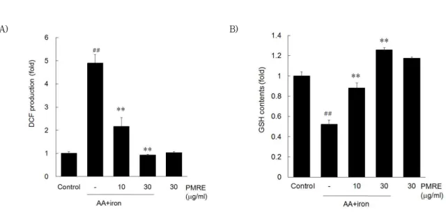
AA + iron에 의한 산화적 스트레스는 과도한 세포 내 활성산소종의 생성을 통해 미토콘드리아의 기능 장애를 유발하여 세포사멸을 유도하는 것으로 알려져 있다. PMRE의 세포 보호효과에 AA + iron에 의한 유도된 산화적 스트레스의 억제가 매개되는지 확인하 기 위하여 DCFH-DA로 염색하여 fluorescence를 microplate reader로 평가하였다.
AA + iron을 처리한 경우 전체 세포 중 H2O2 생 성이 대조군과 비교해 4.9 ± 0.37배로 유의하게 증
가하였고, PMRE를 전처리한 경우에는 H2O2의 생성 이 대조군과 비교하여 유의하게 감소하였다 (Fig. 4A).
또한 세포내 산화적 스트레스의 억제에 연관된 환원 형 GSH의 함량을 측정한 결과, AA + iron을 처리한 경우 GSH 함량이 0.52 ± 0.04배 감소하였고, PMRE를 전처리한 경우, 1.26 ± 0.02배로 유의하게 회복되는 것을 확인하였다 (Fig. 4B). 이와 같은 결 과로 볼 때, PMRE는 AA + iron로 인한 산화적 스 트레스의 억제를 통해 세포 보호 효능을 나타내는 것 으로 생각된다.
A) B)
Fig. 4. Effect of PMRE on AA+iron-induced oxidative stress.
HepG2 cells were treated with PMRE for 1 h, AA (10 μM) for 12 h, followed by iron (5 μM) for 1 h. (A) DCFH-DA (10 μM) was added 0.5 h after iron treatment. DCF fluorescence was measured by automated microplate reader. (B) For the GSH contents, metaphosphoric acid (500 ㎕/well) was added after iron treatment. GSH contents were measured by automated microplate reader at 405 nm wavelength. Data represent the mean ± S.D. of four replicates (significant as compared with vehicle-treated control, ##p<0.01; significant as compared with AA+iron alone, *p<0.05, **p<0.01).
4. PMRE가 CCl4에 의해 유도된 간 손상 모델에 미 치는 영향
CCl4로 유도된 급성 간 손상 모델에서 적출한 간 조직으로부터 immunoblot analysis를 통해 세포자멸 사 관련 단백질의 발현을 확인 해 본 결과, HepG2 cell
모델과 유사하게 CCl4를 처리한 군에서 pro-caspase-3 의 발현 정도가 감소한 것을 관찰할 수 있었으며, PMRE를 처치군에서 그 감소가 완화된 양상을 확인 하였다. 이외에 PMRE를 단독으로 처치하였을 때는 pro-caspase-3의 발현 변화는 관찰되지 않았다 (Fig. 5).
Fig. 5. Effect of PMRE on apoptosis marker protein in CCl4-induced liver damage.
Pro-caspase-3 levels were observed in mice treated with CCl4 with or without PMRE (30 and 100 mg/kg for 4 days) treatment. The immunoblot was assessed in the liver tissues, which were obtained 24 after CCl4 treatment. β-actin was used as a loading control.
Ⅳ. 고찰
다양한 급만성 간질환을 포함한 대사질환 및 면역 질환의 주된 원인인 활성산소는 외부로부터 오는 유 해물질과 미토콘드리아의 산화적 인산화과정에서 생 성되는 산화적 스트레스로 DNA의 변성, 세포막의 파 괴, 암의 발생, 동맥경화, 당뇨, 신경퇴화와 세포노화 를 야기한다20,21). 인체 내에서는 산화억제물질을 생 성하여 대부분의 활성산소를 소멸시키는 방어기전을 가지지만, 이러한 방어기전으로도 제거할 수 없을 만 큼 과도한 활성산소종이 발생할 경우와 체내 항산화 능이 저하되는 경우 산화적 스트레스가 유발된다. 그 러므로, 산화적 스트레스로 인한 독성을 억제할 수 있는 물질의 연구가 다수 수행되었으며, 이전의 연구 결과에서도, HepG2 cell에서 AA와 iron 병용처치를 통해 증가한 활성산소종의 증가를 억제시키는 다양한 본초 추출물 및 본초 유래 활성 성분의 세포보호 효 능이 보고되어져 있다22,23).
何首烏莖葉, 首烏藤, 棋藤 等으로 불리는 夜交藤은 마디풀과(蓼科; Polygonaceae)에 속한 다년생 초본인 何首烏 (Polygonium multiflorum Thunb.)의 덩굴 혹은 잎이 달린 덩굴을 말한다. 夜交藤은 甘微苦平하 며, 주로 心肝經에 작용한다. 養心安神의 효능으로 虛煩不眠, 多夢을 치료하고, 養血通絡의 효능으로 血 虛로 인한 肢體痠痛을 치료하며, 祛風止痒의 효능으 로 風瘡疥癬을 치료한다24).
夜交藤에는 chrysophanol, physcion, stearic acid, emodin, rhein, emodin-8-O-beta-D-glucoside, daucosterol, physcion-8-O-beta-D-glucoside, noreugenin, apigenin, beta-sitosterol, 2,3,4,5’-tetra-hydroxy stibene-2-O- beta-D-glucoside 등의 성분이 함유되어 있는 것으 로 알려져 있으며, catechin 및 epicatechin 단위로 구성된 proanthocyanidin이 고농도로 함유되어 있 고, phenylpropanoid 유도체, stilbene 유도체, anthraquinone 유도체 등도 함유되어 있다13). 특히, 夜交藤 추출물은 염증성 매개물인 nitric oxide와 inducible nitric oxide synthase, COX-2, interleukin-6, tumor necrosis factor-alpha 등을 유의적으로 감소시킴으 로 항염증 효과가 보고되어 있으며 진통작용이 있는 것으로 보고되었다14). 그러나 아직까지 AA + iron의 처치에 의해 유도된 산화적 스트레스에 대한 夜交藤 의 항산화효과 및 간세포 보호 작용에 대해서는 연구
된 바가 없다.
따라서, 본 연구에서는 PMRE의 간보호 효능을 AA 와 iron의 병용처치를 통해 산화적 스트레스를 유도 한 HepG2 cell 독성 모델과 CCl4의 투여에 의해 간 독성이 유도된 간손상 동물모델을 통하여 평가하였다.
본 연구에서 사용한 산화적 스트레스 모델은 AA 와 iron 의 병용처치로서, 다수의 연구에서 AA 와 iron 의 병용 처치는 산화적 스트레스를 매개로 하여 세포자 멸사가 증가되는 것으로 보고되고 있으며7,12,25,26), 또 한 급만성 간질환에서는 간조직 내에 비정상적인 iron 의 축적으로 인해 AA 를 포함한 염증매개물질의 증가가 촉진된다는 것이 보고되어 있다27).
ROS 는 인체 에너지 생산대사에서 생성되며, 적정한 수준에서의 ROS 는 생리적 역할을 하지만28) 과도한 ROS 의 생성은 산화적 스트레스를 유도하여10,29,30), phospolipase A2를 활성화하여 세포막의 지방산과 인지질을 인산화시킴으로써 AA 을 유리하고31,32), AA 는 COX, prostaglandin H2, leukotriene 등을 유도 하여 염증반응에 관여하고33), 또한 에스테르화되지 않은 AA (unesterified AA)는 sphingomyelin 을 ceramide 로 전환시키고, ceramide 는 caspase 를 활성화하여 세포 자멸사를 유도한다10,31). 한편 iron 은 장에서 흡수되 어 주로 간에서 저장되는데, 간에서 iron 은 Fenton 반응을 통해 hydroxyl radical 생성과정을 촉매하고, hydroxyl radical 은 지질과산화 및 세포내 단백질을 손상시켜 세포독성을 유도한다7).
본 연구에서 AA + iron 통해 유도한 세포독성에 대한 PMRE의 보호효능을 관찰한 결과, 이전의 연구
7,25,26)에서와 유사하게 AA와 iron의 병용처치는
control 대비 27.82 ± 2.69 %로 유의한 세포독성을 나타내었고, PMRE는 10, 30, 100 ㎍/ml의 농도에서 각각 79.49 ± 8.92, 116.76 ± 17.54, 109.82 ± 15.03의 세포 생존율을 나타내어, AA + iron 처리군 에 비교하여 유의한 증가를 나타내었다. 또한 AA + iron이 유도하는 세포독성에는 세포자멸사가 개재된 다는 이전의 연구결과들34,35)에 따라, 세포자멸사와 관련된 단백질의 변화를 평가하였다. AA + iron은 pro-caspase-3와 PARP의 발현을 control 대비 0.69 ± 0.04, 0.57 ± 0.14 배로 유의하게 감소시 켰으며, PMRE 10과 30 ㎍/ml은 pro-caspase-3와 PARP 모두에서 AA + iron 처리군에 비교하여 유의 한 증가를 나타내었다.
또한 AA + iron에 의해 유도되는 세포자멸사는 MMP의 증가에서 기인함이 알려져 있다36,37). 본 연구에 서도 control군은 8.35 ± 0.86%의 RN1 fraction을 나 타내었으나, AA + iron군에서는 48.40 ± 5.25%로 유의하게 증가되었고, 이러한 RN1 fraction의 증가는 PMRE 10과 30 ㎍/ml에 의하여 각각 38.57 ± 0.79, 33.84 ± 1.87%로 유의하게 감소되었다. 이와 더불 어, PMRE 10, 30 ㎍/㎖은 AA + iron에 의한 활성 산소의 증가 (4.90 ± 0.37배)를 각각 2.16 ± 0.39, 0.93 ± 0.03 배로 유의하게 감소시켰다.
또한, 산화적 스트레스는 과도한 ROS의 생성뿐만 아니라, 체내 항산화 기능의 저하에서도 초래될 수 있으므로, 체내 항산화 물질중의 하나인 GSH의 함량 을 평가하였다. 세포내 GSH의 고갈은 체내의 항산화 기능의 저하를 초래하여 세포독성을 유도하는 것으로 알려져 있으며38-40), 이미 이전의 연구41,42)에서도 AA + iron은 세포내 GSH의 고갈을 통한 산화적 스트레 스를 유도함이 보고되었다. 본 연구에서도 AA + iron은 0.52 ± 0.04 배로 control 대비 유의한 감소 를 나타내었고, PMRE 10과 30 ㎍/ml은 각각 0.88
± 0.05, 1.26 ± 0.02 배로 유의한 증가를 나타내 어, PMRE가 GSH의 고갈을 유의적으로 억제함을 확 인하였다.
In vitro 모델인 HepG2 cell에서 산화적 스트레스 에 의한 PMRE의 효능을 확인하고, in vivo 모델인 CCl4에 의해 유도된 간손상 마우스에서 PMRE의 효 능을 검증하였다. 대표적인 간독성 유발물질인 CCl4 는 cytochrome p450에 의하여 CCl3۰을 형성하고, CCl3۰는 지방대사에 손상을 유도하여 steatosis를 초 래하고, DNA 손상을 유도하며43), 또한 CCl3۰는 CCl3OO۰ 을 형성하여 지질의 과산화를 유발한다. 지질의 과산 화는 세포막 및 미토콘드리아, DNA 등을 손상시켜 간세포의 손상을 유발하는 것으로43-45) 간질환의 연구 에 빈용되고 있는 동물모델이다. 간 조직으로부터 immunoblot analysis를 통해 세포자멸사 관련 단백 질의 발현을 확인해 본 결과, HepG2 cell 모델과 유 사하게 세포자멸사 관련 단백질의 변화를 억제함을 확인하였다.
따라서, PMRE는 in vitro 및 in vivo 모델에서 산 화적 스트레스에 의한 간조직의 손상을 억제하였으 며, 이러한 간보호효과는 활성산소종 생성억제와 항 산화 효과를 통한 효율적인 간보호 효과가 있음을 확
인하였다.
Ⅴ. 결론
본 연구에서는 AA와 iron에 의해 유도된 산화적 스트레스를 HepG2 in vitro 모델과 CCl4에 의해 유 도된 급성 간손상 in vivo 모델에서 PMRE의 항산화 및 간보호 효과를 평가하였다. In vitro 모델에서는 MTT assay를 통하여 세포 보호효과, immunoblot analysis로 세포자멸사 단백질 발현 및 유세포 분석 을 통한 미토콘드리아 보호효과를 확인하였다. 이와 더불어 In vivo 모델에서도 PMRE의 처치가 세포자 멸사 단백질 발현을 억제하는 것으로 확인되었다. 이 러한 결과는 夜交藤의 처치가 in vitro와 in vivo 모 델에서 항산화 효능 및 세포 보호 효능, 간보호 효과 가 있음을 입증한 것으로 항산화 효과와 더불어 간을 보호하는 유효한 약물 후보로 夜交藤을 급만성 치료 에 활용할 수 있을 것으로 생각된다. 향후 夜交藤의 세포보호 효과에 대한 정확한 분자생물학적 작용기전 을 규명하기 위해서는 후속 연구가 뒷받침되어야 할 것으로 사료된다.
* 이 논문은 2017년도 정부(미래창조과학부)의 재원 으로 한국연구재단의 지원을 받아 수행된 연구임 (No.2012R1A5A2A42671316).
References
1. Weiskirchen R, Tacke F. Cellular and molecular functions of hepatic stellate cells in inflammatory responses and liver immunology. Hepatobiliary Surg Nutr. 2014;3(6):344-363.
2. Yerushalmi B, Dahl R, Devereaux MW, Gumpricht E, Sokol RJ. Bile acid-induced rat hepatocyte apoptosis is inhibited by antioxidants and blockers of the mitochondrial permeability transition.
Hepatology. 2011;33(3):616-626.
3. Muriel P. Role of free radicals in liver diseases.
Hepatol Int. 2009;3(4):526-536.
4. Cichoż.-Lach H, Michalak A. Oxidative stress as a crucial factor in liver diseases. World J Gastroenterol. 2014;20(25):8082-8091.
5. Turner TT, Lysiak JJ. Oxidative stress: a common factor in testicular dysfunction. J Androl.
2008;29(5):488-498.
6. Reuter S, Gupta SC, Chaturvedi MM, Aggarwal BB. Oxidative stress, inflammation, and cancer:
how are they linked?. Free Radic Biol Med.
2010;49(11):1603-1616.
7. Choi SH, Kim YW, Kim SG. AMPK-mediated GSK3beta inhibition by isoliquiritigenin contributes to protecting mitochondria against iron-catalyzed oxidative stress. Biochem Pharmacol. 2010;79(9):
1352-1362.
8. Okuda M, Li K, Beard MR, Showalter LA, Scholle F, Lemon SM, Weinman SA. Mitochondrial injury, oxidative stress, and antioxidant gene expression are induced by hepatitis C virus core protein. Gastroenterology. 2002;122(2):366-375.
9. Ko HL, Jegal KH, Song SY, Kim NE, Kang J, Byun SH, Kim YW, Cho IJ, Kim SC. Water extract of Rosa laevigata Michx. protects hepatocytes from arachidonic acid and iron-mediated oxidative stress. Kor J Herbol. 2015;30(6):7-15.
10. Balboa MA, Balsinde J. Oxidative stress and arachidonic acid mobilization. Biochim Biophys Acta.. 2006;1761(4):385-391.
11. Caro AA, Cederbaum AI. Role of cytochrome P450 in phospholipase A2-and arachidonic acid-mediated cytotoxicity. Free Radic Biol Med. 2006;40(3):
364-375.
12. Shin SM, Kim SG. Inhibition of arachidonic acid and iron-induced mitochondrial dysfunction and apoptosis by oltipraz and novel 1,2-dithiole-3 -thione congeners. Mol Pharmacol. 2009;75(1):
242-253.
13. Kim MO, Park YS, Nho YH, Yun SK, Kim Y, Jung E, Paik JK, Kim M, Cho IH, Lee J. Emodin isolated from Polygoni Multiflori Ramulus inhibits melanogenesis through the liver X receptor-mediated pathway. Chem Biol Interact.
2016;250:78-84.
14. Cha DS, Jeon H. Anti-inflammatory Effect of MeOH extracts of the stem of Polygonum mul
tiflorum in LPS-stimulated mouse peritoneal macrophages. Natural Product Sciences. 2009;
15(2):83-89.
15. Yim TK, Wu WK, Mak DH, Ko KM. Myocardial protective effect of an anthraquinone- containing extract of Polygonum multiflorum ex vivo. Planta. Med. 1998;64(7):607-611.
16. Wang W, Cao CY, Wang DQ, Zhao DZ. Effect of prepared Polygonum multiflorum on striatum extracellular acetylcholine and choline in rats of intracerebral perfusion with sodium azide.
Zhongguo Zhong Yao Za Zhi. 2006;31(9):
751-753.
17. Xiao PG, Xing ST, Wang LW. Immunological aspects of Chinese medicinal plants as antiageing drugs. J Ethnopharmacol. 1993;38(23):167-175.
18. Um MY, Choi WH, Aan JY, Kim SR, Ha TY.
Protective effect of Polygonum multiflorum Thunb on amyloid beta-peptide induced cognitive deficits in mice. J Ethnopharmacol. 2006;
104(1-2):144-148.
19. Li RW, David LG, Myers SP, Leach DN. Anti- inflammatory activity of Chinese medicinal vine plants. J. Ethnopharmacol. 2003;85(1):61-67.
20. Facciorusso A, Villani R, Bellanti F, Mitarotonda D, Vendemiale G, Serviddio G. Mitochondrial signaling and hepatocellular carcinoma: Molecular mechanisms and therapeutic implications. Curr Pharm Des. 2016;22(18):2689-2696.
21. Yang JM, Jun YJ, Nam JY, Son M.Y, Sung JS, Kim DI. The cell protective effects of Dioscoreae Rhizoma by antioxidant activities on HeLa cells. J Orient Obstet Gynecol. 2008;21(2):97-107.
22. Kim JK, Park SM, Jegal KH, Kim YW, Byun SH, Kim SC, Cho IJ. Gleditsia Spina extract pro- tects hepatocytes from oxidative stress through Nrf2 activation. Kor J Herbol. 2015;30(4):
57-64.
23. Jung EH, Kim SC, Cho IJ, Kim YW. Akebiae Caulis inhibits oxidative stress through AMPK activation. J Physiol Pathol Korean Med. 2015;
29(1):18-26.
24. Seo BI, Choi HY. Clinical Herbology. Seoul;
Younglimsa. 2004:713.
25. Kim YW, Lee SM, Shin SM, Hwang SJ, Brooks JS, Kang HE, Lee MG, Kim SC, Kim SG.
Efficacy of sauchinone as a novel AMPK- activating lignan for preventing iron-induced oxidative stress and liver injury. Free Radic Biol Med. 2009;47(7):1082-1092.
26. Choi HY, Jegal KH, Kim YW, Lee JW, Cho SA, Cho IJ, Kim SC. Cytoprotective effects of Artemisia princeps extract through inhibition of mitochondrial dysfunction. Herb Formula Sci. 2013;21(2):63-71.
27. Galaris D, Pantopoulos K. Oxidative stress and iron homeostasis: mechanistic and health aspects. Crit Rev Clin Lab Sci. 2008;45(1):1–23.
28. Janssen-Heininger YM, Mossman BT, Heintz NH, Forman HJ, Kalyanaraman B, Finkel T, Stamler JS, Rhee SG, van der Vliet A. Redox- based regulation of signal transduction: prin- ciples, pitfalls, and promises. Free Radic Biol Med. 2008;45(1):1–17.
29. Thanan R, Oikawa S, Hiraku Y, Ohnishi S, Ma N, Pinlaor S, Yongvanit P, Kawanishi S, Murata M. Oxidative stress and its significant roles in neurodegenerative diseases and cancer.
Int J Molecul Sci. 2014;16(1):193–217.
30. Lee IW, Choi HS, Kim SM. Hepatoprotective ac- tivity of Spatholobi Caulis water extract against acetaminophen-induced toxicity in rats. Kor J Herbol. 2011;26(3):65-73.
31. Chen JK, Capdevila J, Harris RC. Cytochrome p450 epoxygenase metabolism of arachidonic acid inhibits apoptosis. Mol Cell Biol. 2001;21(18):
6322-6331.
32. Roman RJ. P-450 metabolites of arachidonic acid in the control of cardiovascular function.
Physiol Rev. 2002;82(1):131-185.
33. Ehsanian R, Van Waes C, Feller SM. Beyond DNA binding-a review of the potential mechanisms mediating quinacrine's therapeutic activities in parasitic infections, inflammation, and cancers.
Cell Commun Signal. 2011;9:13.
34. Jegal KH, Ko HL, Park SM, Byun SH, Kang KW, Cho IJ, Kim SC. Eupatilin induces Sestrin2- dependent autophagy to prevent oxidative stress.
Apoptosis. 2016;21(5):642-656.
35. Seo HL, Baek SY, Lee EH, Lee JH, Lee SG, Kim KY, Jang MH, Park MH, Kim JH, Kim KJ, Lee HS, Ahn SC, Lee JR, Park SJ, Kim SC, Kim YW.
Liqustri lucidi Fructus inhibits hepatic injury and functions as an antioxidant by activation of AMP-activated protein kinase in vivo and in vitro. Chem Biol Interact. 2017;262:57-68.
36. Kang YK. Lee EA, Park HR. Neuroprotective effect according to reactive oxygen species scavenging activity from extracts of cudrania tricuspidata leaves. Korean J. Food cookery SCI. 2012;28(6):821-828.
37. Seo HL, Lee JH, Jang MH, Kwon YW, Cho IJ, Kim KJ, Park SJ, Kim SC, Kim YW, Byun SH.
Mitochondria protection of Sparganii Rhizoma against oxidative stress in heptocytes. Herb Formula Sci. 2015;23(2):189-198.
38. Han D, Shinohara M, Ybanez MD, Saberi B, Kaplowitz N. Signal transduction pathways in- volved in drug-induced liver injury. Handb Exp Pharmacol. 2010;196:267-310.
39. Yuan L, Kaplowitz N. Glutathione in liver dis- eases and hepatotoxicity. Mol Aspects Med.
2009;30(1-2):29-41.
40. Fisher LJ, Green MD, Harman AW. Levels of acetaminophen and its metabolites in mouse tissues after a toxic dose. J Pharmacol Exp Ther. 1981;219(2):281-286.
41. Dong GZ, Lee JH, Ki SH, Yang JH, Cho IJ, Kang SH, Zhao RJ, Kim SC, Kim YW. AMPK activa- tion by isorhamnetin protects hepatocytes against oxidative stress and mitochondrial dysfunction.
Eur J Pharmacol. 2014;740:634-640.
42. Dong GZ, Jang EJ, Kang SH, Cho IJ, Park SD, Kim SC, Kim YW. Red ginseng abrogates oxi- dative stress via mitochondria protection mediated by LKB1-AMPK pathway. BMC Complement
Altern Med. 2013;13:64.
43. Weber LW, Boll M, Stampfl A. Hepatotoxicity and mechanism of action of haloalkanes: carbon tetrachloride as a toxicological model. Crit Rev Toxicol. 2003;33(2):105-136.
44. Knockaert L, Berson A, Ribault C, Prost PE, Fautrel A, Pajaud J, Lepage S, Lucas-Clerc C, Bégué JM, Fromenty B, Robin MA. Carbon tetra-
chloride-mediated lipid peroxidation induces early mitochondrial alterations in mouse liver.
Lab Invest. 2012;92(3):396-410.
45. Kim TH, Kim YW, Shin SM, Kim CW, Yu IJ, Kim SG. Synergistic hepatotoxicity of N,N- dimethylformamide with carbon tetrachloride in association with endoplasmic reticulum stress.
Chem Biol Interact 2010;184(3):492-501.