Staurosporine에 의해 분화된 망막신경절세포에서 산화 스트레스 유도 세포사멸에 대한 차조기 추출물의 보호 효능
이보경1*․최리라1*․이지인2․이두이2․장선영1,3․김소희1,3․정이숙1,3
1아주대학교 약학대학
2평창 허브나라 농원
3아주대학교 약과학연구소
Protective Effect of Perilla frutescens Extract against Oxidative Stress-Induced Cell Death in a Staurosporine-Differentiated Retinal Ganglion Cell Line
Bo Kyung Lee1*, Lira Choe1*, Ji In Lee2, Doo Yi Lee2, Sun-Young Chang1,3, So Hee Kim1,3, and Yi-Sook Jung1,3
1College of Pharmacy and 3Research Institute of Pharmaceutical Sciences and Technology, Ajou University
2Farm Herbnara
ABSTRACT In this study, we examined the effect of Perilla frutescens extract (PFE) on oxidative stress-induced cell death in RGC-5 cell lines. Staurosporine-differentiated RGC-5 (ssdRGC-5) cells obtained by treating RGC-5 cells with 1 μM staurosporine were incubated with PFE for 30 min and then exposed to buthionine sulfoximine plus glutamate (B/G) for 20 h. Cell death was detected using lactate dehydrogenase release assay and 3-(4,5-dimethylthiazol-2-yl)-2,5- diphenyltetrazolium bromide reduction assay. To investigate the mechanism underlying cell death, we determined cas- pase-3 activity, level of reactive oxygen species (ROS) formation, and expression levels of cytoplasmic cytochrome c and mitochondrial Bax. Treatment of ssdRGC-5 cells with B/G increased intracellular ROS and induced apoptosis with increasing caspase-3 activity. PFE rescued ssdRGC-5 cells from oxidative stress-induced cell death by inhibiting intracellular ROS production and caspase-3 activation and regulating apoptosis-related proteins such as cytochrome c and Bax. These findings suggest that PFE may have a beneficial neuroprotective effect against oxidative stress-induced apoptotic death in ssdRGC-5 cells.
Key words: Perilla frutescens acuta Kudo, retinal ganglion cell, oxidative stress, cell death, glaucoma
Received 10 November 2016; Accepted 23 January 2017 Corresponding author: Yi-Sook Jung, College of Pharmacy, Ajou University, Suwon, Gyeonggi 16499, Korea
E-mail: [email protected], Phone: +82-31-219-3444
*These authors contributed equally to this work.
서 론
눈은 나이가 들면서 기능이 약해져 시력 감소와 시야 결손 등이 심각하게 진행되면 독서, 글쓰기, 운전, 이동과 같은 일상생활을 수행하는 데 어려움을 겪게 될 뿐만 아니라(1) 낙상 및 낙상으로 인한 부상의 위험을 높이게 된다(2,3). 노 화에 따른 중추신경계 감각능력 상실은 시신경병증을 유발 하고, 이로 인한 시신경의 퇴행은 최종적으로 시야 감소 및 비가역적 시력 손상을 유발하는 녹내장을 초래하게 된다(4).
녹내장은 여러 위험요인에 의해 진행이 되지만 병태 생리학 적으로 가장 중요한 요소는 망막신경절세포(retinal gan- glion cell, RGC)의 사멸이다. 망막신경절세포는 세포체가 망막의 안쪽 면에 위치하고 있고 축삭(axon)은 시신경을 형
성하여 망막에 형성된 시각 정보를 뇌 시상의 외측슬상핵 (lateral geniculate nucleus)으로 전달해주는 역할을 한다.
망막신경절세포는 일반 신경세포와 마찬가지로 한 번 손상 되면 회복되지 않을 뿐 아니라 재생도 되지 않으므로 녹내장 역시 알츠하이머 및 파킨슨 질환과 같은 퇴행성 신경질환이 라고 할 수 있다(5,6). 산화적 스트레스는 신경퇴행성 질환 이 가지는 공통적인 병리적 기전으로 알려져 있다. 산화적 스트레스는 반응 산소종의 생성과 제거 사이의 불균형에 의 해 나타나며, DNA, 단백질, 지질 같은 생체 내 분자에 손상 을 주어 신경세포의 손상과 사멸에 이르게 한다. 녹내장과 산화적 스트레스 사이의 연관성이 1980년대에 처음 제시된 이후, 최근에는 녹내장 동물 모델, 임상 연구로부터 그 연관 성을 뒷받침하는 연구들이 보고되고 있다(5-7). 따라서 산 화적 스트레스를 억제하는 항산화 물질은 신경세포의 사멸 을 지연하거나 억제함으로써 신경세포를 보호한다고 할 수 있다. 차조기(차즈기, Perilla frutescens (L.) Britt. var.
acuta Kudo, 꿀풀과)는 천 년 이상 동아시아의 전통 한약재 로 사용되어 왔다. 한방에서 차조기는 발한, 이뇨, 해열, 진
해, 건위 작용을 나타내며, 차조기를 포함한 한약 처방에는 곽향정기산(藿香正氣散), 소자강기탕(蘇子降氣湯), 삼소음 (蔘蘇飮), 향소산(香蘇散), 분심기음(分心氣飮), 반하후박탕 (半夏厚朴湯) 등이 있다(8). 차조기는 항염증 및 항알레르기 (8,9-13), 항우울(14), 항종양(10,15), 항균(16-18), 알츠 하이머 질환(19), 항산화(11,18,20-22) 등 다양한 효능을 나타낸다고 알려져 있다. 차조기의 항산화 효능에 대한 연구 는 차조기 잎을 알코올 또는 물로 추출한 후 다양한 극성을 지닌 용매로 분획하여 total phenolic contents를 구하고 항산화 능력을 측정하는 화학적 분석법이 대부분이며, 실제 눈과 연관된 세포를 이용하여 차조기 추출물의 효능을 세포 수준에서 생물학적 관점으로 접근한 연구는 미비하다. 최근 VDT(visual display terminal) 증후군과 관련하여 차조기 추출물 복용 후 눈의 굴절률 이상이 개선되었다는 연구가 있는데, 이 역시 세포 수준의 기전은 알려진 바 없다(23).
따라서 본 연구팀은 차조기 잎을 차로 마시는 것을 고려하여 산화적 스트레스로 유도한 망막신경절세포의 사멸에 대한 차조기 물 추출물의 보호 효과를 확인하였다.
재료 및 방법
차조기 물 추출물 제조
본 실험에 사용한 차조기는 허브나라 농원으로부터 건조 된 잎 상태로 제공받아 추출하였다. 건조된 차조기 잎 10 g을 증류수 200 mL로 85°C에서 30분씩 2회 추출한 후 여 과한 다음 회전식 감압농축기로 85°C에서 50 mL로 감압 농축하여 300 mg/mL의 차조기 물 추출물(Perilla frutes- cens extract, PFE)을 제조하고 -20°C에 보관하면서 사용 하였다. 0.1~100 μg/mL의 농도로 세포에 처리할 때는 300 mg/mL의 차조기 추출물을 증류수로 100 mg/mL까지 희석 시킨 후, 최대 1:1,000 농도로 세포에 처리하였다.
세포 배양 및 산화 스트레스
본 실험에 사용된 RGC-5는 rat retinal ganglion cell에 서 분리된 세포주로 안재홍(아주대학교 의과대학 안과학교 실) 교수로부터 제공받았다. 세포 배양을 위해 Dulbecco’s modified Eagle’s medium(Invitrogen, Carlsbad, CA, USA)에 10% fetal bovine serum(FBS, Invitrogen), 1%의 penicillin-streptomycin을 첨가하여 37°C, 5% CO2가 유 지되는 세포배양기에서 배양하였다. RGC-5 세포는 대략 10%의 confluence(1.5×10⁴ cells/cm2)로 플라스틱 배양 용기에 분주한 후 80~85%의 confluence로 성장하게 하였 고, 모든 세포는 passage 30 이하에서 사용하였다. RGC-5 를 분화시키기 위해 세포를 1 μM의 staurosporine(Sig- ma-Aldrich Co., St. Louis, MO, USA)에 6시간 동안 노출 시킨 후, culture medium에서 하루 동안 배양하였다.
Staurosporine으로 분화시킨 RGC-5(ssdRGC-5)를 여러 농도(0.1, 1, 10, 100 μg/mL)의 PFE 또는 100 μM Trolox
(Tocris, Ballwin, MO, USA)와 함께 30분 동안 전 처리한 후, 0.5 mM L-buthionine-(S,R)-sulfoximine(BSO, Sigma- Aldrich Co.)과 5 mM glutamate(Sigma-Aldrich Co.)로 24시간 동안 배양하여 산화 스트레스를 유도하였다(24). 산 화 스트레스에 대한 차조기 추출물의 효능은 100 μM Tro- lox와 비교하였다.
Lactate dehydrogenase(LDH) 유리를 통한 세포사멸 측정 ssdRGC-5의 사멸 정도는 배지로 분비되는 LDH를 측정 하여 분석하였다(25). ssdRGC-5를 여러 농도의 PFE 100 μM Trolox로 30분 전 처리한 후 0.5 mM BSO와 5 mM glutamate를 추가하여 20시간 배양한 다음, 배지를 25 μL 씩 취해 96-well plate에 옮긴 후 NADH solution(0.03%
β-NAD[reduced form of disodium salt] in phosphate buffer) 100 μL와 pyruvate solution(22.7 mM pyruvic acid in phosphate buffer) 25 μL를 첨가하여 NADH 소모 량을 340 nm에서 2분간 측정하였다. 실험군마다 평균 흡광 도 변화(ΔA/min)를 계산하고 대조군의 흡광도 변화에 대해 백분율로 표현하였다.
MTT 분석을 통한 세포생존율 측정
ssdRGC-5 세포의 세포생존율 측정을 위하여 3-(4,5- dimethylthiazol-2-yl)-2,5-diphenyltetrazolium bromide (MTT) reduction assay를 실시하였다(26). 24-well plate 에 배양한 ssdRGC-5 세포에 여러 농도의 PFE 또는 100 μM Trolox를 30분 전 처리한 후 0.5 mM BSO와 5 mM glutamate를 첨가하여 20시간 배양한 다음, 각 well에 PBS 완충용액에 녹인 MTT(5 mg/mL) 용액을 50 μL씩 첨가하 여 1시간 동안 다시 배양하였다. 1시간 반응 후 formazan 형성을 확인하고 formazan이 흩어지지 않게 상등액을 완전 히 제거한 다음, 200 μL의 DMSO를 첨가하여 녹인 후 mi- croplate reader(Model 680, BioRad, Hercules, CA, USA) 를 사용하여 540 nm에서 흡광도를 측정하였다. 수치 정량 은 각 실험군에 대해 대조군 세포를 100%로 하였을 때의 상대적인 세포생존율을 계산하였다.
Caspase-3 activity assay
ssdRGC-5 세포를 0.1, 1, 10, 100 μg/mL의 농도로 제조 한 PFE 또는 100 μM Trolox로 30분 전 처리하고, 0.5 mM BSO와 5 mM glutamate를 첨가하여 20시간 동안 배양한 다음, lysis buffer(10 mM Tris/HCl, 0.32 M sucrose, 1 mM PMSF, 1% Triton X-100, 1 μg/mL aprotinin, 10 μg/
mL leupeptin, 5 mM EDTA, 10 mM DTT, pH 8.0)로 용해 한 후 lysate를 원심분리(10,000×g, 4°C, 5분) 하였다. 추 출한 200 μg의 단백질 sample을 200 μM Ac-DEVD-p- NA(Biomol, Plymouth Meeting, PA, USA)와 함께 배양한 후, 효소에 의해 분해되어 생성된 p-NA 양을 microplate reader를 사용하여 405 nm에서 측정하였다(24).
세포 내 superoxide 생성 측정
ssdRGC-5를 0.1, 1, 10, 100 μg/mL의 농도로 제조한 PFE 또는 100 μM Trolox로 30분 전 처리하고, 0.5 mM BSO와 5 mM glutamate를 첨가하여 20시간 동안 배양한 후, 10 μM dihydroethidium dye(DHE, Molecular Probe, Eugene, OR, USA)로 37°C에서 30분간 염색하였다. 염색 된 세포는 HEPES 완충용액으로 3회 세척한 후 즉시 laser scanning confocal microscope(Olympus 5100, Olympus, Tokyo, Japan)으로 평가하였으며, DHE의 형광 intensity는 image-analysis computer software(Axio VisionTM, Carl Zeiss, Oberkochen, Germany)를 사용하여 정량하였다(27).
세포 내 H2O2생성 측정
2′,7′-Dichlorofluorescein diacetate(DCFDA; molec- ular probe)는 살아있는 세포 내의 산화 활성을 측정하는 데 사용되는 ROS-sensitive probe이다. ssdRGC-5를 0.1, 1, 10, 100 μg/mL의 농도로 제조한 PFE 또는 100 μM Trolox로 30분 전 처리하고, 0.5 mM BSO와 5 mM gluta- mate를 첨가하여 20시간 동안 배양한 다음, DCFDA dye (10 μg/mL)로 37°C에서 30분간 처리하였다. 염색된 세포 는 HEPES 완충용액으로 3회 세척한 후 즉시 laser scan- ning confocal microscope(Olympus 5100, Olympus)으 로 평가하였으며, DCFDA의 형광 intensity는 image-anal- ysis computer software(AxioVisionTM, Carl Zeiss)를 사 용하여 정량하였다(24).
세포질과 미토콘드리아 분획의 분리
시약 처리된 세포는 ice-cold PBS(phosphate buffered saline)로 2회 세척한 후 단백질 분해 효소 억제제(10 μg/
mL leupeptin, 10 μg/mL aprotinin)를 첨가한 lysis buf- fer(20 mM HEPES-KOH, pH 7.5, 10 mM KCl, 1.5 mM MgCl2, 1.0 mM EDTA, 1.0 mM EGTA, 1.0 mM DTT, 1 mM PMSF, and 250 mM sucrose)로 재분산하고, glass homogenizer로 파쇄한 후 4°C, 1,000×g에서 15분간 원심 분리 하였다. 상층액은 4°C, 10,000×g에서 15분간 원심분 리 하였고, 얻어진 미토콘드리아 펠렛은 모아서 lysis buffer 에 재분산 하였다. 10,000×g에서 원심분리 하여 얻은 상층 액은 4°C, 100,000×g에서 1시간 동안 1회 더 원심분리 하 였고 상층액을 세포질 분획으로 지정하였다. Cytochrome c와 Bax의 세포 내 이동을 정량하기 위해 미토콘드리아 분 획과 세포질 분획에 대해 western blotting을 수행하였다.
미토콘드리아 표지는 cytochrome oxidase subunit Ⅳ (COXⅣ, Invitrogen), 세포질 분획의 표지는 actin(Sigma- Aldrich Co.)을 사용하였다(26).
Western blot(caspase-3, caspase-6, cytochrome c, Bax)
시약 처리된 세포를 ice-cold PBS로 2회 세척하고, RIPA
buffer(pH 7.4, 150 mM NaCl, 20 mM Tris-HCl, 1% NP- 40, 1% Na-deoxycholate, 1 mM EDTA, protease in- hibitor 10 μg/mL leupeptin, 10 μg/mL aprotinin)에 넣어 세포를 파쇄하였다. 세포는 glass homogenizer로 1회 더 파쇄하였으며, 4°C, 14,000 rpm에서 15분간 원심분리를 하 여 핵과 세포 잔해물은 제거하고 상층액을 모아서 immuno- blotting에 사용하였다. 단백질은 BCATM protein assay (Perce, Rockfor, IL, USA)로 정량하였다. 단백질 시료는 10% sodium dodecyl sulfate-polyacrylamide gel에서 전 기영동으로 분리하고, polyvinylidene difluoride mem- brane으로 blotting 하였다. 5% non-fat dry milk를 넣은 Tris-buffered saline에 blot을 넣고 상온에서 1시간 동안 유지하였고, 1차 항체와 함께 4°C에서 하룻밤 동안 유지하 였다. Blot을 Tris-buffered saline으로 씻어낸 후 horse- radish peroxidase-conjugated 2차 IgG 항체와 함께 1시 간 유지하였다. 결합된 항체는 LAS-1000 Pro(Fujifilm Corporation, Tokyo, Japan)를 사용하여 시각화하고 정량 하였다. Anti-caspase-3, anti-caspase-6, anti-cyto- chrome c antibody, Bax antibody는 Santa Cruz Biotech- nology, Inc.(Santa Cruz, CA, USA)에서 구매하였다(26).
Anti-actin, anti-COXIV antibody는 Cell Signaling(Be- verly, MA, USA)에서 구매하였다.
통계처리
본 실험 결과는 각 항목에 따라 평균과 표준오차를 구하였 고, 평균치 간의 유의성은 Student’s t-test를 이용한 후 P 값이 0.05 미만일 때 유의한 것으로 판정하였다.
결 과
B/G 유도 ssdRGC-5 시신경세포에서 차조기 추출물의 세 포 보호 효과
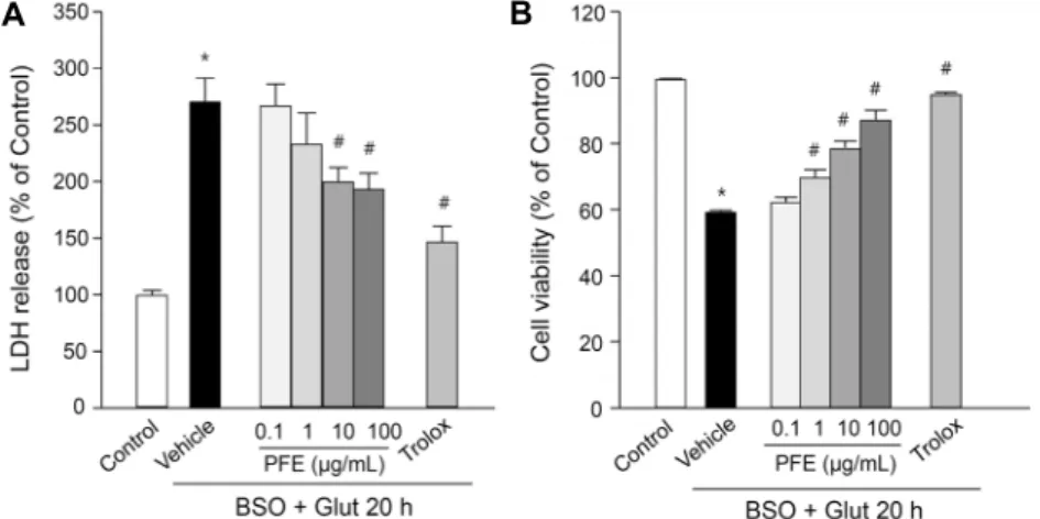
본 연구에서 사용한 RGC-5 세포주는 산화적 스트레스에 대한 세포사멸의 반응 기전을 연구하고 신경보호 기전을 규 명하는 데 유용한 모델이라고 보고된 바 있으며(28), 이를 단백질 인산화효소 억제제인 staurosporine으로 분화시키 면 axon과 neurite가 생성되고 신경세포에 특이적으로 나 타나는 다양한 분자가 발현된다고 알려져 있으므로, 본 실험 에서는 staurosporine으로 분화시킨 RGC-5를 사용하였다 (24,29). BSO와 glutamate(B/G)로 유도한 ssdRGC-5의 LDH 분비는 정상군(100.0±3.8%)과 비교했을 때 대조군 (270.8±20.5%)에서 유의성 있게 증가하였다. 10, 100 μg/
mL PFE는 대조군 대비 유의적으로 LDH 분비가 감소(각각 200.3±12.1%, 193.8±13.5%)하였으며, 항산화제로 알려 진 Trolox를 처리한 양성 대조군(147.1±13.3%)도 LDH 분 비가 유의적으로 감소하였다(Fig. 1A). B/G로 유도한 시신 경 세포의 세포생존율은 정상군 대비 59.8±0.3%로 감소하 였으며, PFE에서는 농도 의존적으로 세포생존율이 증가하
A B Fig. 1. Effects of Perilla frutescens extracts (PFE) on BSO plus glutamate (B/G)-induced cell death in ssdRGC-5. ssdRGC-5 cells were incubated with or without PFE (0.1∼100 μg/
mL) for 30 min before adding 0.5 mM BSO plus 5 mM glutamate, and incubated for 20 h. Trolox (100 μM) was used to compare the protective potency. (A) The effect of PFE on B/G-induced LDH release in ssdRGC-5 cells.
The percentage of LDH release was compared to the control. (B) The effect of PFE on B/G- induced cell death in ssdRGC-5. The percent- age of cell viability was compared to the con- trol. Data represent the mean±SEM of at least three independent experiments. *P<0.05 vs.
untreated control, #P<0.05 vs. vehicle (B/G only, without pretreatment with PFE).
A B
Fig. 2. Effects of PFE on B/G-induced caspase- 3 activation in ssdRGC-5. ssdRGC-5 cells were incubated with or without PFE (1∼100 μg/mL) for 30 min before adding 0.5 mM BSO plus 5 mM glutamate, and incubated for 20 h. Trolox (100 μM) and zVAD (1 μM) was used to com- pare the protective potency. (A) Caspase-3 and caspase-6 expressions were analyzed from the cytosolic fraction of ssdRGC-5 cells after 20 h B/G insult. (B) The percentage of caspase-3 activity was compared to the control. Data rep- resent the mean±SEM of at least three independ- ent experiments. *P<0.05 vs. untreated control,
#P<0.05 vs. vehicle (B/G only, without pre- treatment with PFE).
였다. 대조군과 비교하여 1 μg/mL PFE 농도에서부터 유의 성 있게 세포생존율이 증가하였으며, 100 μg/mL PFE 처리 군(87.7±2.7%)에서 최대 세포생존율이 나타났다(Fig. 1B).
B/G 유도 caspase-3 활성화에서 차조기 추출물의 효과 ssdRGC-5에서 B/G에 의한 caspase-3와 caspase-6 활성을 western blotting으로 측정한 결과, B/G에 의해 caspase-3와 caspase-6의 활성군이 증가하였으며, 100 μg/mL PFE에 의해 정상군 수준으로 억제되었다. 양성대조 군으로 사용한 pan-caspase 억제제인 zVAD와 항산화제인 Trolox에 의해서도 caspase-3와 caspase-6 활성이 정상 군 수준으로 억제되었다(Fig. 2A). PFE의 caspase-3 활성 화를 효소를 이용해 측정한 결과에서도 B/G에 의해 cas- pase-3 활성이 1.6배 증가(158.1±2.3%)하였고, zVAD는 caspase-3 활성을 정상군 수준(93.4±5.3%)으로 억제하였 다. PFE는 농도 의존적으로 caspase-3 활성을 감소시켰으 며, 10, 100 μg/mL(각각 139.2±3.2%, 132.4±1.6%)에서 대조군 대비 유의성 있게 caspase-3 활성이 감소하였다 (Fig. 2B).
B/G 유도 산화적 스트레스에 대한 차조기 추출물의 효과 B/G에 의한 ssdRGC-5 내의 superoxide 생성에 대해서
PFE의 효능이 나타나는지 관찰하기 위해 DHE 염색방법을 이용해 관찰한 결과, B/G에 의한 DHE 발색이 정상군(100.0
±6.3%) 대비 1.4배 증가하였고(143.3±5.7%), 대조군과 비 교하여 100 μg/mL PFE에서 세포 내 DHE 발색이 유의하게 감소하였다(122.9±6.8%)(Fig. 3A). 또한, B/G에 의한 세포 내 hydrogen peroxide 생성에 대해서도 PFE의 효능이 나 타나는지 관찰하기 위해 DCFDA 염색방법을 이용해 관찰한 결과, B/G에 의한 DCFDA 발색이 정상군보다 2배 증가하였 고(202.4±11.4%), 100 μg/mL PFE에 의해 유의하게 감소 하였다(131.4±5.7%)(Fig. 3B).
B/G 유도 미토콘드리아성 시신경 세포사멸 과정에서 차조 기 추출물의 보호 기전
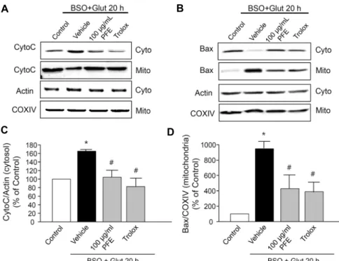
B/G에 의한 ssdRGC-5의 사멸이 미토콘드리아성 세포사 멸 기전을 통해 일어나는지, 만약 그렇다면 PFE가 미토콘드 리아성 사멸기전을 억제하는 것인지 알아보기 위해 미토콘 드리아성 세포사멸기전인 cytochrome c의 세포질로의 방 출과 Bax의 미토콘드리아로의 이동을 western blotting을 통해 관찰하였다. B/G에 의해 cytochrome c가 세포질로 방출되는 것을 관찰하였고, cytochrome c 방출이 100 μg/
mL PFE와 100 μM Trolox에 의해 억제되었다(Fig. 4A).
이를 세포질 분획에서 actin에 대비 정량한 결과 B/G는 정상
A B
Fig. 3. Effects of PFE on B/G-induced ROS accumulation in ssdRGC-5. ssdRGC-5 cells were incubated with or without PFE (0.1∼100 μg/mL) for 30 min before adding 0.5 mM BSO plus 5 mM glutamate, and incubated for 20 h. Trolox (100 μM) was used to compare the protective potency. (A) The amount of intracellular superoxide anion (O2・-) was evaluated using the fluorescent dye DHE and intensity of DHE was quantified. The percentage of O2・- production was compared to the control. (B) The amount of intracellular hydrogen peroxide (H2O2) was evaluated using the fluorescent dye DCF-DA and intensity of DCF was quantified.
The percentage of H2O2 production was compared to the control. Data represent the mean±SEM of at least three independent experiments. *P<0.05 vs. untreated control, #P<0.05 vs. vehicle (B/G only, without pretreatment with PFE).
A B
C D
Fig. 4. Regulation of PFE on B/G-induced mitochondrial death pathway in ssdRGC- 5 cells. (A) Western blots for cytochrome c detected in the cytosolic and mitochon- drial fraction. (B) Western blots for Bax detected in the cytosolic and mitochon- drial fraction. (C) Cytosol cytochrome c signal intensity ratio. The ratio in control was set as 100%. (D) Mitochondrial Bax signal intensity ratio. The ratio in control was set as 100%. Trolox (100 μM) was used to compare the protective potency.
(A, B) Data are shown representative of 3 separate experiments. (C, D) Data are represented as mean±SEM of three in- dependent experiments. *P<0.05 vs. un- treated control, #P<0.05 vs. vehicle (B/G only).
군 대비 1.7배 cytochrome c 방출을 증가시켰고(166.3±
4.3%), 100 μg/mL PFE(104.4±14.5%)와 100 μM Trolox (82.1±19.0%)에 의해 유의성 있게 감소하였다(Fig. 4C).
또한, B/G에 의해 Bax의 미토콘드리아로의 이동이 나타났 고, 이는 100 μg/mL PFE와 100 μM Trolox에 의해 억제되 었다(Fig. 4B). 이를 미토콘드리아 분획에서 COXⅣ 대비 정량한 결과, B/G는 정상군 대비 9.6배 미토콘드리아로의 Bax 이동을 증가시켰고(962.4±94.7%), 100 μg/mL PFE (442.6±178.9%)와 100 μM Trolox(394.4±117.9%)에 의 해 유의성 있게 감소하였다(Fig. 4D).
고 찰
활성산소종(ROS)은 낮은 농도에서는 신호 전달과 같은 세포의 기능을 조절하는 데 필요한 분자이지만, 항산화 체계 가 약해진 상태에서 생성되는 ROS는 오히려 세포의 산화적 스트레스를 일으킨다(6,30). 전기적 흥분 및 시냅스의 복잡 한 구조 때문에 신경세포는 ATP와 ROS를 많이 생성할 수 있는 특이적인 시스템을 가지고 있을 뿐 아니라, 높은 대사속 도와 제한된 세포 재생력 등에 의해 산화적 스트레스는 눈을 포함한 중추신경계 신경 세포사멸에 밀접하게 관여하고 있
다(30). 특히 망막시신경세포(RGC)에는 중추신경계의 다른 신경세포들보다 더 많은 미토콘드리아가 있다는 사실은 산 화적 스트레스를 경감시키는 약물이 RGC 사멸을 지연시켜 RGC를 보호할 수 있다는 것을 의미한다(31). 본 연구에서는 차조기 물 추출물이 B/G로 유도된 산화적 스트레스를 감소 시킴으로써 시신경 세포사멸을 억제함을 규명하였다.
본 연구에서는 BSO와 glutamate를 동시에 처리함으로써 세포의 산화적 스트레스를 유도하였는데, 이는 세포 외부에 서 과산화수소(H2O2)와 같은 ROS를 직접 가하는 것보다 세 포 내부에서 ROS가 증가하도록 하는 것이 in vivo 시스템과 유사하다는 보고가 있어(28), glutathione(GSH) 합성을 억 제하는 BSO와 cystine uptake를 억제하는 glutamate를 함 께 처리하여 GSH 결핍을 유도함으로써 산화적 스트레스를 유발하였다(32,33). ROS는 세포 내 여러 소기관에서 생성 될 수 있지만 가장 주된 장소는 미토콘드리아로 알려져 있다 (34). 특히 O2-는 세포막을 통과할 수 없기 때문에 미토콘드 리아 matrix에 높은 농도로 존재하고, H2O2는 미토콘드리아 전자전달계를 공격하여 미토콘드리아 막 전위를 억제하거 나 막 투과도를 증가시켜 에너지를 고갈시킬 뿐만 아니라 연속적으로 ROS 생성을 증가시켜 세포 내부의 항산화제를 감소시킨다(30). 미토콘드리아 항산화 시스템에서 가장 중 요한 것이 GSH와 GSH와 연관된 여러 항산화 효소들이다.
미토콘드리아 matrix의 glutathione peroxidase(Gpx)1과 막의 Gpx2는 GSH에 의존적인 dithiol 반응을 촉매하여 단 백질의 disulfide를 환원한다(35). 따라서 BSO와 gluta- mate를 처리함으로써 유발하는 GSH 결핍은 미토콘드리아 의 항산화 방어 시스템의 손상으로 인해 강력한 산화적 스트 레스를 유발시키게 된다. 이 내용과 일치하게 본 연구결과에 서도 ssdRGC-5에서 B/G는 O2-과 H2O2와 같은 세포 내 ROS를 증가시키고 세포사멸을 초래하는 것으로 나타났다 (Fig. 1&3). PFE는 100 μg/mL에서 이러한 ROS 증가와 세포사멸을 유의적으로 억제하였고, 이 효과는 PFE 최대 1 mg/mL에서 관찰하였을 때 자체 독성이 나타나지 않았을 뿐 아니라 100 μg/mL PFE에서 나타난 효능보다 더 유의적 인 효능도 나타나지 않았다. 따라서 100 μg/mL PFE의 RGC 보호 효능은 미토콘드리아에 작용하는 강력한 항산화 작용 을 통해 나타나는 것으로 추측할 수 있다. 또한, PFE는 미토 콘드리아에서 일어나는 사멸기전 중 가장 대표적으로 나타 나는 현상인 cytochrome c 방출과 Bax의 이동을 억제하였 다(Fig. 4). Cytochrome c는 미토콘드리아 내막에 있는 단 백질로 전자전달계에서 전자를 전달하는 중요한 역할을 하 는데, ROS에 의해 cytochrome c와 결합하고 있던 인지질 cardiolipin이 과산화되어 구조가 변하게 되면 cytochrome c와의 결합력이 감소하여 미토콘드리아로 이동한 Bax di- mer에 의해 형성된 기공(pore)을 통해 cytochrome c가 세 포로 방출된다고 알려져 있다(36,37). 이 또한 PFE의 항산 화 효능을 포함한 세포 보호 효능이 미토콘드리아에 강력하
게 작용하고 있다는 것을 뒷받침해 주고 있다.
앞선 많은 연구에서 PFE의 성분을 분석하였고, 그 효능을 규명하였다. 최근 연구에서는 차조기를 물로 추출하면 주로 polyphenolic 성분이 검출되고, 이러한 polyphenolic 성분 은 강한 항산화 효능을 가진다고 보고되었다(21,38). 특히 차조기의 polyphenolic 성분 중 rosmarinic acid(R2=0.8477) 와 luteolin(R2=0.4326)은 높은 correlation을 가진다고 알 려져 있다(39). 이 중 rosmarinic acid는 신경세포를 포함한 다양한 세포에서 항균, 항염증뿐 아니라 강력한 항산화 기능 을 가진다고 알려져 있고(40,41), luteolin은 BSO와 gluta- mate를 동시 처리한 시신경세포에 대해 세포 보호 효과 및 항산화 효과를 나타낸다고 알려져 있다(42). 이 같은 연구 결과들을 통해 ssdRGC-5에서 BSO와 glutamate로 유도된 산화적 스트레스에 대한 PFE의 세포 보호 효과는 rosmar- inic acid와 luteolin을 포함한 polyphenolic 성분의 효능일 가능성이 있다. 비록 차조기의 미토콘드리아성 세포 보호에 대한 자세한 분자적 기전과 각 효능 성분에 대한 연구가 추 가로 요구되지만, 산화적 스트레스에 대한 차조기의 시신경 세포 보호 효과를 바탕으로 시신경질환 동물모델에서 차조 기의 시력 보호 효능이 검증된다면 차조기가 눈 건강을 위한 식품 및 의약 소재로 충분히 개발될 가능성이 있는 것으로 생각된다.
요 약
본 연구에서는 산화적 스트레스로 유도한 시신경 세포사멸에 대한 차조기 물 추출물(PFE)의 효과를 확인하였다. Stauro- sporine으로 분화된 ssdRGC-5 세포에 buthionine과 glu- tamate(B/G)로 산화적 스트레스를 유도하였으며, LDH re- lease assay, MTT reduction assay를 통하여 PFE가 농도 의존적으로 B/G에 의한 세포사멸을 억제함을 관찰하였다.
세포사멸의 기전을 연구하기 위해 caspase 활성, 세포 내 ROS 생성량, 세포고사 관련 단백질 발현을 관찰한 결과, B/G에 의해 증가한 ROS 생성량, caspase 활성을 PFE가 억제하였고, 세포질로 방출된 cytochrome c와 미토콘드리 아로 이동한 Bax도 감소함을 확인하였다. 이상의 결과로부 터 차조기는 산화적 스트레스로 유도된 시신경 세포사멸 과 정에서 항산화 효과와 미토콘드리아성 세포사멸을 완화함 으로써 세포 보호 작용을 나타냄을 확인하였다.
감사의 글
본 연구는 2015학년도 아주대학교 일반연구비 지원, 보건 복지부 재원인 한국보건산업진흥원의 보건의료기술 연구개 발사업(과제번호: HI16C0992), 중소기업청 재원인 2016년 도 산학연협력 연구마을 지원사업(과제번호: C0421422)에 의하여 연구되었음.
REFERENCES
1. Voleti VB, Hubschman JP. 2013. Age-related eye disease.
Maturitas 75: 29-33.
2. Patino CM, McKean-Cowdin R, Azen SP, Allison JC, Choud- hury F, Varma R; Los Angeles Latino Eye Study Group.
2010. Central and peripheral visual impairment and the risk of falls and falls with injury. Ophthalmology 117: 199-206.
3. Knudtson MD, Klein BE, Klein R. 2006. Age-related eye disease, visual impairment, and survival: the Beaver Dam Eye Study. Arch Ophthalmol 124: 243-249.
4. Crish SD, Calkins DJ. 2011. Neurodegeneration in glauco- ma: progression and calcium-dependent intracellular mecha- nisms. Neuroscience 176: 1-11.
5. Almasieh M, Wilson AM, Morquette B, Cueva Vargas JL, Di Polo A. 2012. The molecular basis of retinal ganglion cell death in glaucoma. Prog Retin Eye Res 31: 152-181.
6. Chrysostomou V, Rezania F, Trounce IA, Crowston JG. 2013.
Oxidative stress and mitochondrial dysfunction in glaucoma.
Curr Opin Pharmacol 13: 12-15.
7. Izzotti A, Bagnis A, Saccà SC. 2006. The role of oxidative stress in glaucoma. Mutat Res 612: 105-114.
8. Liu J, Steigel A, Reininger E, Bauer R. 2000. Two new pre- nylated 3-benzoxepin derivatives as cyclooxygenase inhibi- tors from Perilla frutescens var. acuta. J Nat Prod 63: 403- 405.
9. Makino T, Furuta Y, Wakushima H, Fujii H, Saito K, Kano Y. 2003. Anti-allergic effect of Perilla frutescens and its active constituents. Phytother Res 17: 240-243.
10. Banno N, Akihisa T, Tokuda H, Yasukawa K, Higashihara H, Ukiya M, Watanabe K, Kimura Y, Hasegawa J, Nishino H. 2004. Triterpene acids from the leaves of Perilla fru- tescens and their anti-inflammatory and antitumor-promot- ing effects. Biosci Biotechnol Biochem 68: 85-90.
11. Son HU, Heo JC, Seo MS, Lee SH. 2010. Effects on Perilla frutescens L. on anti-oxidant and anti-inflammation activity.
Korean J Food Preserv 17: 757-761.
12. Oh HA, Park CS, Ahn HJ, Park YS, Kim HM. 2011. Effect of Perilla frutescens var. acuta Kudo and rosmarinic acid on allergic inflammatory reactions. Exp Biol Med 236: 99- 106.
13. Lim HJ, Woo KW, Lee KR, Lee SK, Kim HP. 2014. Inhibi- tion of proinflammatory cytokine generation in lung inflam- mation by the leaves of Perilla frutescens and its constitu- ents. Biomol Ther 22: 62-67.
14. Nakazawa T, Yasuda T, Ueda J, Ohsawa K. 2003. Antide- pressant-like effects of apigenin and 2,4,5-trimethoxycin- namic acid from Perilla frutescens in the forced swimming test. Biol Pharm Bull 26: 474-480.
15. He YK, Yao YY, Chang YN. 2015. Characterization of an- thocyanins in Perilla frutescens var. acuta extract by ad- vanced UPLC-ESI-IT-TOF-MSn method and their anti- cancer bioactivity. Molecules 20: 9155-9169.
16. Kim MH, Lee NH, Lee MH, Kwon DJ, Choi UK. 2007.
Antimicrobial activity of aqueous ethanol extracts of Perilla frutescens var. acuta leaf. Korean J Food Cult 22: 266-273.
17. Kim DH, Kim YC, Choi UK. 2011. Optimization of anti- bacterial activity of Perilla frutescens var. acuta leaf against Staphylococcus aureus using evolutionary operation facto- rial design technique. Int J Mol Sci 12: 2395-2407.
18. Lee CW, Choi HM, Kim SY, Lee JR, Kim HJ, Jo C, Jung S. 2015. Influence of Perilla frutescens var. acuta water extract on the shelf life and physicochemical qualities of cooked beef patties. Korean J Food Sci An 35: 389-397.
19. Choi SH, Hur JM, Yang EJ, Jun M, Park HJ, Lee KB, Moon E, Song KS. 2008. β-Secretase (BACE1) inhibitors from Perilla frutescens var. acuta. Arch Pharm Res 31: 183-187.
20. Kim MH, Kang WW, Lee NH, Kwoen DJ, Choi UK. 2007.
Antioxidant activities of extract with water and ethanol of Perilla frutescens var. acuta kudo leaf. J Korean Soc Appl Biol Chem 50: 327-333.
21. Hong EY, Park KH, Kim GH. 2010. Phenolic-enriched frac- tions from Perilla frutescens var. acuta: determinating ros- marinic acid and antioxidant activity. J Food Biochem 35:
1637-1645.
22. Jun HI, Kim BT, Song GS, Kim YS. 2013. Structural char- acterization of phenolic antioxidants from purple perilla (Perilla frutescens var. acuta) leaves. Food Chem 148: 367- 372.
23. Jeong KI, Ryu GC. 2016. An effect of extract of Perilla frutescens Britton var. acuta Kudo on changes in refractive error. Kor J Vis Sci 18: 167-174.
24. Yang H, Lee BK, Kook KH, Jung YS, Ahn J. 2012. Protec- tive effect of grape seed extract against oxidative stress-in- duced cell death in a staurosporine-differentiated retinal gan- glion cell line. Curr Eye Res 37: 339-344.
25. Lee BK, Jung YS. 2016. Allium cepa extract and quercetin protect neuronal cells from oxidative stress via PKC-ε in- activation/ERK1/2 activation. Oxid Med Cell Longev 2016:
2495624.
26. Lee BK, Lee S, Yi KY, Yoo SE, Jung YS. 2011. KR-33028, a novel Na+/H+ exchanger-1 inhibitor, attenuates glutamate- induced apoptotic cell death through maintaining mitochon- drial function. Biomol Ther 19: 445-450.
27. Sasaki M, Ozawa Y, Kurihara T, Kubota S, Yuki K, Noda K, Kobayashi S, Ishida S, Tsubota K. 2010. Neurodegener- ative influence of oxidative stress in the retina of a murine model of diabetes. Diabetologia 53: 971-979.
28. Maher P, Hanneken A. 2004. The molecular basis of oxida- tive stress-induced cell death in an immortalized retinal gan- glion cell line. Invest Ophthalmol Vis Sci 46: 749-757.
29. Frassetto LJ, Schlieve CR, Lieven CJ, Utter AA, Jones MV, Agarwal N, Levin LA. 2006. Kinase-dependent differentiation of a retinal ganglion cell precursor. Invest Ophthalmol Vis Sci 47: 427-438.
30. Tezel G. 2006. Oxidative stress in glaucomatous neuro- degeneration: mechanisms and consequences. Prog Retin Eye Res 25: 490-513.
31. Osborne NN, del Olmo-Aguado S. 2013. Maintenance of ret- inal ganglion cell mitochondrial functions as a neuroprotec- tive strategy in glaucoma. Curr Opin Pharmacol 13: 16-22.
32. Griffith OW. 1982. Mechanism of action, metabolism, and toxicity of buthionine sulfoximine and its higher homologs, potent inhibitors of glutathione synthesis. J Biol Chem 257:
13704-13712.
33. Osborne NN, Ji D, Majid AS, Del Soldata P, Sparatore A.
2012. Glutamate oxidative injury to RGC-5 cells in culture is necrostatin sensitive and blunted by a hydrogen sulfide (H2S)-releasing derivative of aspirin (ACS14). Neurochem Int 60: 365-378.
34. Klein JA, Ackerman SL. 2003. Oxidative stress, cell cycle, and neurodegeneration. J Clin Invest 111: 785-793.
35. Ott M, Gogvadze V, Orrenius S, Zhivotovsky B. 2007.
Mitochondria, oxidative stress and cell death. Apoptosis 12:
913-922.
36. Franklin JL. 2011. Redox regulation of the intrinsic pathway in neuronal apoptosis. Antioxid Redox Signal 14: 1437-1448.
37. Asif M. 2012. Phytochemical study of polyphenols in Perilla
frutescens as an antioxidant. Avic J Phytomed 2: 169-178.
38. Meng L, Lozano YF, Gaydou EM, Li B. 2009. Antioxidant activities of polyphenols extracted from Perilla frutescens varieties. Molecules 14: 133-140.
39. Friedman T. 2015. The effect of rosmarinic acid on im- munological and neurological system: A basic science and clinical review. J Resto Med 4: 50-59.
40. Nakamura Y, Ohto Y, Murakami A, Ohigashi H. 1998. Su- peroxide scavenging activity of rosmarinic acid from Perilla
frutescens Britton var. acuta f. viridis. J Agric Food Chem 46: 4545-4550.
41. Lee HJ, Cho HS, Park E, Kim S, Lee SY, Kim CS, Kim DK, Kim SJ, Chun HS. 2008. Rosmarinic acid protects hu- man dopaminergic neuronal cells against hydrogen per- oxide-induced apoptosis. Toxicology 250: 109-115.
42. Maher P, Hanneken A. 2005. Flavonoids protect retinal gan- glion cells from oxidative stress-induced death. Invest Oph- thalmol Vis Sci 46: 4796-4803.