Protective Effects of Fucoidan against UVB-Induced Oxidative Stress in Human Skin Fibroblasts
Mi Jung Ku, Myeong Sook Lee, Hee Jung Moon and Yong Hwan Lee*
Institute of Natural Products for Health Promotion and Department of Preventive Medicine, College of Medicine, Kosin University, 34 Amnam-Dong, Suh-Gu, Busan, 602-702, Korea
Received November 5, 2009 /Accepted November 30, 2009
Ultraviolet-B (UVB) radiation induces the formation of reactive oxygen species (ROS) and depletes stores of cellular antioxidants. Fucoidan, polysaccharides containing L-fucose and sulfate ester groups, are constituents of brown algae. In this study, the protective effects of fucoidan on UVB-induced oxi- dative stress in cultured human skin fibroblast HS68 cells were assessed. Pretreatment with fucoidan significantly reduced malondialdehyde (MDA) content in a dose-dependent manner. With fucoidan pretreatment at a dose of 100 μg/ml, the level of intracellular glutathione was increased by 21.5%, compared to UVB irradiation alone. Fucoidan significantly reduced UVB-induced ROS generation by 40.1% and 68.4% at 10 and 100 μg/ml, respectively, compared to UVB irradiation alone. The positive staining rates of senescence-associated β-galactosidase were reduced by 23.1% and 16.4% with 10 and 100 μg/ml of fucoidan, compared to UVB irradiation alone. Fucoidan may exert a photoprotective ef- fect against UVB radiation-induced oxidative stress.
Key words : Fucoidan, ultraviolet B, antioxidation
*Corresponding author
*Tel:+82-51-990-6459, Fax:+82-51-246-7201
*E-mail : [email protected]
Introduction
Sunlight contains non-photosynthetic wavelengths, in- cluding shorter wavelengths such as ultraviolet-B (UVB, 280-320 nm) radiation. The depletion of the stratospheric ozone levels results in an increase in the amount of solar UVB radiation reaching the earth’s surface. UVB is also known to be damaging to living organisms, as cellular com- ponents such as proteins and nucleic acids absorb this en- ergy-rich radiation [5].
The skin is constantly and directly exposed to the environment. Thus, the exposure of the human skin to UVB radiation results in the depletion of cutaneous antioxidants, including superoxide dismutase (SOD), catalase (CAT), and glutathione (GSH), the deregulation of gene expression and, ultimately, to the development of skin disease [6]. UVB radi- ation generates reactive oxygen species (ROS), which results in the creation of cellular oxidative stress conditions. ROS and free radicals are highly reactive oxygen metabolites, and include the superoxide radical (O
-2), hydrogen peroxide (H
2O
2), and the hydroxyl radical (OH
-). These can induce the peroxidation of cell membrane lipids, in addition to DNA mutation and protein damage [13]. DNA is under con-
stant attack from these reactive species. The interaction of ROS with DNA can cause a multiplicity of products of vary- ing structures and with differing biological impacts.
There is also data to indicate the ROS-induced lipid per- oxidation of cell membranes may represent another different mechanism in the evolution of cell injury. The amount of radicals that actually reach the DNA and cell membranes, thus producing damage, is limited by the activity of cellular antioxidants [2]. Under normal conditions, ROS and free rad- icals are cleared from the cell via both the activities of SOD, CAT, or glutathione peroxidase (GPx), which collectively function as an enzymatic defense system, and via the scav- enging of non-enzymatic antioxidants, including vitamins and flavonoids [13].
In recent years, the development of compounds isolated
from natural products which evidence antioxidative activ-
ities has become the focus of a great deal of attention. Brown
seaweeds have long been a staple of both the Korean and
Japanese diets, and have also been documented as in the
annals of traditional Chinese medicine for more than
1000 years [15]. Extracts prepared from seaweeds have re-
ceived specific attention as the result of their potent pharma-
cological activities, including immuno-stimulation, anti-
tumor, and antioxidant activities [12]. Fucoidan, poly-
saccharides containing L-fucose and sulfate ester groups, are
constituents of brown algae. Within the past decade, fucoi-
dans have been extensively studied, owing to their numer- ous biological activities, including anti-coagulant, an- ti-thrombotic, anti-inflammatory, anti-tumor, and anti-viral effects [4].
The principal objective of this study was to assess the anti- oxidative effects of fucoidan through UVB-induced ma- londialdehyde (MDA), GSH, ROS, and senescence associated β -galactosidase (SA β-gal) assays, using the human skin fi- broblast HS68 cell line.
Materials and Methods Chemicals
Fucoidan, 2',7'-dichlorodihydrofluorescein diacetate (DCF- DA) and 1,1,3,3-tetraethoxypropane (TMP) were purchased from Sigma (St. Louis, MO). The glutathione assay kit was purchased from Bioassaysys (Hayward, CA). The culture medium (Dulbecco’s modified of Eagle’s medium, DMEM), fetal bovine serum (FBS) and penicillin- streptomycin were obtained from Gibco (Grand Island, NY). All other pur- chased chemicals were analytical-grade or reagent-grade.
Cell culture
The normal human newborn foreskin fibroblasts cell lines, HS68 cells (ATCC CRL 1635), were obtained from the American Type Culture Collection (Rockville, MD). The complete culture medium for the HS68 cells was DMEM containing 10% fetal bovine serum, and 1% penicillin- streptomycin. The cells were maintained at 37
oC in a humid incubator with a 5% CO
2atmosphere.
Ultraviolet irradiation and fucoidan treatment
For treatment, the HS68 cells were maintained in culture media without FBS overnight and followed by 24 hr of fucoi- dan pretreatment. The cells were then rinsed twice in phos- phate-buffered saline (PBS), and all UVB irradiations were conducted under a thin layer of PBS. Non-irradiated cells were similarly treated in parallel, and were maintained at room temperature. UVB irradiation was conducted using a Philips TL 20W/12RS fluorescent sun lamp (Amsterdam, Holland), which provided radiation in a range of 285-350 nm with an emission peak at 310-315 nm. The cells were then exposed to a 100 mJ/cm
2dose of UVB light. Following UVB irradiation, the PBS was removed, serum-free medium with fucoidan was applied and the cells were subsequently incubated for 24 hr at 37
oC.
The following parameters were utilized to monitor of the effects of fucoidan on UVB-irradiated human fibroblast cells:
MDA levels, GSH levels, ROS generation, and SA β-gal.
Measurement of MDA levels
The concentration of MDA (nmol/ml) was determined via a modified version of the method developed by Ohkawa et al. [18]. After the application of UVB irradiation, the HS68 cells were cultured for 24 hr in 100 mm tissue culture dishes.
The cells were mixed with thiobarbituric acid (TBA) reagent consisting of 46 mM TBA and 920 mM trichloroacetic acid (TCA) in 20% acetic acid (pH 3.5). The reaction mixtures were then boiled for 30 min at 95
oC. After collecting the su- pernatants of each group, the absorbance was spectrophoto- metrically assessed at 532 nm and compared with a standard curve prepared from different concentrations of 1,1,3,3- tetramethoxypropane.
Measurement of GSH levels
After UVB irradiation, the HS68 cells were cultured for 24 hr in 100 mm tissue culture dishes. The cells were washed twice with PBS, and lysed in 50 mM phosphate buffer (pH 7.0) and 1 mM EDTA. The supernatants of each group were then collected, and the GSH levels were determined using a glutathione assay kit. The supernatant of each group was mixed with 120 μl of reagent A. After vortexing, 200 μl of this mixed solution was mixed with 100 μl of reagent B.
Following 20-25 min of incubation at 25
oC, the absorbance was measured at 412 nm.
Measurement of intracellular ROS generation
The oxidation-sensitive fluorescent probe, DCF-DA, was used for the analysis of intracellular ROS generation, as pre- viously described [1]. The polar, pre-fluorescent DCF-DA undergoes deacetylation by cytosolic esterases to form DCF- DA, which reacts with ROS and gives rise to fluorescein.
In order to measure the levels of ROS production levels, HS68 cells were exposed to UVB irradiation after 24 hr of fucoidan pretreatment. The cells were washed twice with phosphate-buffered saline (PBS) and incubated for 30 min with 20 μM DCF-DA in DMEM at 37
oC.
SA β-gal staining
SA β-gal staining was conducted in accordance with the
methods described by Yang and Hu [19]. In brief, the cells,
which were pretreated with fucoidan and UVB irradiation,
were washed twice in PBS and fixed for 5 min in 3%
formaldehyde. They were then washed and incubated at 37
oC with fresh SA β-gal stain solution (1 mg/ml of X-Gal, 5 mM of pottssium ferricianide, 5 mM of potassium ferrocya- nide, 150 mM of NaCl, 2 mM of MgCl
2in PBS at pH 6.0).
Staining became apparent in 12-16 hr. The cells were count- ed at ×200 magnification.
Statistical analysis
All experiments were conducted three times. The data were expressed as mean±standard deviation (SD). The re- sults were evaluated via unpaired t-test for individual com- parisons, all of which were conducted with the SPSS soft- ware package. The statistical level of significance was set at p<0.05.
Results Effect of fucoidan on the lipid peroxidation
MDA is the principal product of lipid peroxidation, and the MDA content was utilized as an indicator to estimate the effect of fucoidan on lipid peroxidation. The cells were pretreated for 24 hr with various treatment concentrations of fucoidan (1, 10 or 100 μg/ml) followed by UVB irradiation (100 mJ/cm
2). The cells were further incubated for an addi- tional 24 hr. The MDA content was increased by 32.0% with UVB irradiation as compared to the UVB non-irradiated group (Fig. 1). However, fucoidan significantly reduced MDA content by 20.8%, 24.6%, and 31.4% at 1, 10, and 100 μg/ml, respectively, compared to UVB irradiation alone (p<0.05).
Fig. 1. Effect of fucoidan on MDA production induced by UVB in human skin fibroblasts. The cells were pretreated with fucoidan (0, 1, 10, and 100 μg/ml) prior to UVB irradiation (100 mJ/cm2), and harvested after 24 hr later. MDA levels were measured using the thiobarbituric acid method, as described in materials and methods. Each bar represents the mean±SD. *
p
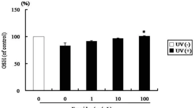
<0.05 versus UVB irradiation alone.Effect of fucoidan on intracellular GSH levels
The intracellular GSH level was reduced to 16.7% by UVB irradiation (100 mJ/cm
2) in HS68 cells, as compared to the UVB non-irradiated group (Fig. 2). However, with fucoidan pretreatment at a dose of 100 μg/ml, the levels of intra- cellular GSH was increased by 21.5% as compared to the UVB irradiation only group (p<0.05).
Effect of fucoidan on ROS generation
ROS production was evaluated via the DCF-DA method.
UVB induced an increase in intracellular ROS as compared to the UVB-non-irradiated group (Fig. 3). Fucoidan sig- nificantly reduced UVB-induced ROS generation by 40.1%
and 68.4% at 10 and 100 μg/ml, respectively, compared to
Fig. 2. Effect of fucoidan on intracellular GSH level induced by UVB in human skin fibroblasts. The cells were pre- treated with fucoidan (0, 1, 10, and 100 μg/ml) prior to UVB irradiation (100 mJ/cm2), and harvested after 24 hr later. The GSH levels were measured using the 5,5-dithiobit (2-nitrobenzoic acid) (DTNB), as described in materials and methods. Each bar represents the mean±SD. *
p
<0.05 versus UVB irradiation alone.Fig. 3. Effect of fucoidan on ROS generation induced by UVB in human skin fibroblasts. The cells were pretreated with fucoidan (0, 1, 10, and 100 μg/ml) prior to UVB irradi- ation (100 mJ/cm2). Followed by incubation for 1 hr, the production of ROS was determined by DCF-DA method.
*
p
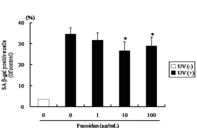
<0.05 versus UVB irradiation alone.Fig. 4. Effect of fucoidan on β-galatosidase activity in human skin fibroblasts. The cells were pretreated with fucoidan (0, 1, 10, and 100 μg/ml) prior to UVB irradiation (100 mJ/cm2), and were stained as described in materials and methods. In all cases a minimum of 400 cells were count- ed for each data point. Each bar represents the mean±
SD. *
p
<0.05 versus UVB irradiation alone.UVB irradiation alone (p<0.05).
Effect of fucoidan on SA β-gal activity
To determine cell senescence as the result of oxidative stress induced by UVB irradiation (100 mJ/cm
2) in the HS68 cells, the SA β-gal activity was assessed. The positive stain- ing rates of SA β-gal were reduced by 23.1% and 16.4% at 10 and 100 μg/ml of fucoidan, respectively, compared to UVB irradiation alone (p<0.05, Fig. 4)
Discussion
UV radiation penetrates readily into dermal tissue and has been confidently and consistently implicated in the ag- ing of skin. Therefore, human dermal skin fibroblasts are considered to be a useful model for investigation of the ef- fects of antioxidants on UV-induced skin photoaging [17].
UVB interacts with cellular chromophores and photo- sensitizers, resulting in the ROS generation, DNA damage, and the activation of cytoplasmic signal transduction path- ways, which are associated with growth, differentiation, rep- licative senescence, and the degradation of connective tissue [9]. According to many researchers, UVB induces a dramatic dose-dependent reduction in intracellular GSH, intra- membrane vitamin E, and membrane fluidity, in addition to an increase in MDA levels [3,16].
In this study, the MDA contents of cells, employed herein as a marker of lipid peroxidation [11], was elevated as the result of UVB irradiation. With fucoidan pretreatment, lipid peroxidation was inhibited during UVB irradiation. MDA,
a by-product of lipid peroxidation, is considered a strongly cytotoxic second messenger which can diffuse within or even escape from the cell, and attack targets distant from the production site. Lipid peroxidation is a complex radical chain reaction, by which unsaturated membrane lipids are oxidized. This process results directly in membrane mod- ifications provoked by reactive lipid peroxidation products such as hydroperosides. Lipid peroxidation has been recog- nized as a critical mechanism in cell injuries occurring dur- ing oxidative stress [2]. Ho et al. [10] previously reported that the MDA level at 150 mJ/cm
2of UVB irradiation was approximately 2.2-fold that of the non-irradiated cells. This result is generally consistent with ours, although the ex- perimental conditions differed.
In this study, fucoidan pretreatment was demonstrated to attenuate DCF fluorescence. The DCF fluorescence is rep- resentative of intracellular activity, and thus an increase in fluorescence is suggestive of intracellular oxidative stress.
The GSH content was reduced significantly in the UVB-irra- diated cells, whereas fucoidan also inhibited the UVB-in- duced decline in GSH content in the skin fibroblasts. The results of the current study suggest that the initial depletion of GSH by UVB induces an increase in ROS production. As GSH is the principal antioxidant system in the cells, the de- pletion of GSH may profoundly affect cell death as the result of ROS accumulation in UVB-irradiated cells [14]. UVB radi- ation is a potent inducer of apoptosis. UVB-mediated nuclear DNA damage has been identified as the primary molecular trigger for the induction of apoptosis. Recently, UVB-gen- erated ROS has been demonstrated to perform an additional function in the execution of apoptotic cell death [20]. In cul- ture and in vivo biopsy, SA β-gal was measured in a variety of cells and tissues in order to assess the onset of cell sen- escence [19]. Eukaryotic β-galactosidase is a hydrolase lo- calized in the lysosome, which cleaves β-D-galactose resi- dues in β-D-galactosides. In 1995, one report suggested that the senescent cells express a specific type of the enzyme β- galactosidase referred to as SA β-gal which has an optimal pH at 6.0 [7]. Thus far, the SA β-gal assay has become one of the most commonly used markers of cell aging in vitro, especially as the result of factors such as sublethal oxidative stress [8]. In this study, fucoidan was shown to reduce SA β -gal activity, and we confirmed that it performs an im- portant function in the retardation of senescent fibroblast ac- cumulation in human skin.
These results suggest that fucoidan may exert a photo-
protective effect against UVB radiation-induced oxidative stress.
References
1. Brandt, R. and A. S. Keston. 1965. Synthesis of diacetyldi- chlorofluorescin: A stable reagent for fluorometric analysis.
Anal. Biochem.
11, 6-9.2. Campo, G. M., A. Avenoso, S. Campo, A. D’Ascola, A. M.
Ferlazzo, and A. Calatroni. 2004. Reduction of DNA frag- mentation and hydroxyl radical production by hyaluronic acid and chondroitin-4-sulphate in iron plus ascorbate-in- duced oxidative stress in fibroblast cultures.
Free Radic. Res.
38, 601-611.
3. Carini, M., G. Aldini, E. Bombardelli, P. Morazzoni, and R. Maffei Facino. 2000. UVB-induced hemolysis of rat eryth- rocytes: protective effect of procyanidins from grape seeds.
Life Sci.
67, 1799-1814.4. Chizhov, A. O., A. Dell, H. R. Morris, S. M. Haslam, R.
A. McDowell, A. S. Shashkov, N. E. Nifant'ev, E. A.
Khatuntseva, and A. I. Usov. 1999. A study of fucoidan from the brown seaweed Chorda filum.
Carbohydr. Res.
320, 108-119.5. Costa, H., S. M. Gallego, and M. L. Tomaro. 2002. Effect of UV-B radiation on antioxidant defense system in sun- flower cotyledons.
Plant Sci.
162, 939-945.6. Darr, D. and I. Fridovich. 1994. Free radicals in cutaneous biology.
J. Invest. Dermatol.
102, 671-675.7. Dimri, G. P., X. Lee, G. Basile, M. Acosta, G. Scott, C.
Roskelly, E. E. Medrano, M. Linskensi, I. Rubeljii, O.
Pereira-Smith, M. Peacocke, and J. Campisi. 1995. A bio- marker that identifies senescent human cells in culture and aging skin in vivo.
Proc. Natl. Acad. Sci.
92, 9363-9367.8. Dumont, P., M. Burton, Q. M. Chen, E. S. Gonos, C.
Frippiat, J. B. Mazarati, F. Eliaers, J. Remacle, and O.
Toussaint. 2000. Induction of replicative senescence bio- markers by sublethal oxidative stresses in normal human fibroblast.
Free Radic. Biol. Med.
28, 361-373.9. Helenius, M., L. Makelainen, and A. Salminen. 1999.
Attenuation of NF-kappaB signaling response to UVB light during cellular senescence.
Exp. Cell Res.
248, 194-202.10. Ho, J. N., Y. H. Lee, J. S. Park, W. J. Jun, H. K. Kim, B.
S. Hong, D. H. Shin, and H. Y. Cho. 2005. Protective effects of aucubin isolated from Eucommia ulmoides against UVB-induced oxidative stress in human skin fibroblasts.
Biol. Pharm. Bull.
28, 1244-1248.11. Jones, S. A., F. McArdle, C. I. Jack, and M. J. Jackson. 1999.
Effect of antioxidant supplementation on the adaptive re- sponse of human skin fibroblasts to UV-induced oxidative stress.
Redox Rep.
4, 291-299.12. Kang, K. A., H. D. Bu, D. S. Park, G. M. Go, Y. Jee, T.
Shin, and J. W. Hyun. 2005. Antioxidant activity of ethanol extract of Callophyllis Japonica
. Phytother. Res.
19, 506-510.13. Lee, B. C., S. Y. Lee, H. J. Lee, G. S. Sim, J. H. Kim, J. H.
Kim, Y. H. Cho, D. H. Lee, H. B. Pyo, T. B. Choe, D. C.
Moon, Y. P. Yun, and J. T. Hong. 2007. Anti-oxidative and photo-protective effects of coumarins isolated from Fraxinus Chinensis.
Arch. Pharm. Res.
30, 1293-130114. Lee, C. S., S. Y. Park, H. H. Ko, and E. S. Han. 2004. Effect of change in cellular GSH levels on mitochondrial damage and cell viability loss due to mitomycin c in small cell lung cancer cells.
Biochem. Pharmacol.
68, 1857-1867.15. McLellan, D. S. and K. M. Jurd. 1992. Anticoagulants from marine algae.
Blood Coagul. Fibrinolysis.
3, 69-77.16. O'Connor, I. and N. O'Brien. 1998. Modulation of UVA light-induced oxidative stress by beta-carotene, lutein and astaxanthin in cultured fibroblasts.
J. Dermatol. Sci.
16, 226-230.17. Offord, E. A., J. C. Gautier, O. Avanti, C. Scaletta, F. Runge, K. Krämer, and L. A. Applegate. 2002. Photoprotective po- tential of lycopene, beta-carotene, vitamin E, vitamin C and carnosic acid in UVA-irradiated human skin fibroblasts.
Free Radic. Biol. Med.
32, 1293-303.18. Ohkawa, H., N. Ohishi, and K. Yagi. 1979. Assay for lipid peroxides in animal tissues by thiobarbituric acid reaction.
Anal. Biochem.
95, 351-358.19. Yang, N. C. and M. L. Hu. 2004. A fluorimetric method using fluorescein di-β-D-galactopyranoside for quantifying the senescence-associated β-galactosidase activity in human foreskin fibroblast Hs68 cells.
Anal. Biochem.
325, 337-343.20. Zeise, E., M. Weichenthal, T. Schwarz, and D. Kulms. 1004.
Resistance of human melanoma cells against the death li- gand TRAIL is reversed by ultraviolet-B radiation via downregulation of FLIP.