Cyanidin-3-O-glucoside Ameliorates Postprandial Hyperglycemia in Diabetic Mice
Kyungha Choi1,2, Sung-In Choi1, Mi Hwa Park2 and Ji-Sook Han1*
1Department of Food Science and Nutrition, and Research Institute of Ecology for the Elderly, Pusan National University, Busan 609-735, Korea
2Department of Food and Nutrition, College of Medical and Life Science, Silla University, Busan 616-735, Korea Received July 29, 2016 /Revised October 10, 2016 /Accepted November 3, 2016
Cyanidin-3-O-glucoside (C3G) shows anti-inflammatory and antioxidant effects; however, its effect on postprandial blood glucose levels remains unknown. Alpha-glucosidase inhibitors regulate post- prandial hyperglycemia by impeding carbohydrate digestion in the small intestine. Here, the effect of C3G on α-glucosidase and α-amylase inhibition and its ability to ameliorate postprandial hyper- glycemia in streptozotocin (STZ)-induced diabetic mice were evaluated. ICR normal and STZ-induced diabetic mice were orally administered soluble starch alone or with C3G or acarbose. The half-max- imal inhibitory concentrations of C3G for α-glucosidase and α-amylase were 13.72 and 7.5 μM, re- spectively, suggesting that C3G was more effective than acarbose. The increase in postprandial blood glucose levels was more significantly reduced in the C3G groups than in the control group for both diabetic and normal mice. The area under the curve for the diabetic mice was significantly reduced following C3G administration. C3G may be a potent α-glucosidase inhibitor and may delay dietary carbohydrate absorption.
Key words : α-Amylase, α-Glucosidase, cyanidin-3-O-glucoside, diabetes, postprandial hyperglycemia
*Corresponding author
*Tel : +82-51-510-2836, Fax : +82-51-583-3648
*E-mail : [email protected]
This is an Open-Access article distributed under the terms of the Creative Commons Attribution Non-Commercial License (http://creativecommons.org/licenses/by-nc/3.0) which permits unrestricted non-commercial use, distribution, and reproduction in any medium, provided the original work is properly cited.
Journal of Life Science 2017 Vol. 27. No. 1. 32~37 DOI : https://doi.org/10.5352/JLS.2017.27.1.32
Introduction
Type 2 diabetes mellitus, which accounts for about 90%
of all diabetes cases, is increasing in incidence worldwide.
The characteristics of type 2 diabetes include postprandial hyperglycemia and atherogenic dyslipidemia. In fact, post- prandial hyperglycemia plays a major role in the develop- ment of type 2 diabetes mellitus and related complications [23]. A postprandial hyperglycemic state is characterized by a rapid and large increase in blood glucose levels. Some studies suggest that postprandial hyperglycemia can cause glucose toxicity, worsen β cell function [14], and induce complications such as cardiovascular disease [3], retinop- athy, and diabetic foot [7]. Therefore, normalizing the post- prandial blood glucose level is important in treating type 2 diabetes.
One of the best ways to lower postprandial glucose levels in the context of hyperglycemia is to inhibit the entry of
glucose into the intestinal endothelial cells by limiting the expression of carbohydrate-hydrolyzing enzymes such as mucosal glucosidases [19]. Intestinal α-glucosidase and pan- creatic α-amylase are the major enzymes of dietary carbohy- drate digestion in humans, and they hydrolyze inner α-1,4- glucosidic linkages in starch and several other poly- saccharides [18]. Oral hypoglycemic agents (e.g., acarbose and voglibose) are able to directly reduce postprandial glu- cose levels [6]. However, these drugs may induce unwanted side effects, including vomiting and diarrhea [15]. Therefore, many researchers have aimed to identify a natural com- pound that can be used for the treatment of diabetes without the induction of major side effects.
As typical antioxidant polyphenols, anthocyanins are wa- ter-soluble pigments that are present in fruits and vegeta- bles, and they range in color from red to purple. The six anthocyanins found in plants and fruits are classified accord- ing to the number and position of the hydroxyl groups on the flavan nucleus [10]; malvindin-3-glucoside, delphini- din-3-glucoside, cyanidin-3-O-glucoside (C3G), and peoni- din-3-glucoside are typical anthocyanins. Moreover, the con- sumption of anthocyanins has beneficial effects in patients with various chronic diseases such as cardiovascular disease, cancer, inflammation, and others [11, 12, 24]. Therefore, eat- ing foods rich in anthocyanins may help individuals remain
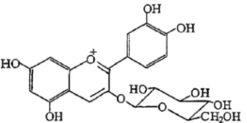
Fig. 1. Chemical structure of cyanidin-3-O-glucoside.
healthy and ameliorate the risk of diverse diseases.
C3G (Fig. 1) is a glycoside within the anthocyanidin group that is abundant in mulberry and red fruits [9]. The molec- ular formula of C3G is C21H21O11+, and its molar mass is 449.3843 g/mol. C3G has the ability to stimulate insulin se- cretion from pancreatic cells in the presence of 4 mM or 10 mM glucose [25] and has also been reported to have anti-in- flammatory and antioxidant effects [8, 28]. Despite these studies, there is no experimental data demonstrating a rela- tionship between postprandial blood glucose levels and C3G. Therefore, in this study, we investigated whether C3G inhibited α-glucosidase and α-amylase, and subsequently de- termined whether it could alleviate postprandial hyper- glycemia in streptozotocin (STZ)-induced diabetic mice.
Materials and Methods
Materials
C3G was purchased from Sigma (St. Louis, MO, USA).
All other chemicals and reagents, including α-glucosidase, α-amylase, and acarbose, were of analytical grade and were purchased from Sigma. All chemicals and reagents were used without any further purification.
Inhibition of α-glucosidase activity by C3G in vitro The α-glucosidase inhibition assay was conducted using a chromogenic method, as described previously (31), with a readily available yeast enzyme. In brief, yeast α-glucosi- dase (0.7 U; Sigma) was dissolved in 100 mM phosphate buf- fer (pH 7.0) containing 2 g/l bovine serum albumin and 0.2 g/l NaN3, and used as an enzyme solution. Five millimolar p-nitrophenyl-α-d-glucopyranoside (pNGP) in the same buf- fer (pH 7.0) was used as the substrate solution. Next, 50 μl of the enzyme solution and 10 μl of the sample dissolved in dimethyl sulfoxide (5 mg/ml) were mixed in each well of a microtiter plate, and the titer was measured by de- termining the absorption at 405 nm at time 0 with a micro- plate reader. After incubation for 5 min, the substrate sol-
ution (50 μl) was added and incubated for another 5 min at room temperature. The increase in absorbance from time 0 was measured. The inhibitory activity was calculated as follows: 100-relative absorbance difference (%) of the test compounds (relative to the absorbance change of the control, where the test solution was replaced by the carrier solvent).
Inhibition of α-amylase activity by C3G
α-Amylase inhibitory activity was assayed in the same way as described for the α-glucosidase inhibition assay, ex- cept that porcine pancreatic amylase (100 U) and p-nitro- phenyl-α-d-maltopentoglycoside (pNPM) were used as the enzyme and substrate, respectively.
Experimental animals
Male ICR mice (4 weeks of age; purchased from Joong Ang Lab Animal Co., Seoul, Korea) were used in this study.
All animals were housed individually in a room with con- trolled light (12 hr on/12 hr off) and temperature, and pel- leted food and water were available ad libitum. After an ad- justment period of approximately 2 weeks, diabetes was in- duced by intraperitoneal injection of STZ (60 mg/kg) freshly dissolved in a citrate buffer (0.1 M, pH 4.5). After 7 days, tail bleeds were performed and animals with a blood glucose concentration above 250 mg/dl were considered diabetic. All procedures for the handling and care of animals were ap- proved by the animal ethics committee of our university (ED-PNU2014-0663).
Measurement of blood glucose level
Normal mice and STZ-induced diabetic mice were fasted overnight and randomly divided into three groups (n=7 mice each). Fasted animals were deprived of food for at least 12 hr but allowed free access to water. After overnight fast- ing, the mice were orally administered soluble starch (2 g/kg body weight), alone or with C3G (10 mg/kg body weight) or acarbose (10 mg/kg body weight). Blood samples were taken from the tail vein at 0, 30, 60, and 120 min. Blood glucose was measured using a glucometer (Roche Diagnos- tics GmbH, Germany). Areas under the curve (AUC) were calculated using the trapezoidal rule [16].
Data and statistical analysis
The data are represented as the mean ± standard devia- tion. Statistical analysis was performed using SAS version 9.1. Differences were evaluated by one-way analysis of var-
Fig. 2. Inhibitory activity of cyanidin-3-O-glucoside on α-gluco- sidase. The inhibitory effect was determined using p-ni- trophenyl-α-glucopyranoside as a substrate. Acarbose was used as a positive control. Each value is expressed as the mean ± standard deviation of triplicate experi- ments. Values with different symbols (*, **, ***) are sig- nificantly different at p<0.05 as analyzed by Duncan's multiple range test. The final concentration of acarbose was 100 μM.
Fig. 3. Inhibitory activity of cyanidin-3-O-glucoside on α-amy- lase. The inhibitory effect was determined using p-nitro- phenyl-α-maltopentoglycoside as a substrate. Acarbose was used as a positive control. Each value is expressed as the mean ± standard deviation of triplicate experi- ments. Values with different symbols (*, **, ***) are sig- nificantly different at p<0.05 as analyzed by Duncan's multiple range test. The final concentration of acarbose was 100 μM.
Table 1. Half-maximal inhibitory concentration (IC50) values of cyanidin-3-O-glucoside on α-glucosidase and α-amylase
Sample IC50 (μM)1
α-glucosidase α-amylase Acarbose
Cyanidin-3-O-glucoside
130.04±8.42 13.72±1.24*
165.12±6.19 7.50±0.58*
1Each value is expressed as the mean ± standard deviation of triplicate experiments.
*p<0.05 compared to the control group.
iance followed by post-hoc Duncan's multiple range tests.
Results
Inhibition effect of C3G on α-glucosidase and α- amylase in vitro
The inhibitory effects of C3G against α-glucosidase were examined using pNGP as a substrate and compared with the effects of the commercial α-glucosidase inhibitor acar- bose (Fig. 2). α-Glucosidase activity was inhibited by C3G in a concentration-dependent manner, exhibiting 30.89±1.77
%, 40.96±2.83%, 53.11±3.08%, and 61.77±2.39% inhibition of activity at concentrations of 5, 10, 25, and 50 μM, respectively.
Moreover, C3G was more effective than acarbose, even at the low concentrations. Next, the inhibitory effects of C3G on α-amylase activity (Fig. 3) were determined using pNPM as a substrate and compared with those of acarbose. The inhibitory effect of C3G against α-amylase increased in a concentration-dependent manner (42.68±1.22%, 57.32±2.15%, 68.54±1.33%, and 74.25±5.14% inhibition of activity at con- centrations of 5, 10, 25, and 50 μM, respectively). C3G also inhibited α-amylase activity more effectively than acarbose.
The half-maximal inhibitory concentration values of C3G against α-glucosidase and α-amylase were 13.72±1.24 μM and 7.50±0.58 μM, respectively, further supporting that C3G had a stronger inhibitory effect than acarbose (Table 1).
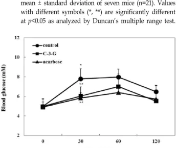
Effect of C3G on blood glucose levels in vivo Next, we investigated the effects of C3G on the blood glu- cose levels after a meal in STZ-induced diabetic and normal mice. The postprandial blood glucose levels of mice consum- ing C3G were significantly lower than those of control dia- betic mice (Fig. 4). Blood glucose levels were increased (19.62±1.75 mM and 20.67±2.39 mM at 30 and 60 min, re- spectively) after a meal and then decreased (17.44±1.20 mM at 120 min) in diabetic mice. However, when C3G was added to the feed, the increase in the postprandial blood glucose level was significantly reduced (16.20±1.36, 16.57±1.16, and 13.39±0.96 mM at 30, 60, and 120 min, respectively; p<0.05).
Postprandial blood glucose levels were also significantly de- creased when normal mice were orally administered starch together with C3G (Fig. 5; p<0.05). The AUC for the glucose response in diabetic mice consuming C3G (1855.05±141.68 mmol·min-1·L-1) was significantly lower (p<0.05) than that
Fig. 4. Blood glucose level after administration of cyanidin-3- O-glucoside (C3G) in streptozotocin-induced diabetic mice. C3G (10 mg/kg), acarbose (10 mg/kg), and dis- tilled water as a control were co-administered orally with starch (2 g/kg). Each value is expressed as the mean ± standard deviation of seven mice (n=21). Values with different symbols (*, **) are significantly different at p<0.05 as analyzed by Duncan's multiple range test.
Fig. 5. Blood glucose level after administration of cyanidin-3-O- glucoside (C3G) in normal mice. C3G (10 mg/kg), acarbose (10 mg/kg), and distilled water as a control were co-administered orally with starch (2 g/kg). Each value is expressed as the mean ± standard deviation of seven mice (n=21). Values with different symbols (*, **) are significantly different at p<0.05 as analyzed by Duncan's multiple range test.
Table 2. Area under the curve (AUC) of postprandial glucose responses of normal and streptozotocin-induced dia- betic mice
Group1 AUC (mmol·min-1·L-1)
Normal mice Diabetic mice Control
Cyanidin-3-O-glucoside Acarbose
861.75±98.55a 736.20±126.75b 708.60±101.55c
2263.05±212.48a 1855.05±141.68b 1880.25±238.05b
1Cyanidin-3-O-glucoside (10 mg/kg), acarbose (10 mg/kg), and control (distilled water) were co-administered orally with starch (2 g/kg). Each value is expressed as the mean ± standard deviation of seven mice (n=42).
a–cValues with different letters are significantly different at p<0.05 as analyzed by Duncan's multiple range test.
in control diabetic mice (2263.05±212.48 mmol·min-1·L-1; Table 2). The AUC values in normal mice were consistent with those in diabetic mice, demonstrating the hypoglycemic effect of C3G.
Discussion
Retaining near normal levels of blood glucose, both in the fasting and postprandial phases, is the main treatment
goal for diabetic patients. During the early stages of type 2 diabetes, postprandial hyperglycemia is the primary con- cern [21]. In the postprandial state, there is a rapid and large increase in blood glucose levels [4]. Elevation of postprandial blood glucose levels leads to enhancement of oxidative stress and endothelial dysfunction, causing diabetic complications such as cardiovascular disease [5, 6]. One attractive strategy for the management of postprandial hyperglycemia is the inhibition of pancreatic α-amylase or intestinal α-glucosidase to slow down the digestion of carbohydrates [26]. Synthetic inhibitors of these enzymes, such as acarbose, miglitol, and voglibose, function directly in reducing carbohydrate hy- drolysis and ameliorating postprandial hyperglycemia [13, 29]. However, most of the currently available anti-diabetic agents cause severe adverse effects [22]. Therefore, identi- fication of new, effective natural agents for the suppression of glucose absorption in the intestine is critical for the treat- ment of postprandial hyperglycemia [27].
α-Glucosidases are located in the intestinal epithelial cell membrane and hydrolyze carbohydrates such as starch and table sugar. α-Amylase also catalyzes the hydrolysis of the glucosidic linkages of starch, glycogen, or other carbohy- drates. Inhibition of intestinal α-glucosidase and pancreatic α-amylase delays carbohydrate digestion and slows down the sharp rise in blood glucose levels. Therefore, the discov- ery of effective, nontoxic inhibitors of α-glucosidase and α- amylase may lead to improved treatment options for diabetes.
In this study, we investigated the effects of C3G on the inhibition of α-glucosidase and α-amylase activities to ex- plore its potential as an anti-hyperglycemic agent. C3G ex- hibited more prominent inhibitory activity against both α-
glucosidase and α-amylase than that observed with the com- mercial inhibitor of carbohydrate digestive enzyme, acarbose.
Indeed, even at low concentrations, C3G had better in- hibitory effects than acarbose.
C3G is a common, naturally occurring anthocyanin and derivative of 2-phenylbenzophyrylium (flavylium) with a bound sugar. One study reported the structural difference between cyanidin, C3G, and cyanidin-3,5-diglucoside, dem- onstrating that C3G had more potent inhibitory activity for pancreatic α-amylase and intestinal sucrase than cyanidin or cyanidin-3,5-diglucoside. Thus, the 3-O-glucoside structure in C3G has more potent inhibitory effects on pancreatic α- amylase than cyanidin or cyanidin-3,5-diglucoside in vitro [2]. Another study demonstrated that introduction of rusi- nose in the 3-O-position of cyanidin-3-rutinoside (C3R) in- creased the level of intestinal sucrase inhibition [1]. Thus, the structure of 3-O-glucoside in C3G may have an im- portant role in the inhibition of α-glucosidase inhibitory activity.
In patients with type 2 diabetes, postprandial hyper- glycemia is a major factor associated with uncontrolled blood glucose levels. In addition, postprandial hyper- glycemia has been reported to contribute to cardiovascular complications [17]. Thus, we also investigated the anti-hy- perglycemic effects of C3G in STZ-induced diabetic and nor- mal mice after the consumption of starch. After consumption of C3G, the increase in postprandial blood glucose levels was significantly suppressed in both STZ-induced diabetic mice and normal mice. In this experiment, C3G alleviated postprandial hyperglycemia almost as well as acarbose. This effect on postprandial hyperglycemia may be due to the in- hibition of carbohydrate digestive enzymes by C3G in epi- thelial cells of the small intestine. C3G also reduced the AUC of the blood glucose response curve. These results demon- strated that absorption of glucose may be delayed by C3G, leading to attenuation of the increase in postprandial blood glucose level.
Several studies have described the connection between postprandial hyperglycemia and cardiovascular disease [20].
Thus, controlling postprandial hyperglycemia is important for improving diabetic symptoms and preventing related complications. Many synthetic products are already avail- able as therapies for controlling postprandial hyperglycemia.
However, these products have unwanted side effects.
Therefore, researchers have attempted to identify natural substances for alleviating postprandial hyperglycemia. Our
current study showed that C3G may be effective for improv- ing postprandial hyperglycemia and preventing diabetic complications.
References
1. Adisakwattana, S., Yibchok-Anun, S., Charoenlertkul, P.
and Wongsasiripat, N. 2010. Cyanidin-3-rutinoside alle- viates postprandial hyperglycemia and its synergism with acarbose by inhibition of intestinal α-glucosidase. J. Clin.
Biochem. Nutr. 49, 36-41.
2. Akkarachiyasit, S., Charoenlertkul, P., Yibchok-anun, S. and Adisakwattana, S. 2010. Inhibitory activities of cyanidin and its glycosides and synergistic effect with acarbose against intestinal α-glucosidase and pancreatic α-amylase. Int. J.
Mol. Sci. 11, 3387-3396.
3. Baron, A. D. 1998. Postprandial hyperglycaemia and al- pha-glucosidase inhibitors. Diabetes Res. Clin. Pract. 40, S51- 55.
4. Borona, E. and Muggeo, M. 2001. Postprandial blood glu- cose as a risk factor for cardiovascular disease in type 2 diabetes: the epidemiological evidence. Diabetologia 44, 2107-2114.
5. David, Bell., O’Keefe, J. and Jellinger, P. 2008. Postprandial dysmetabolism: the missing link between diabetes and car- diovascular events? Endocr. Pract. 14, 112-124.
6. Derosa, G. and Maffioli, P. 2012. α-Glucosidase inhibitors and their use in clinical practice. Arch. Med. Sci. 8, 899-906.
7. Ergul, A. 2011. Endothelin-1 and diabetic complications: fo- cus on the vasculature. Pharmacol. Res. 163, 477-482.
8. Fu, Y., Wei Z., Zhou, E., Zhang, N. and Yang, Z. 2014.
Cyanidin-3-O-β-glucoside inhibits lipopolysaccharide-in- duced inflammatory response in mouse mastitis model. J.
Lipid Res. 21, 1111-1119.
9. Galvano, F., La Fauci, L., Vitaglione P., Fogliano, V., Vanella, L. and Felgines, C. 2007. Bioavailability, antioxidant and bio- logical properties of the natural free-radical scavengers cya- nidin and related glycosides. Ann. Ist. Super Sanita. 43, 382- 393.
10. Ghosh, D. and Konishi, T. 2007. Anthocyanins and antho- cyanin-rich extracts: role in diabetes and eye function. Asia Pac. J. Clin. Nutr. 16, 200-208.
11. Graf, D., Seifert, S., Jaudszus, A., Bub, A. and Watzl, B. 2013.
Anthocyanin-rich juice lowers serum cholesterol, leptin, and resistin and improves plasma fatty acid composition in fischer rats. PLoS. One 18, e66690.
12. Huang, W. Y., Liu, Y. M., Wang, J., Wang, X. N. and Li, C. Y. 2014. Anti-inflammatory effect of the blueberry antho- cyanins malvidin-3-glucoside and malvidin-3-galactoside in endothelial cells. Molecules 21, 12827-12841.
13. Yamagishi, S., Nakamura, K. and Takeuchi, M. 2004.
Inhibition of postprandial hyperglycemia by acarbose is a promising therapeutic strategy for the treatment of patients with the metabolic syndrome. Med. Hypotheses 65, 152-154.
14. Jovanovic, L. 1999. Rationale for prevention and treatment
초록:당뇨 마우스에서 cyanidin-3-O-glucoside의 식후 고혈당 완화 효과
최경하1,2․최성인1․박미화2․한지숙1*
(1부산대학교 식품영양학과, 2신라대학교 식품영양학과)
Cyanidin-3-O-glucoside (C3G)는 오디와 붉은색의 과일에 풍부하게 함유되어 있으며, 항염증과 항산화 효과와 관련하여 보고되어있다. 그러나, C3G의 식후 혈당에 관한 연구 결과는 보고되지 않았다. α-glucosidase 억제제는 소장에서 탄수화물 소화의 속도를 방해함으로써 식후 고혈당을 조절한다. 본 연구에서는 C3G가 α-글루코시다아 제와 α-아밀라아제에 미치는 억제효과 및 스트렙토조토신(STZ)이 유발하는 당뇨병 생쥐의 식후고혈당에 미치는 완화 효과를 조사하였다. ICR 마우스와 streptozothocin (STZ)으로 유도된 당뇨병 마우스에 수용성 전분(2 g/kg body weigh)으로 경구부하 후 C3G (10 mg/kg body weight) 또는 acarbose (10 mg/kg body weight)를 단독 또는 함께 투여하였다. 혈액 샘플은 꼬리에서 0, 30, 60, 120분 간격으로 채취하였다. α-글루코시다아제와 α-아밀라 아제에 대한 C3G의 IC50 값은 각각 13.72와 7.5 μM의 결과값을 나타내어, 양성대조군인 acarbose보다 더 효과적이 었다. STZ으로 유발된 당뇨 쥐의 식후 혈당 수치는 대조군에 비해 C3G 투여시 유의적으로 더 낮았다. 게다가, C3G 투여는 당뇨병 흰쥐에서 포도당 반응에 대한 곡선하면적 감소와 관련이 있었다. 그러므로, C3G는 α-글루코 시다아제의 강력한 억제제이며 식이 탄수화물의 흡수를 지연시킬 수 있음을 나타낸다.
of postprandial glucose-mediated toxicity. Endocrinologist 9, 87-92.
15. Katahira, H. and Ishida, H. 2002. Indication and side effect of alpha glucosidase inhibitor. Nihon Rinsho 60, 399-408.
16. Kim, J. S. 2004. Effect of Rhemanniae radix on the hyper- glycemic mice induced with streptozotocin. J. Kor. Med. Sci.
33, 1133-1138.
17. Laakso, M. 1999. Hyperglycemia and cardiovascular disease in type 2 diabetes. Diabetes 48, 937-942.
18. Lebovitz, H. E. 1997. Alpha-Glucosidase inhibitors. Endocri- nol. Metab. Clin. North. Am. 26, 539-551.
19. Lee, B. H., Eskandari R., Jones, K., Reddy, K. R., Roberto, Q. C., Nichols, B. L., David, R. R., Hamaker, B. R. and Mario Pinto, B. 2012. Modulation of starch digestion for slow glu- cose release through “Toggling” of activities of Mucosal α- Glucosidases. J. Biol. Chem. 287, 31929-31938.
20. Madsbad, S., Brock, B., Schmitz, O. and Ugeskr, L. 2003. Post- prandial blood glucose fluctuations, cardiovascular disease and late diabetic complications. Ugeskr. Laeg. 165, 3149-3153.
21. Moradi-Afrapoli, F., Asghari, B., Saeidnia, S., Ajani, Y., Mirjani, M., Malmir, M., Bazaz, R. D., Hadjiakoondi, A., Salehi, P., Hamburger, M. and Yassa, N. 2012. In vitro α- glucosidase inhibitory activity of phenolic constituents from aerial parts of Polygonum hyrcanicum. DARU 20, 37.
22. Sama, K., Murugesan, K. and Sivaraj, R. 2012. In vitro alpha amylase and alpha glucosidase inhibition activity of crude ethanol extract of Cissus arnottiana. Asian J. Plant Sci. Res.
14, 550-553.
23. Sheng, Z., Dai, H., Pan, S., Wang, H., Hu, Y. and Ma, W.
2014. Isolation and Characterization of an α-Glucosidase Inhibitor from Musa spp. (Baxijiao) Flowers. Molecules 9, 10563-10573.
24. Special, A., Cimino, F., Saija, A., Canali, R. and Virgili, F.
2014. Bioavailability and molecular activities of anthocya
nins as modulators of endothelial function. Genes. Nutr. 9, 404.
25. Sun, C. D., Zhang, B., Zhang, J. K., Xu, C. J., Wu, Y. L., Li, X. and Chen, K. S. 2012. Cyanidin-3-glucoside-rich ex- tract from Chinese bayberry fruit protects pancreatic β cells and ameliorates hyperglycemia in streptozotocin-induced diabetic mice. J. Med. Food 15, 288-298.
26. Tarling, C. A., Woods, K., Zhang, R., Brastianos, H. C., Brayer, G. D., Andersen, R. J. and Withers, S. G. 2008. The search for novel human pancreatic alpha-amylase inhibitors:
high-throughput screening of terrestrial and marine natural product extracts. Chembiochem 15, 433-438.
27. Toma, A., Makonnen, E., Mekonnen, Y., Debella, A. and Addi- sakwattana, S. 2014. Intestinal α-glucosidase and some pan- creatic enzymes inhibitory effect of hydroalcholic extract of Moringa stenopetala leaves. BMC Complement Altern. Med. 14, 180.
28. Tsuda, T., Horio, F. and Osawa, T. 1998. Dietary cyanidin 3-O-β-D-glucoside increases ex vivo oxidation resistance of serum in rats. Lipids 33, 583-588.
29. Van de Laar, F. A., Lucassen, P. L., Akkermans, R. P., Van de Lisdonk, E. H., Rutten Guy, E. and Van Weel, C. 2005.
Alpha-glucosidase inhibitors for patients with type 2 dia- betes: results from a Cochrane systematic review and meta-analysis. Diabetes Care 28, 154-163.
30. Wang, L. S., Kuo, C. T., Cho, S. J., Seguin, C., Siddiqui, J., Stoner, K., Weng, Y. I., Huang, T. H., Tichelaar, J., Yearsley, M., Stoner, G. D. and Huang, Y. W. 2013. Black rasp- berry-derived anthocyanins demethylate tumor suppressor genes through the inhibition of DNMT1 and DNMT3B in colon cancer cells. Nutr. Cancer 65, 118-25.
31. Watanabe, J., Kawabata, J., Kurihara, H. and Niki, R. 1997.
Isolation and identification of alpha-glucosidase inhibitors from tochucha (Eucommia ulmoides). Biosci. Biotechnol.
Biochem. 61, 177-178.