Alleviating Effects of Mulberry Fruit Extract on Postprandial Hyperglycemia in Streptozotocin-induced Diabetic Mice
Kyung Ha Choi1,2, Ji-Hye Kang1 and Ji-Sook Han1*
1Department of Food Science and Nutrition, and Research Institute of Ecology for the Elderly, Pusan National University, Busan 609-735, Korea
2Department of Food and Nutrition, College of Medical and Life Science, Silla University, Busan 616-735, Korea Received April 1, 2016 /Revised April 8, 2016 /Accepted April 11, 2016
Postprandial hyperglycemia is an early defect of type 2 diabetes and one of primary anti-diabetic targets. The alpha-glucosidase inhibitors regulate postprandial hyperglycemia by impeding the rate of carbohydrate (such as starch) digestion in the small intestine. This study was designed to investigate the inhibitory actions of mulberry fruit extract (MFE) on α-glucosidase and α-amylase activities, and its alleviating effect on postprandial hyperglycemia activities in vitro and in vivo. Male four-week old ICR mice and streptozotocin (STZ)-induced diabetic mice were treated with mulberry fruit extract.
MFE showed strong inhibitory effects against α-glucosidase and α-amylase activities, with half-max- imal inhibitory concentration (IC50) values of 0.16 and 0.14 mg/ml, respectively, and was more effec- tive than acarbose, which was used as a positive control. The increase in postprandial blood glucose levels was more significantly attenuated in the MFE-administered group mice than in the control group mice of both STZ-induced diabetic and normal mice. Moreover, the area under the glucose re- sponse curve significantly decreased following MFE administration in diabetic mice. These results in- dicate that MFE may be a potent inhibitor of α-glucosidase and α-amylase, and helpful in suppressing postprandial hyperglycemia in diabetic mice. The mulberry fruit extracts may be considered as a po- tential candidate for the management of diabetes.
Key words : α-Amylase, α-Glucosidase, diabetic mice, mulberry fruit extract, postprandial hyperglycemia
*Corresponding author
*Tel : +82-51-510-2836, Fax : +82-51-583-3648
*E-mail : [email protected]
This is an Open-Access article distributed under the terms of the Creative Commons Attribution Non-Commercial License (http://creativecommons.org/licenses/by-nc/3.0) which permits unrestricted non-commercial use, distribution, and reproduction in any medium, provided the original work is properly cited.
Journal of Life Science 2016 Vol. 26. No. 8. 921~927 DOI : http://dx.doi.org/10.5352/JLS.2016.26.8.921
Introduction
The prevalence of diabetes mellitus, a metabolic disease characterized by hyperglycemia resulting from defects in both regulations of insulin secretion and/or insulin action, is increasing worldwide [9]. The possibility that postprandial hyperglycemia is associated with the development of dia- betic complications has recently received much attention [30]. An acute hyperglycemia after a meal may result in en- dothelial dysfunction and oxidative stress [11]. Moreover, studies have shown an association between postprandial glucose levels and the pathogenesis of atherosclerosis [32].
Therefore, management of postprandial hyperglycemia is considered a major therapeutic strategy for type 2 diabetes treatment, and can be achieved by delaying the release of glucose through the inhibition of carbohydrate hydrolyzing
enzymes such as α-amylase and α-glucosidase in the diges- tive tract [26].
α-glucosidase inhibitors such as acarbose and miglitol are hypoglycemic agents that slow the digestion and absorption of carbohydrates in the small intestine and hence, reduce the increase in blood glucose levels after a meal. However, it is well acknowledged that administration of such drugs is frequently associated with adverse effects including flat- ulence, abdominal pain, and diarrhea [5, 29]. Thus, many natural products having minimal side effects have been in- vestigated with respect to their inhibition of glucose pro- duction from carbohydrates in the gut or glucose absorption from the intestine [3].
Mulberry (Morus alba L.) is a genus of flowering plants in the family Moraceae, and its fruits and leaves have been used as traditional oriental medicines to treat diabetes [4].
The mulberry fruit, called Oddi in Korea, is a 2- to 3 cm long multiple fruit. Immature fruits are white, green, or pale yellow. The fruits turn pink and then red while ripening, and finally dark purple or black with a sweet flavor when fully ripe. Mulberry fruits contain large amounts of antho- cyanins. Cyanidin-3-glucoside (C3G) and cyanidin-3-rutino- side (C3R) are the most important active components of mul-
berry, having many potentially beneficial effects on human health [22]. According to traditional oriental medicine, mul- berry fruit can protect the liver and kidney from damage, strengthen the joints, improve eyesight, and possess anti-ag- ing effects [21]. Furthermore, recent studies have demon- strated that mulberry fruit exhibits inhibitory effects on CYP3A activity [12], as well as neuroprotective [10], im- munomodulatory [18], anti-inflammatory [19], hypolipi- demic, and antioxidant properties [35].
However, no information is currently available about the inhibitory effect of mulberry fruits on intestinal α-glucosi- dase, either in vitro or in vivo. Therefore, we investigated whether mulberry fruit extracts may inhibit α-glucosidase and α-amylase activities, and alleviate postprandial hyper- glycemia in streptozotocin (STZ)-induced diabetic mice.
Materials and Methods
Materials
Mulberry (M. alba L.) fruits were collected from the coast of Sangju-si Gyeongsangbuk-do, South Korea. The fruits were individually washed with tap water, rinsed carefully with fresh water, and freeze-dried. The dried sample was ground and sifted through a 50 mesh standard testing sieve.
The sample was extracted three times with ten volumes of 80% methanol for 12 hr at room temperature. The filtrate was then vacuum-evaporated to obtain the extract. After the mulberry fruit extract (MFE) was thoroughly dried, it was stored in a deep freezer (-80°C). The composition of antho- cyanins in the MFE was analyzed by high-performance liq- uid chromatography (HPLC), and C3G and C3R were found to be the major anthocyanins. The levels of C3G and C3R in the MFE were 34.1 and 14.3 mg/1.5 g, respectively [4].
Inhibition assay for α-glucosidase activity in vitro The α-glucosidase inhibition assay was conducted by the chromogenic method described by Watanabe et al. (1997) us- ing a readily available yeast enzyme [33]. Briefly, yeast α -glucosidase (0.7 units, Sigma, St. Louis, MO, USA) was dis- solved in 100 mM phosphate buffer (pH 7.0) containing 2 g/l bovine serum albumin and 0.2 g/l NaN3 to form the enzyme solution. The substrate solution was prepared by dissolving p-nitrophenyl-α-D-glucopyranoside (5 mM) in the same buffer. Next, 50 μl of enzyme solution and 10 μl of sample dissolved in dimethyl sulfoxide (5 mg/ml) were mixed in a well of a microtiter plate and the absorbance
at 405 nm was measured with a microplate reader at time zero. After incubation for 5 min, the substrate solution (50 μl) was added and the mixture was incubated for another 5 min at room temperature. The increase in absorbance from time zero was measured. The inhibitory activity of MFE at varying concentrations was expressed as 100 minus the ab- sorbance change of test compounds relative to the absorb- ance change of the control (%), where the test solution was replaced by the carrier solvent. The measurements were per- formed in triplicate and the IC50 value (the concentration of MFE that results in 50% inhibition of maximal activity) was determined.
Inhibition assay for α-amylase activity in vitro The α-amylase inhibition assay was performed in the same way as described for the α-glucosidase inhibition assay except that porcine pancreatic amylase (100 units, Sigma) and p-nitrophenyl-α-D-maltopentoglycoside were used as the enzyme and substrate, respectively.
Experimental animals
Four-week-old, male ICR mice (Orient Bio Inc., Seong- nam, Korea) were used as test animals and were housed individually in a light (12 hr on/12 hr off) and temper- ature-controlled room with ad libitum access to pelleted food and water. After a 2-week adjustment period, diabetes was induced as described below. All procedures were approved by the animal ethics committee of our university. All animal experiments were treated in accordance with guidelines of the Pusan National University for the care and use of labo- ratory animals (ED-PNU2014-0663).
Induction of diabetes
To induce diabetes, mice were fasted for 18 hr before ad- ministration of a single intraperitoneal (i.p.) injection of 60 mg/kg STZ prepared in 0.1 M sodium citrate buffer (pH 4.5). One week after injection of STZ, fasting blood glucose levels were periodically measured using a glucometer (Roche Diagnostics GmbH, Mannheim, Germany). Mice with blood glucose values of 250 mg/dl or higher were consid- ered diabetic.
Measurement of blood glucose level
Normal mice and STZ-induced diabetic mice fasted over- night (deprived of food for at least 12 hr but allowed free access to water) were each randomly divided into 3 groups
de
cd
c
b
a
e
d
cd cd
b
0 20 40 60 80
0.05 0.10 0.15 0.20 0.50
Inhibitory effect (%)
Concentration (mg/ml) MTE
Acarbose
Fig. 1. Inhibitory effect of mulberry fruit extract (MFE) on α- glucosidase. Each value represents the mean ± SD of triplicate experiments. a–eValues denoted by different letters are significantly different at p<0.05 as analyzed by Duncan’s multiple range test. Acarbose was used as positive control.
cd
c
bc
b
a
e
e d cd
bc
0 20 40 60 80
0.05 0.10 0.15 0.20 0.50
Inhibitory effect (%)
Concentration (mg/ml) MTE
Acarbose
Fig. 2. Inhibitory effect of mulberry fruit extract (MFE) on α- amylase. Each value represents the mean ± SD of triplicate experiments. a–eValues denoted by different letters are significantly different at p<0.05 as analyzed by Duncan’s multiple range test. Acarbose was used as positive control.
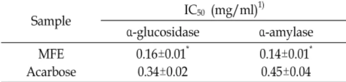
Table 1. IC50 values of the inhibitory effect of MFE on α-glucosi- dase and α-amylase
Sample IC50 (mg/ml)1)
α-glucosidase α-amylase
MFE Acarbose
0.16±0.01* 0.34±0.02
0.14±0.01* 0.45±0.04
1IC50 value is the concentration of sample required for 50%
inhibition. Each value represents the mean ± SD of triplicate experiments.
*p<0.05 compared to the control group.
of 7 mice (a total 6 groups) and treated as follows: 1) control:
mice received oral administration of soluble starch (2 g/kg body weight [BW]) alone; 2) MFE: mice received oral admin- istration of starch with MFE (300 mg/kg BW); 3) acarbose:
mice received oral administration of starch with acarbose (100 mg/kg BW). Blood samples were taken from the tail vein at 0, 30, 60, and 120 min after oral administration. Blood glucose level was measured using a glucometer (Roche Diagnostics GmbH) and the area under the glucose response curve (AUC) was calculated using the trapezoidal rule [13].
Data and statistical analysis
The data are represented as the mean ± standard devia- tion (SD) of triplicate experiments. The statistical analysis was performed using SAS software ver. 9.1 (SAS Institute Inc., Cary, NC, USA). Differences among groups were eval- uated by one-way analysis of variance (ANOVA) followed by Duncan’s multiple range test. Differences between two groups were compared using Student’s t-test. Values of p less than 0.05 were considered statistically significant.
Results and Discussion
Inhibitory effect of MFE on α-glucosidase and α- amylase in vitro
The inhibitory effect of MFE against α-glucosidase was determined using p-nitrophenyl-α-glucopyranoside as the substrate. α-glucosidase activity was inhibited by MFE in a dose-dependent manner, with 28.57, 38.10, 48.15, and 59.26%
inhibition at MFE concentrations of 0.05, 0.10, 0.15, and 0.20 mg/ml, respectively (Fig. 1). The α-glucosidase inhibitory ac- tivity of the MFE was significantly higher than that of the positive control, acarbose. The α-amylase inhibitory activity of MFE on the other hand, was determined using p-nitro- phenyl-α-maltopentoglycoside as the substrate. Similarly, the inhibitory activity of MFE against α-amylase was also dose-dependent (34.52, 40.74, 53.70, and 61.57% inhibition at MFE concentrations of 0.05, 0.10, 0.15, and 0.20 mg/ml, respectively) (Fig. 2). Compared with the commercial in- hibitor, acarbose, MFE was more effective in inhibiting α- amylase activity. The IC50 values of MFE against α-glucosi- dase and α-amylase were 0.16 and 0.14 mg/ml, respectively, which demonstrated its stronger inhibitory activities over those of acarbose (Table 1).
Postprandial hyperglycemia is a prominent and early de- fect in the development of diabetes and an important factor
that contributes to complications associated with diabetes, such as micro- and macrovascular diseases. Therefore, allevi- ating hyperglycemia in the postprandial state is a treatment goal for patients with diabetes [27]. Starch is primarily di-
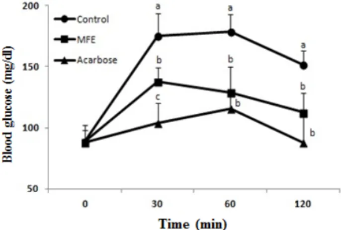
Fig. 3. Blood glucose levels after the administration of mulberry fruit extract (MFE) in STZ-induced diabetic mice. Distil- led water (as control), MFE (300 mg/kg), and acarbose (100 mg/kg) were co-administered orally with starch (2 g/kg). Each value represents the mean ± SD of seven mice (n = 21). a–cValues denoted by different letters are significantly different at p<0.05 as analyzed by Duncan’s multiple range test.
Fig. 4. Blood glucose levels after the administration of mulberry fruit extract (MFE) in normal mice. Distilled water (as control), MFE (300 mg/kg), and acarbose (100 mg/kg) were co-administered orally with starch (2 g/kg). Each value represents the mean ± SD of seven mice (n = 21).
a–cValues denoted by different letters are significantly different at p<0.05 as analyzed by Duncan’s multiple range test.
gested in the small intestine through the action of pancreatic α-amylase, producing both linear maltose and branched iso- maltose oligosaccharides, which are further hydrolyzed by intestinal α-glucosidase to yield absorbable monosaccharides [17]. α-glucosidase catalyzes the final step in the digestive process of carbohydrates; hence, its inhibitors could impede the absorption of dietary carbohydrates to reduce post- prandial hyperglycemia [24]. Thus, controlling postprandial hyperglycemia by inhibiting α-glucosidase and α-amylase is one of the key goals in diabetic therapy [1].
In the present study, the inhibitory activities of MFE on α-glucosidase and α-amylase were investigated to examine the possible use of MFE as an anti-hyperglycemic functional food. MFE has higher inhibitory activities on the afore- mentioned enzymes than the commercial carbohydrate di- gestive enzyme inhibitor, acarbose; and this can be attrib- uted to the active compounds present in MFE. Mulberry fruits contain abundant anthocyanins, particularly C3G, C3R, pelargonidin 3-O-glucoside, and pelargonidin 3-O-rutino- side [20, 25].
These anthocyanins, belonging to the flavonoid class of polyphenolic compounds, are known to bind to a variety of proteins. The hydroxyl groups in polyphenolic com- pounds may link and form complexes with enzymes, thus inhibiting their activity [14, 31]. McDougall et al. [23] re- ported that anthocyanins from berry extracts have been im- plicated in the inhibition of α-glucosidase [23]. Furthermore, Iwai et al. [8] demonstrated that C3G contained in mulberry fruit significantly inhibited the activities of carbohydrases such as sucrose, α-glucosidase, and α-amylase [8]. Thus, we assume that MFE may possess similar inhibitory effects on α-glucosidase and α-amylase owing to the presence of these anthocyanins.
Effect of MFE on blood glucose level in vivo The effect of MFE on postprandial blood glucose levels was determined in STZ-induced diabetic and normal mice.
In diabetic mice, postprandial blood glucose levels of the MFE-administered group were lower than those of the con- trol were (Fig. 3). Blood glucose levels of the control group increased to 402.5 mg/dl at 60 min after starch loading, be- fore decreasing to 379.67 mg/dl at 120 min. On the other hand, postprandial blood glucose levels were noticeably lower (p<0.05) when diabetic mice were fed with MFE (351.0, 369.7, and 352.0 mg/dl at 30, 60, and 120 min, respectively).
Similarly in normal mice, MFE significantly reduced (p<0.05)
postprandial hyperglycemia where the increase in post- prandial blood glucose level was suppressed to 137.6, 129.2, and 112.6 mg/dl at 30, 60, and 120 min, respectively (Fig.
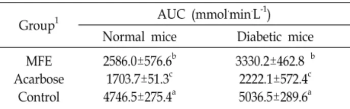
4). In diabetic mice, the AUC of glucose response was sig- nificantly lower in the MFE-administrated group (3330.2±
462.79 mmol.min.L-1) than in the control group (5036.5±289.6 mmol.min.L-1) (Table 2), further supporting the hypo- glycemic effect of MFE.
Table 2. Area under the curve (AUC) of postprandial glucose responses of normal and streptozotocin-induced dia- betic mice
Group1 AUC (mmol.min.L-1)
Normal mice Diabetic mice
MFE Acarbose
Control
2586.0±576.6b 1703.7±51.3c 4746.5±275.4a
3330.2±462.8 b 2222.1±572.4c 5036.5±289.6a
1MFE (300 mg/kg), acarbose (100 mg/kg), and distilled water (as control) were co-administered orally with starch (2 g/kg).
Each value represents the mean ± SD of 7 mice (n = 42).
a–cValues denoted by different letters are significantly different at p<0.05 as analyzed by Duncan’s multiple range test.
In diabetes, the postprandial phase is typified by a rapid and large increase in blood glucose levels, and the possibility that these postprandial “hyperglycemic spikes” may have a negative influence on the pathophysiology of late diabetes complications has recently received much attention [30].
Postprandial hyperglycemia reduces not only insulin secre- tion due to the dysfunction of the pancreas [2] but also in- sulin sensitivity [15, 16], resulting in a deteriorated diabetic state and late diabetes complications. Therefore, maintaining near-normal levels of glycemic control, both in the fasting and postprandial states, is a treatment goal for patients with type 2 diabetes [27]. In this study, we investigated the an- ti-hyperglycemic effects of MFE in STZ-induced diabetic mice and normal mice after the consumption of starch to- gether with MFE. Postprandial blood glucose levels were significantly decreased in STZ-induced diabetic mice.
Consistent with these findings, Ritta et al. [28] reported that dietary anthocyanins in berries significantly reduced post- prandial glucose levels. Therefore, the results of our study suggest that anthocyanin contained in MFE may delay the absorption of dietary starch, thus attenuating the increase in postprandial blood glucose levels. This alleviating effect on postprandial hyperglycemia may be due to the inhibition of carbohydrate digestive enzymes in the small intestine of diabetic mice. Inone et al. (1997) showed that medication that flattens the peak of the postprandial blood glucose levels reduces the AUC of the blood glucose response curve [7].
In this study, MFE significantly decreased blood glucose lev- el at the peak time point and hence, the AUC.
Diabetes is caused by alterations in carbohydrate metabo- lism leading to resistance to insulin secretion, thus resulting in a state of hyperglycemia [34, 36]. Postprandial hyper- glycemia plays an important role not only in the develop-
ment of type 2 diabetes but also in complications associated with the condition, including microvascular and macro- vascular diseases [2]. A large number of intervention trials have demonstrated that improving hyperglycemia achieves considerable reduction of these complications [6]. Although many synthetic compounds such as gliclazide, metformin, and voglibose have been used in the treatment of diabetes, they have in general been associated with marked toxic or undesirable side effects [5]. Therefore, mulberry fruit, con- taining abundant anthocyanins, may possibly be an excellent source of natural anti-diabetic agents. Our current findings suggest that MFE could potentially be developed as a natu- ral functional food to improve postprandial hyperglycemia and prevent diabetic complications.
In conclusion, MFE inhibited α-glucosidase and α-amylase activities and attenuated the rise in blood glucose levels, leading to a reduction in postprandial hyperglycemia.
Moreover, MFE may delay the absorption of dietary carbo- hydrates in the gut, hence suppressing the increase in blood glucose levels after a meal. Thus, MFE may be a useful food source to control diabetes and helpful in improving post- prandial hyperglycemia.
Acknowledgement
This work was supported by a 2-Year Research Grant of Pusan National University.
References
1. Abrahamson, M. J. 2004. Optimal glycemic control in type 2 diabetes mellitus: fasting and postprandial glucose in context. Arch. Intern. Med. 164, 486-491.
2. Baron A. D. 1998. Postprandial hyperglycemia and α-gluco- sidase inhibitors. Diabetes Res. Clin. Pract. 40, 51-55.
3. Bhandari, M. R., Jong-Anurakkun, N., Hong, G and Kawa- bata, J. 2007. α-glucosidase and α-amylase inhibitory activ- ities of nepalese medicinal herb pakhanbhed (Bergenia ciliata, Haw.). Food Chem. 106, 247-252.
4. Choi, S. J., Jeon, H. J., Lee, C. U., Yoon, S. H., Bae, S. K., Chin, Y. W. and Yoon, K. D. 2015. Isolation and develop- ment of quantification method for cyanidin-3-glucoside and cyanidin-3-rutinoside in mulberry fruit by high-perform- ance countercurrent chromatography and high-performance liquid chromatography. Nat. Prod. Sci. 21, 20-24.
5. Hanefeld, M. 1998. The role of acarbose in the treatment of non-insulin-dependent diabetes mellitus. J. Diabetes Complications 12, 228-237.
6. Holman, R. R., Paul, S. K., Bethel, M. A., Matthews, D. R.
and Neil, H. A. 2008. 10-year follow-up of intensive glucose
control in type 2 diabetes. N. Engl. J. Med. 359, 1577-1589.
7. Inoue, I., Takahashi, K., Noji, S., Awata, T., Negishi, K. and Katayama, S. 1997. Acarbose controls postprandial hyper- proinsulinemia in non-insulin dependent diabetes mellitus.
Diabetes Res. Clin. Pract. 36, 143-151.
8. Iwai, K., Kim, M. Y., Onodera, A. and Matsue, H. 2006.
Alpha-glucosidase inhibitory and antihyperglycemic effects of polyphenols in the fruit of Viburnum dilatatum Thunb. J.
Agric. Food Chem. 54, 4588-4592.
9. Jung, J. K. and Park, Y. K. 2010. Antioxidative effect of so-dang-tang in streptozotocin-diabetic rats. J. Life Sci. 20, 691-696.
10. Kang, T.H., Hur, J. Y., Kim, H. B., Ryu, J. H. and Kim, S.
Y. 2006. Neuroprotective effects of the cyanidin-3-O-beta -d-glucopyranoside isolated from mulberry fruit against cer- ebral ischemia. Neurosci. Lett. 391, 122-126.
11. Kim, J. W., Cha, J. Y., Heo, J. S., Jin, H. J. and Cho, Y. S.
2008. CMS-1 hot water extract on streptozotocin-induced di- abetic rats. J. Life Sci. 18. 1584-1591.
12. Kim, H., Yoon, Y. J., Shon, J. H., Cha, I. J., Shin, J. G. and Liu, K. H. 2006. Inhibitory effects of fruit juices on CYP3A activity. Drug Metab Dispos. 34, 521-523.
13. Kim, J. S. 2004. Effect of Rhemanniae radix on the hyper- glycemic mice induced with streptozotocin. J. Kor. Med. Sci.
33, 1133-1138.
14. Kim, K. Y., Nam, K. A., Kurihara, H. and Kim, S. M. 2008.
Potent α-glucosidase inhibitors purified from the red alga Grateloupia elliptica. Phytochemistry 69, 2820-2825.
15. Koivisto, V. A. 1993. Insulin therapy in type II diabetes.
Diabetes Care 16, 29-39.
16. Lebovitz, H. E. 1998. Postprandial hyperglycaemic state: im- portance and consequences. Diabetes Res. Clin. Pract. 40, 27- 28.
17. Lee, H. A., Song, Y. O., Jang, M. S. and Han, J. S. 2013.
Alleviating effects of baechu kimchi added Ecklonia cava on postprandial hyperglycemia in diabetic mice. Prev. Nutr.
Food Sci. 18, 163-168.
18. Lee, J. S., Synytsya, A., Kim, H. B., Choi, D. J., Lee, S., Lee, J. S., Kim, W. J., Jang, S. J. and Park, Y. I. 2013. Purification, characterization and immunomodulating activity of a pectic polysaccharide isolated from korean mulberry fruit oddi (Morus alba L.). Int. Immunopharmacol. 17, 858-866.
19. Liu, C. J. and Lin, J. Y. 2013. Anti-inflammatory effects of phenolic extracts from strawberry and mulberry fruits on cytokine secretion profiles using mouse primary splenocytes and peritoneal macrophages. Int. Immunopharmacol. 16, 165- 170.
20. Liu, L.K., Lee, H. J., Shih, Y. W., Chyau, C. C. and Wang, C. J. 2008. Mulberry anthocyanin extracts inhibit LDL oxida- tion and macrophage-derived foam cell formation induced by oxidative LDL. J. Food Sci. 73, 113-121.
21. Li, S. Z. 1982. Compendium of materia medica. People’s Medical Press, Beijing. 2066-2067.
22. Masahito, T., Seiya, I., Fumihiko, H. and Takanori, T. 2014.
Dietary anthocyanin-rich bilberry extract ameliorates hyper- glycemia and insulin sensitivity via activation of AMP-acti- vated protein kinase in diabetic mice. J. Nutr. 140, 527-533.
23. McDougall, G. J., Shpiro, F., Dobson, P., Smith, P., Blake, A. and Stewart, D. 2005. Different polyphenolic components of soft fruits inhibit α-amylase and α-glucosidase. J. Agric.
Food Chem. 53, 2760-2766.
24. Nishioka, T., Kawabata, J. and Aoyama, Y. 1998. Baicalein, an alpha-glucosidase inhibitor from Scutellaria baicalensis. J.
Nat. Prod. 61, 1413-1415.
25. Pawlowska, A. M., Oleszek, W. and Braca, A. 2008. Quali- quantitative analyses of flavonoids of Morus nigra L. and Morus alba L. (Moraceae) fruits. J. Agric. Food Chem. 56, 3377- 3380.
26. Priscilla, D. H., Roy, D., Suresh, A., Kumar, V. and Thir- umurugan, K. 2014. Naringenin inhibits α-glucosidase activ- ity: A promising strategy for the regulation of postprandial hyperglycemia in high fat diet fed streptozotocin induced diabetic rats. Chem. Biol. Interact. 210, 77-85.
27. Ratner, R. E. 2001. Controlling postprandial hyperglycemia.
Am. J. Cardiol. 88, 26-31.
28. Ritta, T., Essi, S., Niina, T., Elina, H., Kyllikki, K. and Leo, N. 2010. Berries modify the postprandial plasma glucose re- sponse to sucrose in health subjects. Br. J. Nutr. 103, 1094- 1097.
29. Scott, L. J. and Spencer, C. M. 2000. Miglitol: a review of its therapeutic potential in type 2 diabetes mellitus. Drugs 59, 521-549.
30. Sirichai, A., Sirintorn, Y. A., Piyawan, C. and Natthakarn, W. 2011. Cyanidin-3-rutinoside alleviates postprandial hy- perglycemia and its synergism with acarbose by inhibition of intestinal α-glucosidase. J. Clin. Biochem. Nutr. 49, 36-41.
31. Stern, J. L., Hagerman, A. E., Steinberg, P. D. and Mason, P. K. 1996. Phlorotannin-protein interactions. J. Chem. Ecol.
22, 1877-1899.
32. Temelkova-Kurktschiev, T. S., Koehler, C., Henkel, E., Leon- hardt, W., Fuecker, K. and Hanefeld, M. 2000. Postchallenge plasma glucose and glycemic spikes are more strongly asso- ciated with atherosclerosis than fasting glucose or HbA1c level. Diabetes Care 23, 1830-1834.
33. Wanatabe, J., Kawabata, J., Kurihara, H. and Niki, R. 1997.
Isolation and identification of α-glucosidase inhibitors from tochu-cha (Eucommia ulmoides). Biosci. Biotechnol. Biochem. 61, 177-178.
34. Yadav, S., Vats, V., Dhunnoo, Y. and Grover, J. K. 2002.
Hypoglycemic and antihyperglycemic activity of Murraya koenigii leaves in diabetic rats. J. Ethnopharmacol. 82, 111-116.
35. Yang, X., Yang, L. and Zheng, H. 2010. Hypolipidemic and antioxidant effects of mulberry (Morus alba L.) fruit in hyper- lipidaemia rats. Food Chem. Toxicol. 48, 2374-2379.
36. Youn, J. Y., Park, H. Y. and Cho, K. H. 2004. Anti-hyper- glycemic activity of Commelina communis L.: Inhibition of α- glucosidase. Diabetes Res. 66, 149-155.
초록:STZ으로 유도된 당뇨 마우스에서 오디열매추출물의 식후 고혈당 완화 효과
최경하1,2․강지혜1․한지숙1*
(1부산대학교 식품영양학과, 2신라대학교 식품영양학과)
식후 고혈당은 제2형 당뇨병 초기 진단과 항당뇨병의 중요한 바이오마커 중 하나이다. α-글루코시다아제 억제 제는 소장에서 탄수화물 소화의 속도를 방해함으로써 식후 고혈당을 조절한다. 본 연구에서는 오디열매추출물이 α-글루코시다아제와 α-아밀라아제에 미치는 억제효과 및 스트렙토조토신(STZ)이 유발하는 당뇨병 생쥐의 식후고
혈당에 미치는 완화 효과를 조사하였다. α-글루코시다아제와 α-아밀라아제에 대한 오디열매추출물(MFE)의 IC50
값은 각각 0.16과 0.14 mg/ml의 결과값을 나타내어, 양성대조군인 acarbose보다 더 효과적이었다. STZ으로 유발 된 당뇨병 흰쥐의 식후 혈당 수치는 정상 대조군에 비해 오디열매추출물에서 유의적으로 더 낮았다. 더욱이, 오디 열매추출물 투여는 당뇨병 흰쥐에서 포도당 반응에 대한 곡선하면적 감소와 관련이 있었다. 결론적으로, 오디열매 추출물은 α-글루코시다아제와 α-아밀라아제 활성의 강력한 억제제이며 식후 고혈당을 완화할 수 있다는 내용이 다. 따라서 오디열매추출물은 당뇨병 치료제 및 기능성식품의 소재로 가치가 높다고 사료된다.