Sargassum yezoense Extract Inhibits Carbohydrate Digestive
Enzymes In Vitro and Alleviates Postprandial Hyperglycemia in
Diabetic Mice.
Jae-Eun Park, Ji-Hee Lee, and Ji-Sook Han
Department of Food Science and Nutrition, Pusan National University, Busan 46241, Korea pISSN 2287-1098ㆍeISSN 2287-8602
Received 7 March 2017; Accepted 21 April 2017; Published online 30 September 2017 Correspondence to Ji-Sook Han, Tel: +82-51-510-2836, E-mail: [email protected]
Copyright © 2017 by The Korean Society of Food Science and Nutrition. All rights Reserved.
This is an Open Access article distributed under the terms of the Creative Commons Attribution Non-Commercial License (http://creativecommons.org/licenses/by-nc/4.0) which permits unrestricted non-commercial use, distribution, and reproduction in any medium, provided the original work is properly cited.
ABSTRACT: In this study, we investigated whether Sargassum yezoense extract (SYE) could inhibit α-glucosidase and α- amylase activities, and alleviate postprandial hyperglycemia in streptozotocin (STZ)-induced diabetic mice. Freeze-dried S. yezoense was extracted with 80% ethanol and concentrated for use in this study. The hypoglycemic effect was deter-mined by evaluating the inhibitory activities of SYE against α-glucosidase and α-amylase as well as its ability to decrease postprandial blood glucose levels. The half-maximal inhibitory concentrations of SYE against α-glucosidase and α-amylase were 0.078±0.004 and 0.212±0.064 mg/mL, respectively. SYE was a more effective inhibitor of α-glucosidase and α- amylase activities than the positive control, acarbose. The increase in postprandial blood glucose levels was significantly alleviated in the SYE group compared with that in the control group of STZ-induced diabetic mice. Furthermore, the area under the curves significantly decreased with SYE administration in STZ-induced diabetic mice. These results suggest that SYE is a potent inhibitor of α-glucosidase and α-amylase activities and alleviates postprandial hyperglycemia caused by dietary carbohydrates.
Keywords: Sargassum yezoense, α-glucosidase, α-amylase, postprandial hyperglycemia, diabetic mice
INTRODUCTION
Diabetes mellitus is a serious chronic disease that is in-creasing with obesity and aging in the worldwide pop-ulation (1). Diabetes patients have high blood glucose levels because either they cannot produce enough in-sulin or the cells of their liver, muscle, and fat tissue do not respond to insulin normally (2). In diabetes, the postprandial phase is accompanied by rapid and large in-creases in blood glucose levels. Postprandial hyperglyce-mia negatively affects the development of type-2 dia-betes and causes diabetic complications, including dysli-pidemia and hypertension (3). In addition, postprandial hyperglycemia was reported as a direct risk factor for cardiovascular diseases, and most cardiovascular risk factors are affected by acute blood sugar increases (4). Therefore, controlling postprandial hyperglycemia is the most important factor for treating patients with diabetes to reduce the risks of diabetic complications (5).
One therapeutic approach to suppressing postprandial hyperglycemia is to retard absorption of glucose through inhibition of carbohydrate hydrolyzing enzymes, such as
α-glucosidase and α-amylase in digestive organs (6). Syn-thetic α-glucosidase inhibitors, such as acarbose, migli-tol, and voglibose, are available to treat postprandial hy-perglycemia in patients with type-2 diabetes. However, chronic use of these agents can result in side effects, such as flatulence, abdominal cramping, vomiting, and diar-rhea; therefore, their use may be limited (7-10). There-fore, many studies have been performed to identify more effective and safer inhibitors of α-glucosidase and α-amy-lase from natural sources.
Marine macroalgae, or seaweeds, are one of nature’s most biologically active resources and contain a wealth of bioactive compounds. Seaweed extracts have demon-strated various biological activities, such as antioxidant (11), anti-inflammatory (12), anticoagulant (13), and ap-optotic activities (14). Sargassum yezoense is a brown al-gae belonging to the Sargassum genus. It is one of the most abundant marine algae on the east coast of Korea and is popular in Korea and Japan as a food ingredient and marine herb (15). S. yezoense extract (SYE) showed various bioactivities including inflammatory, anti-bacterial, and anti-atopy activities (16-19). Most notably,
SYE has strong antioxidant and peroxisome proliferator- activated receptor (PPAR)α and PPARγ stimulating ef-fects in 3T3-L1 cells because it contains biologically ac-tive substances, such as sargaquinoic acid and sargahy-droquinoic acid (20).
However, there are presently no studies reporting the ability of SYE to alleviate postprandial hyperglycemia through the inhibition of carbohydrate digestive enzymes in diabetic mice. Therefore, this study was conducted to determine whether SYE inhibits α-glucosidase and α- amylase activities in vitro and alleviates postprandial hy-perglycemia in diabetic mice in vivo.
MATERIALS AND METHODS
Materials and preparation of SYE
The brown algae, S. yezoense, were collected from the east coast of Korea. After collection of S. yezoense, the sam-ples were washed three times under running tap water to remove foreign substances and sand on the surface, carefully rinsed with fresh water, and then freeze-dried. The lyophilized samples were homogenized with a grind-er prior to extraction. The sample was extracted three times with ten volumes of 80% ethanol for 12 h at room temperature. The filtrate was then evaporated at 40oC using a rotary evaporator (N-1300VW, EYELA, Tokyo, Japan). After the solvent had been completely removed from the SYE, it was stored in a deep freezer (−80oC). Inhibition assay for α-glucosidase activity in vitro
The α-glucosidase inhibitory assay was based on the chro-mogenic method developed by Watanabe et al. (21), and it was performed using a readily available yeast enzyme. Briefly, yeast α-glucosidase (0.7 U, Sigma-Aldrich Co., St. Louis, MO, USA) was dissolved in 100 mM phosphate buffer (pH 7.0) containing 2 g/L bovine serum albumin and 0.2 g/L NaN3 and used as the enzyme solution. Five
mM p-nitrophenyl-α-D-glucopyranoside in the same buf-fer (pH 7.0) was used as the substrate solution. Fifty μL enzyme solution and 10 μL SYE [5 mg/mL in dimethyl sulfoxide (DMSO)] were mixed in a well, and the absorb-ance at 405 nm was measured as time zero using a mi-croplate reader. After incubation for 5 min, the substrate solution (50 μL) was added, and the incubation contin-ued for another 5 min at room temperature. The increase in absorbance from the zero time point was then meas-ured. The inhibitory activities of varying concentrations of SYE were expressed as 100 minus the absorbance dif-ference (%) of the test compounds relative to the absorb-ance change of the negative control (i.e., DMSO used as the test solution). The measurements were performed in triplicate, and the IC50 value (i.e., the concentration of
SYE that result in 50% inhibition of maximal activity)
was determined.
Inhibition assay for α-amylase activity in vitro
The α-amylase inhibitory activity was assayed in the same manner as described previously for α-glucosidase (21), except that porcine pancreatic amylase (100 U, Sigma- Aldrich Co.) and blocked p-nitrophenyl- α-D-maltopen-toglycoside (Sigma-Aldrich Co.) were used as the enzyme and substrate, respectively.
Measurement of cytotoxicity
Cell viability was assessed using the 3-(4,5-dimethylthia-zol-2-yl)-2,5-diphenyltetrazolium bromide (MTT) assay, and 3T3-L1 cells were purchased from the Korean Cell Line Bank (Seoul, Korea). 3T3-L1 cells were seeded at 1×10⁴ cells/well in 96-well plates and pre-incubated in a humidified atmosphere containing 5% CO2 at 37oC for
24 h. Afterward, the cells were treated with various con-centrations (0.1, 0.5, 1, and 2 mg/mL) of SYE, and in-cubated for 20 h. After completion of the treatment, the cells were incubated for 3 h at 37oC with filtered MTT (Sigma-Aldrich Co.) solution, which was added to each well at a final concentration of 0.5 mg MTT/mL. The su-pernatants were carefully aspirated, 200 μL of DMSO was added to each well, and the plates were agitated to dis-solve the crystal product. The absorbance of DMSO sol-utions was measured spectrophotometrically at 540 nm. Experimental animals
Four-week old male mice (ICR, Central Laboratory Ani-mal Inc., Seoul, Korea) were housed individually in a temperature-controlled room (25∼30oC) with a relative humidity of 45∼55% and 12 h on/12 h off light/dark cycles. The animals were provided pelleted food and tap water ad libitum. After an adjustment period of 2 weeks, diabetes was induced by an intraperitoneal injection of streptozotocin [STZ; 60 mg/kg body weight (b.w)] that was freshly dissolved in citrate buffer (0.1 M, pH 4.5) (22). After seven days, tail bleeds were performed, and animals with a blood glucose concentration above 250 mg/dL were considered to be diabetic. The procedures for the handling and care of mice adhered to the guide-lines that comply with current international laws and pol-icies (NIH guide for the Care and Use of Laboratory Ani-mals), and all procedures were approved by the animal ethics committee at Pusan National University (PNU- 2016-1273).
Measurement of blood glucose levels
Both normal mice and STZ-induced diabetic mice were fasted overnight and randomly divided into three groups of 7 mice. Before testing blood glucose levels, animals were kept in a fasting state for at least 12 h, but had free access to water. The mice were orally administered as
Table 1. IC50values of the inhibitory effect of Sargassum ye-zoense extract (SYE) against α-glucosidase and α-amylase ac-tivities Sample IC50 (mg/mL)1) α-Glucosidase α-Amylase SYE 0.078±0.004* 0.212±0.064* Acarbose 0.189±0.013 0.262±0.037
Each value is expressed as mean±SD in triplicate experiments. *Significantly different from acarbose at P <0.05.
1)IC
50 value is the concentration of sample required for 50% inhibition.
Fig. 2. Inhibitory activity of Sargassum yezoense extract (SYE) against α-amylase. Each value is expressed as mean±SD in triplicate experiments. Values with different letters (a-e) are significantly different at P <0.05 as analyzed by Duncan’s multi-ple range test. The concentration of acarbose used as a positive control was 0.25 mg/mL.
Fig. 1. Inhibitory activity of Sargassum yezoense extract (SYE) against α-glucosidase. Each value is expressed as mean±SD in triplicate experiments. Values with different letters (a-c) are significantly different at P <0.05 as analyzed by Duncan’s multi-ple range test. The concentration of acarbose used as a positive control was 0.25 mg/mL.
follows: control, mice received starch orally (2 g/kg b.w); SYE, mice received starch with SYE orally (300 mg/kg b.w); acarbose, mice received starch with acarbose orally (100 mg/kg b.w). Blood samples were taken from the tail vein at 0, 30, 60, and 120 min. Blood glucose was measured using a glucometer (Roche Diagnostics GmbH, Mannheim, Germany). The areas under the curve (AUC) were calculated using the trapezoidal rule (23).
Data statistical analysis
The data are presented as mean±standard deviation (SD). Statistical analysis was performed using SAS ver-sion 9.1 (SAS Institute Inc., Cary, NC, USA). The Stu-dent’s t-test was used for comparisons between the con-trol and treatment groups. Differences were evaluated by one-way analysis of variance (ANOVA), followed by post-hoc Duncan’s multiple range tests (P<0.05).
RESULTS AND DISCUSSION
Inhibitory effect of SYE on α-glucosidase and α-amylase in vitro
The inhibitory effects of SYE against α-glucosidase activ-ity were determined using p-nitrophenyl- α-D-glucopy-ranoside as the substrate, and were compared to the ef-fects of a commercial α-glucosidase inhibitor, acarbose. SYE inhibited α-glucosidase activity in a dose-dependent manner by 39.76±2.38, 57.83±1.43, 75.90±1.51, and 78.92±2.11% at concentrations of 0.05, 0.10, 0.25, and 0.50 mg/dL, respectively (Fig. 1). Acarbose, an α-gluco-sidase inhibitor used as an oral hypoglycemic agent, in-hibited the enzyme activity by 58.03±3.22% at a concen-tration of 0.25 mg/dL. The α-glucosidase inhibitory ac-tivity of SYE was significantly higher than that of acar-bose at the same concentration (0.25 mg/dL).
As shown in Fig. 2, the inhibitory effects of SYE on α- amylase increased in a dose-dependent manner by 29.03 ±3.20, 38.71±2.59, 53.76±2.60, and 64.52±2.75% at concentrations of 0.05, 0.10, 0.25, and 0.50 mg/dL, res-pectively. SYE also inhibited α-amylase activity more ef-fectively than acarbose. The IC50 values of SYE against α-
glucosidase and α-amylase were 0.078±0.004 and 0.212 ±0.064 mg/mL, respectively. The IC50 values of SYE
against α-glucosidase and α-amylase were significantly lower than those of acarbose, indicating that SYE has stronger inhibitory effects than the positive control (Ta-ble 1). These results indicate that SYE may prove useful as a natural, postprandial anti-hyperglycemic agent ow-ing to its inhibitory effects against α-glucosidase and α- amylase.
The treatment aim for diabetic patients is to maintain normal levels of glycemic control, both in fasting and postprandial states. Postprandial hyperglycemia is the first sign that appears in diabetes mellitus-related meta-bolic abnormalities. α-Amylase is the key enzyme for cat-alytic hydrolysis of the α−1,4 glycosidic linkages of starch to form maltose, which is further hydrolyzed to glucose prior to absorption in the small intestine (24). α-Gluco-sidases are a family of membrane-bound enzymes in the
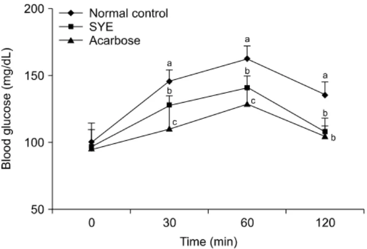
Fig. 4. Blood glucose levels after the administration of Sargas-sum yezoense extract (SYE) in streptozotocin-induced diabetic mice. Each value is expressed as mean±SD of seven mice. Values with different letters (a-c) are significantly different at each time (P <0.05) as analyzed by Duncan’s multiple range test. Control, mice received starch orally (2 g/kg b.w); SYE, mice received starch with Sargassum yezoense extract orally (300 mg/kg b.w); Acarbose, mice received starch with acarbose or-ally (100 mg/kg b.w).
Fig. 3. Cytotoxic effect of Sargassum yezoense extract (SYE) in 3T3-L1 cells. 3T3-L1 cells were treated with various concen-trations (0.1, 0.5, 1.0, and 2.0 mg/mL) of SYE for 20 h, and cell viability was measured via MTT assay. Each value is expressed as mean±SD in triplicate experiments. NS: non-significant.
intestine that are involved in the digestion and uptake of carbohydrates into the bloodstream. Monosaccharides such as glucose and fructose, are absorbed directly in the small intestine. Disaccharides such as sucrose, and poly-saccharides such as starch, must be degraded before be-ing absorbed. This digestive process is performed by the α-glucosidases (10).
Since the activity of α-glucosidase in the small intes-tine of diabetic patients is higher than that of normal peo-ple, the intake of carbohydrates in food significantly in-creases postprandial blood glucose levels (25). Inhibition of α-amylase and α-glucosidase attenuates the increase in postprandial blood glucose after ingestion of carbohy-drates to manage postprandial blood glucose in type 2 diabetic patients (10). α-Glucosidase inhibitors are typi-cally compounds such as acarbose, which inhibit the α- glucosidase in the small intestine, thereby reducing the degradation of polymeric sugar compounds and suppres-sing the rapidly increasuppres-sing blood glucose levels that oc-cur after ingestion of carbohydrate (26). However, acar-bose, which is used as a commercial carbohydrate inhibi-tor, has side effects such as flatulence, abdominal cramp-ing, and diarrhea when used long-term (7,8). Thus, sci-entists have long considered alternative effective and non-toxic inhibitors of α-glucosidase and α-amylase.
In this study, we investigated the inhibitory effect of the natural product, SYE, against α-glucosidase and α- amylase to elucidate its possible use as an anti-postpran-dial hyperglycemic agent. SYE afforded significantly high-er inhibitory activities against both α-glucosidase and α-amylase than the commercial inhibitor, acarbose. It al-so did not show any cytotoxicity (Fig. 3). SYE contains various bioactive compounds, such as sargaquinoic acid and sargahydroquinoic acid, which are types of plasto-quinones. According to another study, sargaquinoic acid and sargahydroquinoic acid have remarkable α-glucosi-dase inhibition ability (25). Plastoquinones contain
droxyl groups, and Tadera et al. (27) reported that hy-droxyl substitution on the phenyl ring structure could be effective in inhibiting the enzyme activity. Thus, the inhibitory effect of SYE on α-glucosidase and α-amylase appears to be due to the plastoquinones in SYE.
Effects of SYE on blood glucose levels in vivo
The effects of SYE on blood glucose levels after a meal were investigated in normal and STZ-induced diabetic mice. The postprandial blood glucose levels of the SYE administered mice were lower than those of the diabetic control mice (Fig. 4). Blood glucose levels in the diabetic control mice increased to 378.7±27.9 mg/dL at 30 min and 412.0±16.5 mg/dL at 60 min after a meal, and then decreased to 381.0±18.6 mg/dL at 120 min. However, when SYE was added to starch, the increase in post-prandial blood glucose levels was significantly suppressed (351.5±19.8, 384.5±23.7, and 335.5±20.4 mg/dL at 30, 60, and 120 min, respectively; P<0.05). The peak post-prandial blood glucose levels also significantly decreased when the normal mice were orally administered starch with SYE (Fig. 5). Thus, this confirms that SYE can sup-press the postprandial hyperglycemia that is caused by starch in normal mice. The AUC for the glucose re-sponse in diabetic mice administered SYE (706.7±43.9 mg・h/dL) was significantly lower (P<0.05) than that in diabetic control mice (764.1±45.1 mg・h/dL) (Table 2).
The ability to control postprandial hyperglycemia is important in achieving the tight glycemic control that is targeted in diabetes treatment (28). In addition, post-prandial hyperglycemia increases the risk of cardiovas-cular disease, increases free radical production, induces vasoconstriction, and plays a negative role in type 2
dia-Table 2. Areas under the curve (AUC) of the postprandial glu-cose responses of normal and streptozotocin-induced diabetic mice
Group1)
AUC (mg・h/dL)
Normal mice Diabetic mice
Control 287.5±20.4a 764.1±45.1a
SYE 247.6±13.9b 706.7±43.9b
Acarbose 228.4±23.7c 660.6±39.4c
Each value is expressed as the mean±SD of seven mice. Different letters (a-c) in a column are significantly different at P <0.05 using Duncan’s multiple range tests.
1)Control, mice received starch orally (2 g/kg b.w); SYE, mice received starch with Sargassum yezoense extract orally (300 mg/kg b.w); Acarbose, mice received starch with acarbose or-ally (100 mg/kg b.w).
Fig. 5. Blood glucose levels after the administration of Sargas-sum yezoense extract (SYE) in normal mice. Each value is ex-pressed as mean±SD of seven mice. Values with different let-ters (a-c) are significantly different at each time (P <0.05) as analyzed by Duncan’s multiple range test. Control, mice re-ceived starch orally (2 g/kg b.w); SYE, mice rere-ceived starch with Sargassum yezoense extract orally (300 mg/kg b.w); Acarbose, mice received starch with acarbose orally (100 mg/kg b.w).
betes; therefore, controlling postprandial hyperglycemia plays an important role in diabetic patients (29).
Thus, we determined the anti-postprandial hyperglyce-mic effect of SYE in diabetic and normal hyperglyce-mice after con-sumption of starch. The increase in postprandial blood glucose levels was suppressed significantly in both dia-betic and normal mice when treated with SYE. These re-sults show that SYE may delay the absorption of dietary carbohydrates, resulting in suppression of the increase in postprandial blood glucose levels. Inoue et al. (30) re-ported that medications flatten the peak of postprandial blood glucose and decrease the AUC of the blood glucose response curve. In this study, SYE was shown to reduce both blood glucose levels at the peak time point and the AUC in diabetic mice. The AUCs in normal mice were also lowered by SYE, paralleling that observed in diabetic mice. As shown in Fig. 4 and Fig. 5, postprandial hyper-glycemia was significantly alleviated after ingestion of
starch supplemented with SYE in both diabetic and nor-mal mice. This may be due to inhibition of the activity of carbohydrate degrading enzymes (e.g., pancreatic α-amy-lase and intestinal α-glucosidase) by SYE, thereby delay-ing the absorption of dietary carbohydrates in the epithe-lial cells of the small intestine.
Recently, marine algae have been recognized as a good resource for anti-diabetic materials derived from nature (31). Results from our investigation suggest that SYE from brown algae is helpful in preventing postprandial hyperglycemia and diabetic complications, as assessed by the anti-hyperglycemic effects of SYE in both diabetic and normal mice. This study demonstrates that SYE may prove useful as an effective, natural anti-diabetic sub-stance.
In conclusion, SYE inhibited α-glucosidase and α-amy-lase activities, suppressing the formation of glucose from starch, and resulting in a reduction in postprandial hyp-erglycemia. Furthermore, SYE may delay the absorption of dietary carbohydrates in the intestine, resulting in sup-pression of increased blood glucose levels after a meal. Thus, SYE may be used as a functional food to alleviate postprandial hyperglycemia.
ACKNOWLEDGEMENTS
This work was supported by a 2-Year Research Grant of Pusan National University.
AUTHOR DISCLOSURE STATEMENT
The authors declare no conflict of interest.
REFERENCES
1. Zimmet P, Alberti KGMM, Shaw J. 2001. Global and societal implications of the diabetes epidemic. Nature 414: 782-787. 2. Muoio DM, Newgard CB. 2006. Obesity-related derange-ments in metabolic regulation. Annu Rev Biochem 75: 367-401. 3. Baron AD. 1998. Postprandial hyperglycaemia and
α-gluco-sidase inhibitors. Diabetes Res Clin Pract 40: S51-S55. 4. Ceriello A. 2005. Postprandial hyperglycemia and diabetes
complications: is it time to treat?. Diabetes 54: 1-7. 5. UK Prospective Diabetes Study (UKPDS) Group. 1998.
In-tensive blood-glucose control with sulphonylureas or insu-lin compared with conventional treatment and risk of com-plications in patients with type 2 diabetes (UKPDS 33). Lancet 352: 837-853.
6. Saito N, Sakai H, Suzuki S, Sekihara H, Yajima Y. 1998. Effect of an α-glucosidase inhibitor (voglibose), in combination with sulphonylureas, on glycaemic control in type 2 diabetes pa-tients. J Int Med Res 26: 219-232.
7. Lebovitz HE. 2002. Treating hyperglycemia in type 2 diabe-tes: new goals and strategies. Cleve Clin J Med 69: 809-820. 8. Carroll MF, Gutierrez A, Castro M, Tsewang D, Schade DS.
2003. Targeting postprandial hyperglycemia: a comparative study of insulinotropic agents in type 2 diabetes. J Clin Endo-crinol Metab 88: 5248-5254.
9. Fonseca V. 2003. Clinical significance of targeting postpran-dial and fasting hyperglycemia in managing type 2 diabetes mellitus. Curr Med Res Opin 19: 635-641.
10. Hanefeld M. 1998. The role of acarbose in the treatment of non-insulin-dependent diabetes mellitus. J Diabetes Complica-tions 12: 228-237.
11. Yuan YV, Walsh NA. 2006. Antioxidant and antiproliferative activities of extracts from a variety of edible seaweeds. Food Chem Toxicol 44: 1144-1150.
12. Kang JY, Khan MN, Park NH, Cho JY, Lee MC, Fujii H, Hong YK. 2008. Antipyretic, analgesic, and anti-inflammatory ac-tivities of the seaweed Sargassum fulvellum and Sargassum thunbergii in mice. J Ethnopharmacol 116: 187-190.
13. Pushpamali WA, Nikapitiya C, De Zoysa M, Whang I, Kim SJ, Lee J. 2008. Isolation and purification of an anticoagulant from fermented red seaweed Lomentaria catenata. Carbohydr Polym 73: 274-279.
14. Kwon MJ, Nam TJ. 2006. Porphyran induces apoptosis re-lated signal pathway in AGS gastric cancer cell lines. Life Sci 79: 1956-1962.
15. Hong IS, Kim GA, Park JK, Boo SM. 2008. Morphology and phenology of Sargassum yezoense (Sargassaceae, Phaeophyceae). Korean Journal of Nature Conservation 2: 132-139.
16. Nakai M, Kageyama, N, Nakahara K, Miki W. 2006. Phloro-tannins as radical scavengers from the extract of Sargassum ringgoldianum. Mar Biotechnol 8: 409-414.
17. Reddy P, Urban S. 2009. Meroditerpenoids from the southern Australian marine brown alga Sargassum fallax. Phytochemistry 70: 250-255.
18. Jung M, Jang KH, Kim B, Lee BH, Choi BW, Oh KB, Shin J. 2008. Meroditerpenoids from the brown alga Sargassum sili-quastrum. J Nat Prod 71: 1714-1719.
19. Seo Y, Park KE, Kim YA, Lee HJ, Yoo JS, Ahn JW, Lee BJ. 2006. Isolation of tetraprenyltoluquinols from the brown alga Sargassum thunbergii. Chem Pharm Bull 54: 1730-1733. 20. Kim SN, Choi HY, Lee W, Park GM, Shin WS, Kim YK. 2008.
Sargaquinoic acid and sargahydroquinoic acid from Sargassum yezoense stimulate adipocyte differentiation through PPARα/γ activation in 3T3-L1 cells. FEBS Lett 582: 3465-3472. 21. Watanabe J, Kawabata J, Kurihara H, Niki R. 1997. Isolation
and identification of α-glucosidase inhibitors from tochu- cha (Eucommia ulmoides). Biosci Biotechnol Biochem 61: 177-178. 22. Zheng J, He J, Ji B, Li Y, Zhang X. 2007. Antihyperglycemic activity of Prunella vulgaris L. in streptozotocin-induced dia-betic mice. Asia Pac J Clin Nutr 16: 427-431.
23. Kim JS. 2004. Effect of Rhemanniae Radix on the hyperglycemic mice induced with streptozotocin. J Korean Soc Food Sci Nutr 33: 1133-1138.
24. Prashanth D, Padmaja R, Samiulla DS. 2001. Effect of certain plant extracts on α-amylase activity. Fitoterapia 72: 179-181. 25. Lee EH, Ham J, Ahn HR, Kim MC, Kim CY, Pan CH, Um BH,
Jung SH. 2009. Inhibitory effects of the compounds isolated from Sargassum yezoense on α-glucosidase and oxidative stress. Kor J Pharmacogn 40: 150-154.
26. Lebovitz HE. 1992. Oral antidiabetic agents: the emergence of α-glucosidase inhibitors. Drugs 3: 21-28.
27. Tadera K, Minami Y, Takamatsu K, Matsuoka T. 2006. Inhib-ition of α-glucosidase and α-amylase by flavonoids. J Nutr Sci Vitaminol 52: 149-153.
28. Koskinen P, Mänttäri M, Manninen V, Huttunen JK, Heinonen OP, Frick MH. 1992. Coronary heart disease incidence in NIDDM patients in the Helsinki Heart Study. Diabetes Care 15: 820-825.
29. Ceriello A, Davidson J, Hanefeld M, Leiter L, Monnier L, Owens D, Tajima N, Tuomilehto J; International Prandial Glucose Regulation Study Group. 2006. Postprandial hyper-glycaemia and cardiovascular complications of diabetes: an update. Nutr Metab Cardiovasc Dis 16: 453-456.
30. Inoue I, Takahashi K, Noji S, Awata T, Negishi K, Katayama S. 1997. Acarbose controls postprandial hyperproinsuline-mia in non-insulin dependent diabetes mellitus. Diabetes Res Clin Pract 36: 143-151.
31. Stern JL, Hagerman AE, Steinberg PD, Mason PK. 1996. Phlo-rotannin-protein interactions. J Chem Ecol 22: 1877-1899.