CR 시스템에서 IP 잠상의 소거 후 Unexposed Image의 평가
*연세대학교 보건과학대학 방사선학과, †연세대학교 보건과학연구소
임보연*ㆍ박혜숙*†ㆍ김주혜*ㆍ박광현*ㆍ김희중*†
Computed Radiography (CR) 시스템에서 영상의 신뢰성과 재현성을 높이기 위해서는 영상 획득 과정의 마지막 단계에서 이루어지는 나트륨램프의 소거에 의한 Image Plate (IP)의 완전한 초기화가 매우 중요하다. 본 연구에서는 CR (Agfa CR 25; Agfa, BELGIUM)을 이용하여 잠상의 소거 후 지연시간에 따른 노이즈의 평가, 소거량에 따른 잠상의 소거 후 노이즈 의 평가, 그리고 American Association of Physicist in Medicine (AAPM) Report 93 (2006)의 CR acceptance test 중 erasure thoroughness 평가를 시행하였다. 임상에서 주로 쓰이는 Chest postero-anterior (PA), Hand PA, L-spine lateral을 촬영 후 스캔하였다. Chest PA와 L-spine lateral은 Chest phantom (3D-torso; CIRS, USA)을 이용하여 촬영했고 Hand PA는 피검자 를 통해 촬영되었다. 실험 결과 Hand PA를 제외한 나머지 unexposed image에서 지연시간이 길어질수록 전반적으로 ghost image를 비롯한 noise가 증가하였으며, overexposure된 부분을 따라 ghost image가 나타났다. 지연시간에 의해 야 기된 noise에 의한 영향은 육안으로 확인되지 않았다. 픽셀 값의 standard deviation (SD)은 overexposure 된 부분이 상대 적으로 높게 측정되었다. 잠상의 소거 정도에 따른 영상의 noise평가 결과 Hand PA, Chest PA는 소거 정도 값이 높아져 도 noise는 증가하지 않았다. 그러나 상대적으로 조사선량이 큰 L-spine lateral은 기준 소거 정도보다 낮을 경우 noise가 발생하였다. Erasure thoroughness 평가 결과 학과 내 CR 장비의 잠상소거능력은 기준치에 부합함을 확인하였다. 본 연 구가 IP 초기화에 기초적인 자료를 제공함으로써 신뢰성 있는 영상을 획득하는데 기여할 것으로 기대한다.
중심단어: Image Plate, Erasure level, Erasure thoroughness
이 논문연구는 2009학년도 연세대학교 학술연구비의 지원에 의하 여 이루어진 것임.
이 논문은 2009년 8월 4일 접수하여 2009년 11월 9일 채택되었음.
책임저자:김희중, (220-710) 강원도 원주시 흥업면 매지리 234번지 연세대학교 보건과학대학 방사선학과
Tel: 033)760-2475, Fax: 033)760-2815 E-mail: [email protected]
서 론
CR (Computed Radiography, CR)이 등장한 이후로 지난 몇 년 동안 CR 시스템은 빠른 발전을 하였다. 1970년대 효 율적인 집광 시스템을 위한 연구가 오늘날 CR 발전의 시 작이라고 볼 수 있다. 상업적인 CR 시스템을 사용한 것은 1981년 Fuji사의 CR 시스템 도입이 시작이었다. 그 이후로 휘진성 형광(Photostimulable luminescence, PSL) 효과는 의료 영상을 포함한 여러 분야에서 이용되고 있다. CR은 screen/
film 형식에 비해 관용도(exposure latitude)가 크며,1,2) 의료 영상 저장 및 전송 시스템(Picture Archiving and Communi- cation System, PACS)의 도입과 더불어 영상의 후처리 기능 이 가능하고 데이터처리가 용이하다는 장점이 있다.3,4) 또
한 필름은 1회용인 반면에 CR 시스템의 Image Plate (IP)는 획득된 영상을 소거하고 초기화하여 반영구적으로 사용할 수 있다.5,6) 디지털 처리기술로 인해 screen/film 방식이 영 상의 획득 및 표시기능을 모두 담당했던 것과는 달리 디지 털 엑스선 영상 진단기기는 영상의 획득과 표시기능이 분 리되고 획득된 영상데이터는 디지털화 되어 저장된다.1,7) 디지털 영상을 얻기 위해서 기존의 screen/film 시스템과 동 일한 엑스선 조사 장치 사용이 가능하다는 점에서 도입이 용이하다는 장점도 있다.8,9) Cassette 기반의 시스템이라는 점 에서(이동촬영 등), CR은 앞으로도 발전 가능성이 크다.10,11) CR 시스템의 영상획득 원리는 3단계로 설명할 수 있다.
IP에 엑스선을 조사하고, 레이저 빔으로 조사된 잠상을 읽 어내고, 소거와 동시에 IP를 초기화 시키는 것이다. 동일한 IP를 반복해서 사용하는 CR 시스템에서 신뢰성 있는 영상 의 획득을 위해서는 잠상의 소거에 의한 IP의 초기화가 선 행되어야 하며, 현상 후 지연시간에 의한 noise를 최소화해 야한다. 이에 본 연구에서는 IP 스캔의 마지막에서 행해지 는 소거의 정도가 noise에 영향을 주는지 평가하고, 현상 후 지연시간과 noise와의 연관성을 분석하고자 하였다. 또
Table 1. Specification of the image reader & image plate.
Imaging system (image reader)
Phosphor Image size Digital Pixel size
screen (inch) matrix (μm)
Agfa AD CR 25 Agfa MD 4.0 14×17 2,320×2,828 150 8×10 1,950×2,460 100
Table 2. Specification of the x-ray tube.
X-ray tube E7239; TOSHIBA
Focus size 1.2/0.6 mm Inherent filtration 2.7 mmAl Additional filtration 21 mmAl Target material/angle Rhenium-Tungsten-faced
Molybdeneum/12o
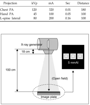
Fig. 1. Illustration of experimental set-up for noise evaluation of
CR images respect to delay time after erasure of latent image and its effect.Table 3. Experimental setup for Chest PA, Hand PA, L-spine lateral projection.
Projection kVp mA Sec Distance
Chest PA 120 320 0.01 180
Hand PA 45 100 0.05 100
L-spine lateral 80 200 0.16 100
한 현상 후 지연에 의한 잠상이 다음 엑스선 촬영 후 현상 에 미치는 영향을 평가하였다. 이와 함께 AAPM Report 93 (2006)12)의 CR acceptance test 중 erasure thoroughness를 측 정하여 CR 시스템의 성능을 평가하였다.
재료 및 방법
본 연구에서는 엑스선 영상 획득을 위해서 엑스선 발생 장치(TE-E7252X; Toshiba, JAPAN)와 CR 시스템(Agfa CR 25; Agfa, BELGIUM), 그리고 IP (Agfa MD 4.0; Agfa, BELGIUM)와 Chest phantom (3D-torso; CIRS, USA)을 사용 하였다(Table 1, 2).
1. 잠상의 소거 후 지연시간에 따른 노이즈의 평가 임상에서 주로 쓰이는 Chest postero-anterior (PA), Hand PA, L-spine lateral을 촬영 후 스캔하였다. Chest PA와 L- spine lateral은 3D-torso phantom을 이용하였으며 Hand PA는 피검자를 통해 촬영되었다. 촬영된 IP는 스캔 후 0, 1, 3, 6, 9, 12시간의 지연시간을 가진 후 동일한 IP의 unexposed im- age를 획득하였다. 각 영상들은 재현성을 높이기 위해 4장 씩 획득하였다. Chest PA, Hand PA, L-spine lateral의 촬영조 건은 Table 3과 같다.
Ghost image가 존재할 경우, 다음 촬영에 미치는 영향을 측정하기 위해 Fig. 1과 같은 영상을 획득하였다. Chest phantom을 이용하여 각 부위를 촬영 후 스캔하였다. 스캔 된 IP는 스캔 후 0, 1, 3, 6, 9, 12시간의 지연시간을 갖은 후 55 kVp, 100 mA, 0.05 sec의 조건으로 선속 조절 없이 개조
사야(open field)에 조사되었다. IP 위에 5 mm 두께의 알루 미늄 3개를 Fig. 1과 같이 배치하였다.
촬영 후 tibia AP 후처리를 적용한 후 raw 데이터를 획득 하였다. 후처리는 필요에 따라 표준 영상의 특정부위를 강 조 또는 억제하여 원하고자 하는 영상으로 변환시키는 처 리를 말한다. Agfa CR 시스템은 Multi-Scale Image Contrast Amplification (MUSICA) 알고리즘을 통해 raw data를 자동 으로 촬영별로 이미지 처리를 하는 것이다. 각 촬영별로 후 처리를 적용시킨다. MUSICA의 영상처리는 multi scale 접 근방식을 이용함으로써 하나의 영상에서 다양한 주파수 대 역이 생기게 한다. 영상은 라플라스 변환에 의해 분해 혹은 재조합된다. MUSICA parameter에는 MUSI 대비(MUSI-con- trast), 가장자리 대비(edge enhancement), 관용도 감소(lati- tude reduction), 잡음감소(noise reduction)가 있다. MUSI 대 비는 세부적인 대조도를 정하는데 이는 강조하는 부위의 농도 영역을 조절한다. 가장자리 대비는 영상의 선예도를 증가시고 관용도 감소는 세기가 큰 부분을 감쇠시켜 중간 이하의 농도 부위를 강조시킨다. 잡음감소는 노이즈를 감
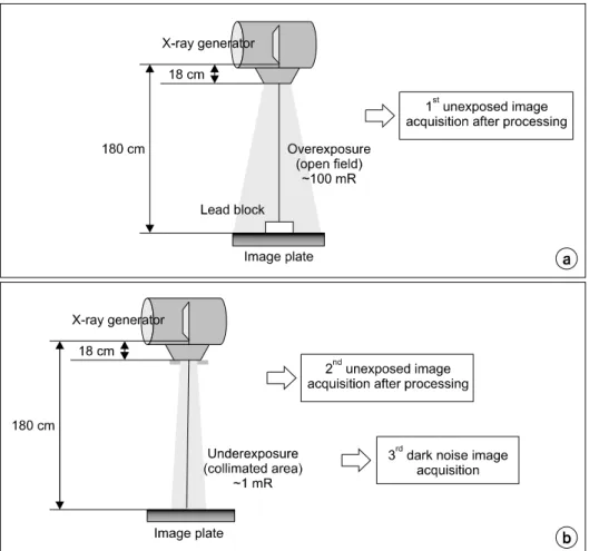
Fig. 2. Illustration of experimental
set-up for erasure thoroughness eva- luation: overexposure on lead block (a), underexposure without lead blo- ck (b).Table 4. Criteria for erasure thoroughness.
Qualitative criteria (underexposure image)
Absence of a ghost image of the lead block Quantitative criteria
(unexposed image)
Log Median Exposure (lgM)=0.28 Scan Average Level (SAL)<130 Pixel Value (PV)<630
Pixel Value Standard Deviation (PVSD)<5
소시킨다. 영상이 Digital Imaging and Communications in Medicine (DICOM) 서버에 보내지기 전에, 영상각각의 MU- SICA parameter는 0에서 6까지 바꿀 수 있다.13) Tibia AP의 MUSICA 알고리즘 값은 MUSI 대비(MUSI-contrast)=3.50, 가장자리 대비(edge enhancement)=2.50, 관용도 감소(latitude reduction)=2.10, 잡음감소(noise reduction)=1.30으로 설정되 어 있다. 획득된 영상에서 각각의 알루미늄의 중심에 300×
300 matrix 크기의 관심영역(region of interest, ROI)을 설정 하여 평균 픽셀 값, standard deviation (SD), profile을 평가하 였다.
2. 잠상의 소거 정도에 따른 영상의 noise 평가
Table 2와 동일 조건하 Chest PA, Hand PA, L-spine lateral 을 3D-torso phantom을 이용하여 촬영하였다. 스캔 전 MU- SICA 알고리즘에서 소거 정도를 100, 300, 500, 750 mili Rembert (mR)으로 나트륨램프의 세기를 임의 조절하여 각 영상에 적용시켰다. 최초의 소거 정도는 Chest PA의 경우 100, Hand PA는 100, L-spine lateral은 300 mR으로 설정되
어 있었다.
3. Erasure thoroughness 평가
IP에서 잠상의 소거가 올바로 되지 않았거나 충분히 되 지 않으면 소거하기 전 영상의 잔상이 보이는 artifact가 생 길 수 있다.12,14) 본 연구에 사용된 CR 시스템의 잠상소거능 력을 입증하기 위해 AAPM Report 93 (2006)의 CR accept- ance test의 평가 중 erasure thoroughness 평가를 시행하였다.
IP의 중심에 2 mm 두께의 납 블록을 놓고 open field에서
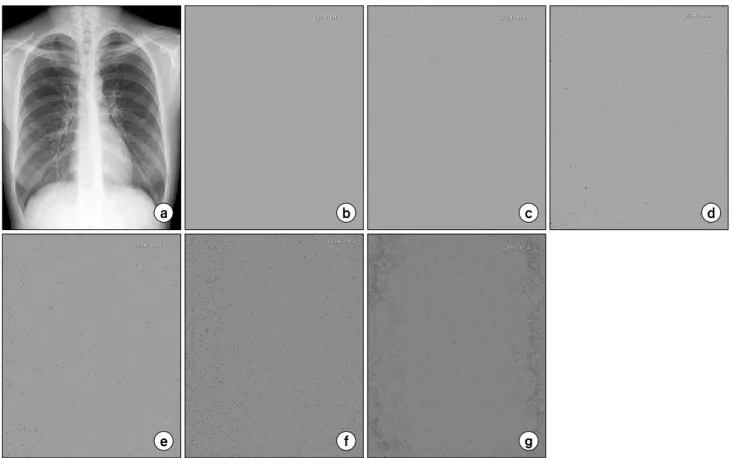
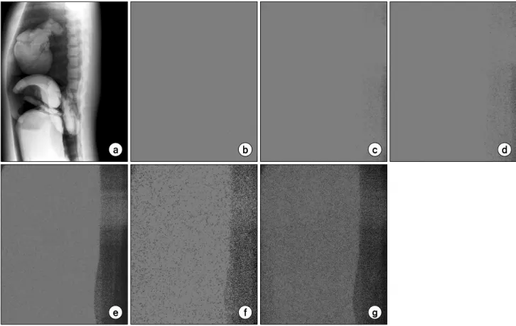
Fig. 3. Chest PA image (a), undelayed unexposed image (b), delayed unexposed image after1 hour (c), 3 hours (d), 6 hours (e), 9
hours (f), 12 hours (g).100 mR의 선량으로 overexposure 한 후 스캔하여 첫 번째 영상을 획득하였다. 동일한 IP를 이용하여 납 블록을 없애 고, 150×200 mm 크기의 조사야에 1 mR의 선량으로 under- exposure 한 후 스캔하여 두 번째 영상을 획득하였다. 동일 한 IP의 unexposed image (dark noise image)를 획득하여 평 가하였으며 촬영 거리는 180 cm를 유지하였다(Fig. 2). 조 사선량을 측정하기 위하여 ion chamber (Radical 9095;
Radical, USA)를 사용하였다. 획득된 영상은 MUSICA 파라 미터를 모두 0으로 하여 후처리를 적용시키고, sensitometry 는 linear를 적용하였다.7)
AAPM Report 93 (2006)의 CR acceptance test의 erasure thoroughness평가 항목 및 기준은 Table 4와 같다. Quantita- tive criteria의 경우 획득된 모든 영상의 80%내에서 평가하 도록 한다. Log median exposure (lgM)은 Agfa 장비 고유의 엑스선 조사 지표로써, 상대적인 엑스선 조사 값을 나타낸 다. 이는 누적된 엑스선 조사가 영향을 미친다. Agfa CR 시 스템은 조사 지표로써 lgM을 사용한다. lgM은 raw 히스토 그램의 조사량의 로그 값이다. LgM 값은 IP에 실제로 조사
된 선량을 나타내는데 이는 SAL과 수식으로 연관되어있으 며 이는 식 2와 같다.12)
(1)
SAL은 gray scale의 평균값이다. CR과 같은 디지털 장비 에서는 조사량이 적거나 과다조사 되었을 경우에도 영상이 밝거나 어둡게 나오지 않는데, 이는 exposure index (EI)와 관련이 있다. 대부분의 CR 시스템은 EI를 detector에 얻어 지는 신호의 양이나 조사량으로 판단한다. Agfa의 CR 시스 템의 경우 EI는 lgM에 기반을 둔다. LgM은 영상의 ex- posure level을 기본으로 설정된 exposure value와 비교한다.
EI는 10을 진수로 하는 로그의 지표이다. 지수함수 방식은 CR 시스템의 조사 범위가 클 경우 이를 처리 가능한 범위 로 압축할 수 있게 한다.15)
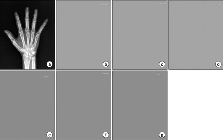
Fig. 4. Hand PA image (a), undelayed unexposed image (b), delayed unexposed image after 1 hour (c), 3 hours (d), 6 hours (e),
9 hours (f), 12 hours (g).결 과
1. 잠상의 소거 후 지연시간에 따른 노이즈의 평가 1) Chest PA, Hand PA: 영상 소거 후 획득된 un- exposed image는 지연시간 0, 1시간일 때 영상 내 픽셀 값 은 모두 같은 값이며, noise는 발생하지 않았다. 지연시간 3 시간 후부터 ghost image가 관찰되었다. 지연시간이 길수록 뚜렷한 ghost image가 확인되었다(Fig. 3).
Hand PA의 경우 지연시간에 따른 영상의 ghost image는 관찰되지 않았다(Fig. 4).
2) L-spine lateral: 3D-torso phantom을 이용하여 L-spi- ne lateral 촬영 후 스캔하였다. Fig. 5는 잠상의 소거 후 0, 1, 3, 6, 9, 12의 지연시간을 두고 획득한 L-spine lateral의 unexposed image이다. 지연시간이 길어질수록 ghost image 가 뚜렷해짐을 보였다. 특히 overexposure된 부분의 ghost image가 뚜렷하게 관찰되었다.
Ghost image의 발생에 따라 생기는 픽셀 값의 변화를 영
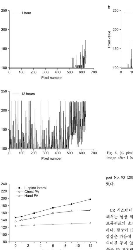
상의 profile를 통해 평가하였다. Fig. 6에서 지연시간이 길 어질수록 unexposed image에서의 전체 변동(fluctuation)은 증가하였다. 팬텀이 있는 영역에 비해 overexposure된 영역 의 픽셀 값의 fluctuation이 더 증가하였다.
Chest PA, Hand PA, L-spine lateral 영상들의 평균 픽셀 값을 비교한 결과 지연시간에 따라 증가하였다. 특히 고 선 량으로 촬영을 하는 L-spine lateral의 경우 상대적으로 두드 러진 증가를 나타내었다(Fig. 7).
Chest PA, L-spine lateral의 경우 ghost image를 나타내었 다. 다음 영상에 미치는 영향을 평가하기 위해 5 mm 두께 의 알루미늄을 3개 놓고, 55 kVp, 100 mA, 5 mAs와 같은 조건으로 underexposure하여 평가하였다. Chest PA의 경우 각 알루미늄에 설정된 ROI의 픽셀 값 및 SD 변화는 미약 하였다. L-spine lateral의 경우 underexposed image의 over- exposure된 부분 위에 놓인 알루미늄 ROI의 평균 픽셀 값 과 SD가 증가하였다. 이는 지연시간이 길수록 SD도 높게 측정되었다. 알루미늄 영상을 육안 관찰했을 경우 큰 영향 은 없었지만, underexposure한 촬영을 할 경우 문제가 될 가
Fig. 5. L-spine lateral image (a), undelayed unexposed image (b), delayed unexposed L-spine lateral image after 1 hour (c), 3 hours
(d), 6 hours (e), 9 hours (f), 12 hours (g).능성이 보였다.
촬영부위마다 정도의 차이는 있었지만 지연시간이 길수 록 unexposed image에서 noise가 증가하는 양상을 보였다.
IP는 엑스선 뿐만 아니라 자외선, 감마선 등의 전자파, 알 파선, 베타선, 전자선등의 입자선에도 민감하게 반응하는 고감도의 센서라고 할 수 있다. 이는 자연 방사성 동위원소 와 지구 위에 내리는 우주선 등의 영향을 받는 것을 의미 한다. 이에 소거한 IP을 장시간 방치할 경우 적은 양의 방 사선이 IP에 기록되게 된다.
2. 잠상의 소거 정도에 따른 노이즈의 평가
Chest PA와 hand PA의 unexposed image는 소거 정도의 증감에 따른 변화가 없었다. L-spine lateral의 경우 설정된 소거 정도 300 mR보다 작은 100 mR에서 overexposed 부분 에 ghost image가 발생하였다. 설정된 잠상의 소거 정도 300 mR 및 설정된 소거 정도보다 높은 500 mR과 750 mR 의 경우 ghost image는 관찰되지 않았다(Fig. 8).
선량이 상대적으로 많은 L-spine lateral의 경우 선량의 영
향이 더 컸으며, ghost image도 뚜렷하게 관찰되었다. 이는 선량이 ghost image 생성에 큰 영향을 끼치는 것을 알 수 있다. IP의 잠상을 제거하는 나트륨램프의 세기가 촬영부 위마다 적정치가 있으며 부족한 소거 정도는 ghost image를 유발할 수 있다.
3. Erasure thoroughness 평가
획득된 3장의 영상 80%내에서 quantitative criteria를 측정 하였다. 본교 CR 장비의 acceptance test 중 erasure thorough- ness 결과 lgM=0.28, 50<SAL<90, PV=255, PVSD=0으로 측정되었다. Table 3과 비교해본 결과 평가 기준에 부합하 였다.
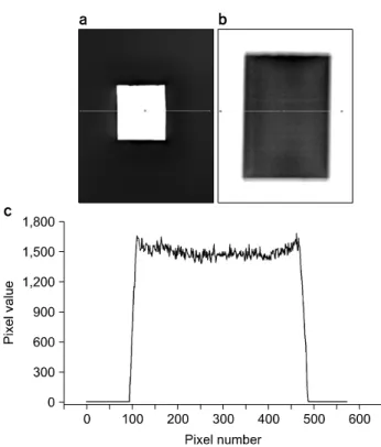
납 블록을 놓고 overexposure한 첫 번째 영상을 획득한 후 납 블록을 제거한 후 underexposure 하여 두 번째 영상을 획득하였다. Fig. 9(a, b)에서 영상을 보여주고 있으며, 두 번째 영상에서 납 블록의 ghost image 유무를 확인하였다.
Fig. 9(c)은 두 번째 영상의 profile이며, ghost image가 존재 하지 않음을 평가하였다. 실험대상의 CR장비는 AAPM re-
Fig. 6. (a) pixel value of delayed unexposed L-spine lateral
image after 1 hour, (b) 3 hours, (c) 12 hours.Fig. 7. Average pixel values of unexposed images (Chest PA,
Hand PA, L-spine lateral) according to delay time after erasure of latent image.port No. 93 (2006)의 CR acceptance test 평가 기준에 부합하 였다.
고찰 및 결론
CR 시스템에서 신뢰성 있는 영상의 재현성을 높이기 위 해서는 영상 획득 과정의 마지막 단계에서 이루어지는 나 트륨램프의 소거에 의한 IP의 완전한 초기화가 매우 중요 하다. 잠상이 100% 소거되는 것은 불가능 하지만 남아있는 잠상은 다음에 조사될 양에 비해 작은 것이 사실이므로 큰 의미를 두지 않기도 한다. 하지만 IP의 사용횟수가 증가할 수록 IP 초기화의 재현성을 유지하기가 어려워진다. IP에 남아있는 잠상뿐만 아니라 자연 방사선 또한 IP에 영향을 미칠 수 있다. 따라서 IP를 오래 사용하지 않았을 경우 잠 상의 소거 후 사용하는 것이 바람직하다.
본 연구를 통해 촬영조건이 unexposed image인 경우 노
Fig. 9. Image of overexposed lead block image (a), under-
exposed image for qualitative criteria of erasure thoroughness (b), profile of underexposed image (c).Fig. 8. L-spine lateral image (a), unexposed image of erasure level at 100 mR (b), 300 mR (c),
500 mR (d), 750 mR (e).이즈에 미치는 영향을 알 수 있었다. 촬영조건 중 선량이 높을수록, 상대적으로 overexposure된 부분을 따라 ghost image가 나타났다. 이는 지연시간이 길어질수록 뚜렷해지 는 결과를 보였다. 지연시간이 길어진 overexposed image는 이후 촬영한 영상에서 육안으로 보이지는 않지만, under- exposure하는 인체부위를 촬영할 경우 문제가 될 수 있음을 입증하였다. 또한 erasure thoroughness 평가는 본 실험에 이 용한 CR 장비가 AAPM report No. 93 (2006)의 CR accept- ance test의 평가 기준 범위 내에 있음을 보여 주었다. 본 연 구의 결과는 IP 초기화의 최적화에 기초적인 자료를 제공 함으로써 신뢰성 있는 영상을 획득하는데 기여할 것으로 판단된다.
참 고 문 헌
1. Ralph Schaetzing: Computed radiography technology. Pro- ceeding of Radiological Society of North America. Chicago (2003), pp. 10-13
2. Ehsan Samei: An experimental comparison of detector per- formance for computed radiography systems. Med Phys 29:
447-459 (2002)
3. Flynn MJ, Samei E: Experimental comparison of noise and
Evaluation of Unexposed Images after Erasure of Image Plate from CR System
Bo-Yeon Lim*, Hye-Suk Park*†, Ju-Hye Kim*, Kwang-Hyun Park*, Hee-Joung Kim*†
*Department of Radiological Science, College of Health Science,
†Research Institute of Health Science, Yonsei University, Wonju, Korea
It is important to initialize Image Plate (IP) completely for removing residual latent image by sodium lamp for reliability and repeatability of computed radiography (CR) system. The purpose of this study was to evaluate latent images of computed radiography (CR) images respect to delay time after erasure of foregone latent image and its effect, and erasure level. Erasure thoroughness for CR acceptance test from American Association of Physicist in Medicine (AAPM) Report 93 (2006) was also evaluated. Measurements were made on a CR (Agfa CR 25;
Agfa, BELGIUM) system. Chest postero-anterior (PA), Hand PA, L-spine lateral radiographs were chosen for evaluation. Chest phantom (3D-torso; CIRS, USA) was used for Chest PA and L-spine lateral radiography. For Hand PA radiography, projections was done without phantom. Except Hand PA radiographs, noise was increased with delay time, and ghost image was appeared on overexposed area. Effect of delay after erasure on latent image was not seen on naked eye, but standard deviation (SD) of pixel value on overexposed area was relatively higher than that of other areas. On Hand PA and Chest PA radiographs, noise were not occurred by adjustment of erasure level. On L-spine lateral images at lower erasure level than standard level, noise including ghost image were occurred because of high tube current. Erasure thoroughness of CR system in our department was to be proved by these evaluation. The results of this study could be used as a baseline for IP initialization and reliability of CR images.
Key Words: Image Plate, Erasure level, Erasure thoroughness resolution for 2k and 4k storage phosphor radiography systems.
Med Phys 26:1612-1623 (1999)
4. KONICA MINOLTA Technical Report: A New CsBr Photostimulable Phosphor plate. KONICA MINOLTA Medical &
Graphic Inc, Yasushi Nakano, Takehiko Shoji, Takafumi Yanagita, Shigetami Kasai, Kuniaki Nakno, Hidehito Nanto (1988) 5. Pongnapang N: Practical guidelines for radiographers to im- prove computed radiography image quality. Biomed Imaging Interv J 1:18-22 (2005)
6. Anders Tingberg, David sjöström: Optimisation of image plate radiography with respect to tube voltage. Radiation Protec- tion Dosimetry 114:286-293 (2005)
7. 권덕문, 김성수, 김영근 등: Analog & DigitalㆍPACS 의료영상정 보학. 개정 5판, 대학서림, 서울 (2004), pp. 334-336
8. 정지영, 박혜숙, 조효민 등: CR 시스템의 종류와 I.P 크기에 따 른 정량적 영상특성 평가. 한국의학물리학회 19:63-72 (2008) 9. Lu ZF, Nickoloff EL, So JC, et al: Comparison of comput-
ed radiography and film/screen combination using a con- trast-detail phantom. J Appl Clin Med Phys 4:91-98 (2003) 10. Richard Whittington, Peter Bloch, Della Hutchinson, et
al: Verification of prostate treatment setup using computed ra- diography for portal imaging. J Appl Clin Med Phys 3:88-96 (2002)
11. Edward L. Siegel, Larry T. Cook, Michael B. Parsa:
Conventional screen/film vs Reduced exposure photostimulable phosphor plate imaging in lower extremity venography. Am J Roentgenol 156:1095-1099 (1991)
12. AAPM Report 93: Acceptance Testing and Quality Control of Photostimulable Storage Phosphor Imaging Systems.
American Association of Physicist in Medicine, J. Anthony Seibert, Terese M.Boguki, Ted Ciona, et al. (2006)
13. Nina Kowalczyk, Elizabeth Comer: Exposure indicator degradation from CR plate processing delays. Radiologic Tech- nology 80:401-403 (2009)
14. Rampado O, Isoardi P, Ropolo R: Quantitative assessment of computed radiography quality control parameters. Phys Med Biol 51:1577-1593 (2006)
15. Moore CS, Liney GP, Beavis AW, et al: A method to opti- mize the processing algorithm of a computed radiography sys- tem for chest radiography. Br J Radiol 80:724-730 (2007)