Received: 30 May, 2015 Revised: 20 June, 2015 Accepted: 22 June, 2015 Corresponding author: Don Mo Choi
Physical Therapy Team, SOL Rehabilitation Hospital, 9 Geumnanghwa-ro, Gangseo-gu, Seoul 157-851, Republic of Korea Tel: 82-2-2064-7575 Fax: 82-2-2064-7534 E-mail: misoul01@naver.com
This is an Open-Access article distributed under the terms of the Creative Commons Attribution Non-Commercial License (http://creativecommons.org/licens es/by-nc/4.0) which permits unrestricted non-commercial use, distribution, and reproduction in any medium, provided the original work is properly cited.
Copyright © 2015 Korean Academy of Physical Therapy Rehabilitation Science
http://dx.doi.org/10.14474/ptrs.2015.4.1.55 Phys Ther Rehabil Sci
pISSN 2287-7576 2015, 4 (1), 55-59
eISSN 2287-7584 www.jptrs.org
The clinical efficacy of thoracolumbar fascia release for shoulder pain
Don Mo Choi
a, Ji Hye Jung
baPhysical Therapy Team, SOL Rehabilitation Hospital, Seoul, Republic of Korea
bPhysical Therapy Team, Hangeoleum Rehabilitation Hospital, Seoul, Republic of Korea
Objective: This study aimed to elucidate the effects of thoracolumbar fascia release (TLFR) on the degree of pain and disability in patients with shoulder pain.
Design: Randomized control trial.
Methods: Thirty subjects with shoulder pain participated in this study. They were allocated to TLFR group (n=15) and manual physical therapy (MPT) group (n=15). Shoulder pain and disability index (SPADI) and the score on the visual analogue scale (VAS) were measured before and after TLFR.
Results: In the TLFR group, the degree of shoulder pain as indicated by SPADI measured after the intervention significantly dif- fered from that before the intervention (p<0.05); moreover, in the MPT group, the degree of shoulder pain was significantly lower (p<0.05). The data of the 2 groups before the intervention significantly differed from those after the intervention (p<0.05). SPADI significantly differed within the groups (p<0.05), but not between the groups. The sum of SPADI did not differ significantly be- tween the groups. The VAS scores of shoulder pain measured before the intervention significantly differed from those measured after the intervention (p<0.05) in the both groups. After the intervention, shoulder pain decreased significantly in the TLFR group as compared to that in the MPT group.
Conclusions: TLF release was effective in reducing shoulder pain. The results of this study can be applied in clinical practice for TLFR performed to reduce shoulder pain. Further studies will need to be performed to elucidate the effects of TLFR on functional recovery.
Key Words: Myofascial, Shoulder pain, Thoracolumbar fascia
Introduction
Patients with shoulder pain show a high incidence of mus- culoskeletal disorders that account for approximately one- third of the reasons for visiting a doctor [1,2]. Shoulder pain occurs in 7%-36% of the population [3].
Various approaches such as electrical stimulation therapy, acupuncture, manual physical therapy (MPT), and therapeutic exercises, have been proposed to resolve musculoskeletal disorders [4]. Among therapeutic massages and joint mobi- lization techniques, MPT that includes manipulation in- creases the range of motion and improves joint functionality
as well as reduces shoulder pain [5].
One of the main fascia, the connective tissue of the mus-
culoskeletal system, protects the visceral organs of the body
and, at the same time, forms a three-dimensional network
throughout the body. The fascia has been thought to play a
passive role in inactive structures, such as the cushioning
system [6,7], and considered to be less important than the
fascia in other tissues [8]. However, a decrease in the func-
tion of the fascia is likely to cause a potential stress to the
structures surrounded by the fascia, and in turn affect the
functioning of the whole body [9]. Disruption in the three-
dimensional alignment of the fascia reduces biomechanical
Table 1. General characteristics of the subjects (N=30) Characteristic TLFR group
(n=15)
MPT group
(n=15) p
Age (y) 46.93 (8.55) 48.40 (10.33) 0.675
Sex (male/female) 5/10 3/12 0.426
Weight (kg) 64.00 (8.58) 59.20 (6.38) 0.093 Height (cm) 162.93 (8.98) 165.40 (6.31) 0.391 Values presented as mean (SD) or number only.
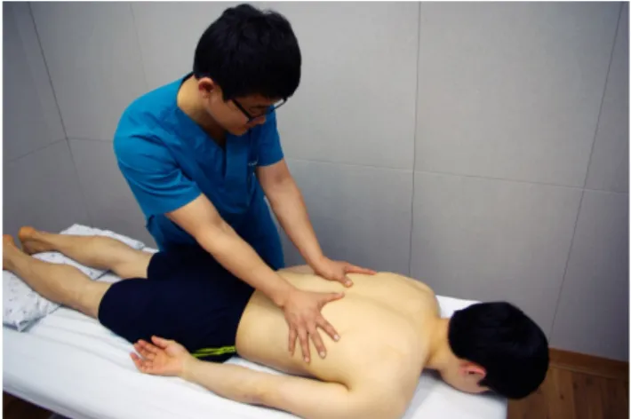
TLFR: thoracolumbar fascia release, MPT: manual physical therapy. Figure 1. Application of thoracolumbar fascia release technique.
functions [10].
Myofascial release (MFR), performed as part of osteop- athy techniques for resolving problems in the fascia by re- ducing the adhesion of the fascia, is widely used for the re- storation or optimization of the fascia movement in an acute or chronic condition [11-15]. As reported previously, MFR and decrease in pain represent the effects on the quality of life of patients [16,17].
Myofascia receives sensory innervations, and sensory peptides may be a potential cause of chronic pain [18-20]. In addition, the fascia is closely related to the autonomic nerv- ous system [21]. Believed to arise from the autonomic nerv- ous system, myelinated and demyelinated fibers were found in the fascia [22].
A major connective tissue structure, the thoracolumbar fascia (TLF), covers the deep muscles in the back of the spine and abdominal muscles. TLF acts as a force-trans- mitting structure; the latissimus dorsi, gluteus maximus, and other muscles in the region are connected with a number of muscles involved in the movement of the proximal limbs.
Moreover, previous experimental studies showed noticeable effects of TLF on pain. Hypertonic saline administered to the targeted muscles and subcutaneous tissue in healthy adults caused pain and discomfort is greater than the amount injected was found not to be spread to the waist [23]. Recent electrophysiological studies showed that the lumbar dorsal horn receives pain signals from the TLF, indicating that the TLF could be the cause of back pain [19].
We think that the TLF release (TLFR) approach should be considered when the upper extremities are affected. However, studies using TLFR for the upper extremities have been limited. This study aimed to apply TLFR for patients with shoulder pain, and to evaluate the effect of TLFR on pain and function in the shoulder joint.
Methods Subjects
Our study was approved from Sahmyook University in- stitutional review board. For this study included men and women (30 people) who agreed to participate in the experi- ment; a detailed explanation about the purpose and methods of the study was given to the participants. The participants were outpatients with shoulder pain who visited SOL Rehabilitation Hospital located in Gangseo-gu, Seoul, Korea. Of the 30 patients selected, 12 were excluded. The in- clusion criteria of this study were as follows: (1) fracture or dislocation of the shoulder without subluxation; (2) no his- tory of a surgery for the shoulder joint; and (3) no symptoms of cervical spine problems. The general characteristics of the study participants are presented in Table 1.
Procedures
In the present study, we applied TLFR, mainly performed as part of osteopathy techniques, which, for the fascia, can be divided as direct or indirect. The selection of the direct or the indirect technique depends on the problem to be re- solved. In this study, we used the direct technique, which is called Still's technique (Figure 1). This technique has lim- ited applications, in that it is used to induce relaxation of the fascia. The case when the functional barrier to tissue in-lim- ited in application to determine a three-dimensional pressure or pulling force and maintain 60-90 seconds until the tissue relaxes [24].
The patients were placed in the prone position on a table,
with both arms extended downward and the head in a neutral
position. The therapist, positioned on the pelvic side of the
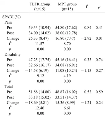
Table 2. Comparison of change in the SPADI by group (N=30) TLFR group
(n=15)
MPT group
(n=15) ta p
SPADI (%) Pain
Pre 59.33 (10.94) 54.80 (17.62) 0.84 0.41 Post 34.00 (14.02) 38.00 (12.78)
Change 25.33 (8.47) 16.80 (7.47) −2.92 0.01
tb 11.57 8.70
p 0.00 0.00
Disability
Pre 47.25 (17.75) 45.16 (16.41) 0.33 0.74 Post 32.66 (16.17) 34.08 (16.91)
Change −14.58 (6.19) 11.08 (10.24) −1.13 0.27
tb 9.12 4.19
p 0.00 0.00
Total
Pre 51.88 (14.80) 48.87 (16.02) 0.53 0.59 Post 33.18 (15.02) 33.51 (14.37)
Change −18.69 (5.81) 15.36 (8.99) −1.21 0.24
tb 12.46 6.61
p 0.00 0.00
Value are presented as mean (SD).
SPADI: shoulder pain and disability index, TLFR: thoracolumbar fascia release, MPT: manual physical therapy.
aIndependent t-test, bpaired t-test.
patient, placed both the hands on the TLF area and stretched the fascia by applying adequate pressure. The therapist per- formed palpations slowly and applied pressure on the deep fascia in upward and downward, and left and right move- ments, in a three-dimensional pattern of rotation. The pres- sure point was felt when resistance occurred at the end of the resistance 90-second maintenance. The procedure was re- peated 2 or 3 times in some cases.
The patients in the TLFR group and MPT group under- went MPT. MPT techniques were used for joint mobilization and relaxation, and therapeutic massage was administered to induce muscle relaxation. In some cases, therapeutic mas- sage was administered to the pectoralis major, pectoralis mi- nor, rotator cuff, latissimus dorsi, and teres major and minor muscles; joint mobilization and shoulder flexion, abduction, and internal or external rotation of the restriction point were performed. Furthermore, the participants performed traction and gliding movements. The TLFR group and the MPT group were administered MPT for 40 minutes, and the TLFR was administered additional TLFR for 10 minutes.
Groups were administered therapy 3 times a week for 4 weeks.
Outcome measures
Shoulder pain and disability index (SPADI) and visual an- alogue scale (VAS) scores were measured before and after the intervention. SPADI is an assessment tool developed by Roach et al. [25] in 1991 to evaluate the degree of shoulder pain and disability. Eight items including two parts are sepa- rated by a total of five questions to evaluate the scale and ac- tivities of daily living disability related to pain in the upper limbs, a total of 13 evaluation items made 10 cm VAS form.
The individual items were presented as percentage (%) values. Score means a higher score is made and severe pain and disability 100 points in the 0 states. The average of the score on 13 evaluation items was determined. SPADI has been reported to be a valid and reliable tool [26,27]. A more than 10-point decrease in the score was defined as a clin- ically minimal important change [28]. Reliability coeffi- cients of intraclass correlation coefficient ≥0.89 in variety of patients and SPADI demonstrates good construct validity that correlating well other specific shoulder questionnaire [29].
VAS score was used to assess the degree of pain. A score of 0 indicated absence of pain and a score of 100 indicated very severe pain. Test-retest reliability and validity has been shown to be good in musculoskeletal pain conditions [30].
Data analysis
The general characteristics of the subjects were analyzed using descriptive statistics; groups were compared before and after the paired t-test for dependent variables. The in- dependent t-test was performed to determine the differences in dependent variables between the groups. The statistical significance level was set at 0.05 for all the data.
Results
In the TLFR group, the degree of shoulder pain as in- dicated by SPADI measured after the intervention signifi- cantly differed from that before the intervention (p<0.05);
moreover, in the MPT group, the degree of shoulder pain was significantly lower (p<0.05). The data of the 2 groups before the intervention significantly differed from those af- ter the intervention (p<0.05). SPADI significantly differed within the groups (p<0.05), but not between the groups. The sum of SPADI did not differ significantly between the groups (Table 2).
The VAS scores of shoulder pain measured before the in-
Table 3. Comparison of change in the VAS by group (N=30) TLFR group
(n=15)
MPT group
(n=15) ta p
VAS (score)
Pre 5.48 (2.37) 4.38 (1.69) −1.45 0.15
Post 2.57 (1.62) 2.97 (1.29)
Change −2.91 (1.76) 1.44 (0.93) −2.85 0.01
tb 6.37 5.99
p 0.00 0.00
Value are presented as mean (SD).
VAS: visual analogue scale, TLFR: thoracolumbar fascia release, MPT: manual physical therapy.
tervention significantly differed from those measured after the intervention (p<0.05) in the both groups. After the inter- vention, the shoulder pain decreased significantly in the TLFR group as compared to that in the MPT group (p<0.05;
Table 3).
Discussion
Since the origins of the osteopathic progression, the fascia is regarded as very important for achieving the best treat- ment outcomes [31]. Osteopathic diagnostic palpation in the case of a musculoskeletal injury or bone modifications, de- termination of the function of the fascia, and detection of pathophysiological conditions are necessary to determine the causes of physical dysfunction [32].
MFR facilitates mechanical and neural responses, thereby enabling a hearing physiological adaptation of the fascia through the interface system [33].
TLF is the most widely present in humans. TLF autonom- ic nerve fibers may be adjusted in advance by the tension of the fascia and the contraction of the smooth muscles [34].
Autonomic nerve fibers may indirectly affect the proprio- ceptive sense by reducing the blood flow to the muscles [35].
Proprioceptive sensory damage reduces the muscle sensi- tivity [36].
After MFR in the TLF of patients with chronic low back, TLF thickness increased and remained constant for 24 hours, as observed on ultrasonography [37]. A recent study showed that the TLF consists of a dense network of nerve fi- bers, including the nociceptive fibers, which could play a major role in back pain. The finding that most calcitonin gene-related peptide and substance P immunoreactivity fi- bers are located in the outer layers of the subcutaneous tissue or fascia may explain the occurrence of pain during a passive
manual approach for the fascia and subcutaneous tissue [20].
Previous studies on MFR showed that TLF affects the up- per extremities. In this study, TLFR was performed in 30 pa- tients with shoulder pain to study its effects on shoulder pain and disability. MPT was effective in the reduction of pain VAS score, shoulder pain, and SPADI in both the groups;
however, the effects were greater in the TLFR group than in the MPT group. The extent of shoulder disability as in- dicated by SPADI reduced in both the groups, and the differ- ence between the SPADI measured before the intervention and that measured after the intervention was significant;
however, the differences between the groups were insignifi- cant.
Why was TLFR effective in reducing shoulder pain? TLF is widely distributed in the dorsal part of our body, and is connected to the latissimus dorsi fascia. TLF was considered to play a role in the relaxation of the shoulder muscles. In ad- dition, stimulation of the autonomic nervous system, which originates in the fascia was thought to reduce shoulder pain.
Limitations of this study are a small number of study sub- jects and application of the technique for only a short period, imbalance of gender ratio, whether the effects of TLFR on shoulder pain and disability are long lasting could not be confirmed. Thus, we did not apply to classify the shoulder injury.
However, the effects of the application of TLFR on shoulder pain could be applied in clinical practice. Further studies will need to be performed to elucidate the effects of TLFR on functional recovery.
Conflict of Interest
The authors declared no potential conflicts of interest with respect to the authorship and/or publication of this article.
References
1. Greving K, Dorrestijn O, Winters JC, Groenhof F, van der Meer K, Stevens M, et al. Incidence, prevalence, and consultation rates of shoulder complaints in general practice. Scand J Rheumatol 2012;41:150-5.
2. Tekavec E, Jöud A, Rittner R, Mikoczy Z, Nordander C, Petersson IF, et al. Population-based consultation patterns in patients with shoulder pain diagnoses. BMC Musculoskelet Disord 2012;13:238.
3. Green S, Buchbinder R, Hetrick S. Physiotherapy interventions for shoulder pain. Cochrane Database Syst Rev 2003;(2):CD004258.
4. Brox JI. Regional musculoskeletal conditions: shoulder pain.
Best Pract Res Clin Rheumatol 2003;17:33-56.
5. Ho CY, Sole G, Munn J. The effectiveness of manual therapy in the management of musculoskeletal disorders of the shoulder: a systematic review. Man Ther 2009;14:463-74.
6. Standring S. Gray's anatomy: The anatomical basis of clinical practice. 39th ed. Edinburgh: Churchill Livingstone; 2004.
7. Tozzi P. Selected fascial aspects of osteopathic practice. J Bodyw Mov Ther 2012;16:503-19.
8. Benjamin M. The fascia of the limbs and back--a review. J Anat 2009;214:1-18.
9. Greenman PE. Principles of manual medicine. Baltimore (MD):
Williams and Wilkins; 1989.
10. Rolf IP. Rolfing: the integration of human structures. Santa Monica (Calif): Dennis-Landman; 1977.
11. Sucher BM. Myofascial manipulative release of carpal tunnel syndrome: documentation with magnetic resonance imaging. J Am Osteopath Assoc 1993;93:1273-8.
12. Barnes JF. Myofascial release for craniomandibular pain and dysfunction. Int J Orofacial Myology 1996;22:20-2.
13. Walton A. Efficacy of myofascial release techniques in the treat- ment of primary Raynaud's phenomenon. J Bodyw Mov Ther 2008;12:274-80.
14. Martin MM. Effects of the myofascial release in diffuse systemic sclerosis. J Bodyw Mov Ther 2009;13:320-7.
15. Tozzi P, Bongiorno D, Vitturini C. Fascial release effects on pa- tients with non-specific cervical or lumbar pain. J Bodyw Mov Ther 2011;15:405-16.
16. LeBauer A, Brtalik R, Stowe K. The effect of myofascial release (MFR) on an adult with idiopathic scoliosis. J Bodyw Mov Ther 2008;12:356-63.
17. Lukban J, Whitmore K, Kellogg-Spadt S, Bologna R, Lesher A, Fletcher E. The effect of manual physical therapy in patients di- agnosed with interstitial cystitis, high-tone pelvic floor dysfunc- tion, and sacroiliac dysfunction. Urology 2001;57(6 Suppl 1):
121-2.
18. Barker PJ, Hapuarachchi KS, Ross JA, Sambaiew E, Ranger TA, Briggs CA. Anatomy and biomechanics of gluteus maximus and the thoracolumbar fascia at the sacroiliac joint. Clin Anat 2014;27:234-40.
19. Hoheisel U, Taguchi T, Treede RD, Mense S. Nociceptive input from the rat thoracolumbar fascia to lumbar dorsal horn neurones.
Eur J Pain 2011;15:810-5.
20. Tesarz J, Hoheisel U, Wiedenhöfer B, Mense S. Sensory in- nervation of the thoracolumbar fascia in rats and humans. Neuro- science 2011;194:302-8.
21. Barnes MF. The basic science of myofascial release: morpho- logic change in connective tissue. J Bodyw Mov Ther 1997;1:
231-8.
22. Schleip R. Fascial plasticity - a new neurobiological explanation:
part 1. J Bodyw Mov Ther 2003;7:11-9.
23. Schilder A, Hoheisel U, Magerl W, Benrath J, Klein T, Treede RD. Sensory findings after stimulation of the thoracolumbar fas- cia with hypertonic saline suggest its contribution to low back pain. Pain 2014;155:222-31.
24. Pilat A. Inducción miofascial. Madrid: McGraw-Hill/Interamer- icana de España, DL; 2003.
25. Roach KE, Budiman-Mak E, Songsiridej N, Lertratanakul Y.
Development of a shoulder pain and disability index. Arthritis Care Res 1991;4:143-9.
26. Bot SD, Terwee CB, van der Windt DA, Bouter LM, Dekker J, de Vet HC. Clinimetric evaluation of shoulder disability question- naires: a systematic review of the literature. Ann Rheum Dis 2004;63:335-41.
27. McConnell J. A novel approach to pain relief pre-therapeutic exercise. J Sci Med Sport 2000;3:325-34.
28. Williams JW Jr, Holleman DR Jr, Simel DL. Measuring shoulder function with the Shoulder Pain and Disability Index. J Rheumatol 1995;22:727-32.
29. Angst F, Goldhahn J, Pap G, Mannion AF, Roach KE, Siebertz D, et al. Cross-cultural adaptation, reliability and validity of the German Shoulder Pain and Disability Index (SPADI). Rheuma- tology (Oxford) 2007;46:87-92.
30. Boonstra AM, Schiphorst Preuper HR, Reneman MF, Posthumus JB, Stewart RE. Reliability and validity of the visual analogue scale for disability in patients with chronic musculoskeletal pain.
Int J Rehabil Res 2008;31:165-9.
31. Lee RP. Still's concept of connective tissue: lost in "translation"?
J Am Osteopath Assoc 2006;106:176-7; author reply 213-4.
32. Allen TW. The glossary review committe of the educational counsil on osteopathic principle. In: American Osteopathic As- sociation, editor. Yearbook and directory of osteopathic physi- cians. Chicago: American Osteopathic Association; 1993.
33. Manheim CJ. The Myofascial Release Manual. 3rd ed. New Jersey (USA): Slack Incorperated; 2001.
34. Staubesand J, Li Y. Zum Feinbau der fascia cruris mit besonderer Berucksichtigung epi-und intrafaszialer nerven. Manullen Medizine 1996;34:196-200.
35. Thomas GD, Segal SS. Neural control of muscle blood flow dur- ing exercise. J Appl Physiol (1985) 2004;97:731-8.
36. Roatta S, Windhorst U, Ljubisavljevic M, Johansson H, Passatore M. Sympathetic modulation of muscle spindle afferent sensitivity to stretch in rabbit jaw closing muscles. J Physiol 2002;540:237-48.
37. Blanquet M, Domingo T, Ortiz JC. Ultrasound study of thor- acolumbar fascia and surrounding tissues in chronic low back pain before and after spinal manipulative therapy. LA: 7th Interdisciplinary World Congress on Low Back and Pelvic Pain, Nov 9-12; 2010.