ISSN 1225-6552, eISSN 2287-7630 http://dx.doi.org/10.7853/kjvs.2015.38.1.37
< Original Article >
Veterinary Service
Available online at http://kjves.org
*Corresponding author: Okjin Kim, Tel. +82-63-850-6668, Fax. +82-63-850-7308, E-mail. [email protected]
Comparison of the non-invasive diagnostic methods, stool antigen test and PCR assay, for Helicobacter felis detection in dogs
Sunhwa Hong1, Hak-Yong Lee2, Tae-wan Kim3, Okjin Kim1*
1Center for Animal Resources Development, Wonkwang University, Iksan 570-749, Korea
2Huvet Co. Ltd., Iksan 570-749, Korea
3College of Veterinary Medicine, Kyungpook National University, Daegu 702-701, Korea (Received 24 February 2015; revised 18 March 2015; accepted 20 March 2015)
Abstract
The aim of the present study was to compare the non-invasive methods for the diagnosis of H. felis with HpSA kit-based detection method and H. felis-specific PCR assay with dog’s stool samples without sacrifice. Male Beagle dogs (n=6) were infected with H. felis ATCC 49179 (1.0×10⁹ CFU/dog) by intra- gastric inoculation two times at 3-day intervals, and the stool specimens of dogs were collected 1, 3, 5, 7, 14, 21 days after infection to submit to HpSA test and H. felis-specific PCR. As the results, the sensitivity of the HpSA and the PCR analysis was 50.0%, 83.3% respectively. Although HpSA test is less sensitive, it could be used for rapid, cheap and easy screening assay for H. felis infection in dog and cats. We suggest that the H. pylori stool antigen kit, HpSA, is useful and effective for monitoring H. felis infection. If HpSA test would be made with H. felis antibodies in the future, its sensitivity could be increased. Also, PCR assay could be successfully used to detect the H. felis in stools. Applying the H. pylori stool antigen kit and PCR assay may be the recommended non-invasive strategy to identify H. felis in dog and cats.
Key words : Helicobacter felis, H. pylori stool antigen kit, Non-invasive, HpSA, Dog
INTRODUCTION
The discovery of the association of H. pylori with gastritis, peptic ulcers, and gastric neoplasia has led to fundamental changes in the understanding of gastric dis- ease in humans (Kim et al, 2006; Lee et al, 2007). Like H. pylori, other species of Helicobacter have also been shown to colonize the stomach and cause disease in animals. Gastric colonizers include H. felis, H. mustelae, H. acinonychis, H. bizzozeronii, H. heilmannii, H. salo- monis, and a recently isolated novel Helicobacter sp. of dolphins (Hodzic et al, 2001). H. felis is a gram-neg- ative, spiral-shaped bacterium originally isolated from the stomachs of cats and dogs (Lee et al, 1990). This organism has been shown by 16S rRNA gene sequenc-
ing to be very closely related to H. pylori (Fox et al, 1995). Also, H. felis has been shown to colonize gnoto- biotic and CV mice and elicit host reactions and histo- pathology which are very similar to those seen in H.
pylori infections of humans (Lee et al, 1990).
In human, H. pylori is a major Helicobacter species.
H. pylori produce large amounts of urease to catalyze urea hydrolysis. Urease neutralizes stomach acid by gen- erating ammonia from urea, which is essential for sur- vival of H. pylori in the host (Hu and Mobley, 1990;
Eaton et al, 1991). Thus, H. pylori diagnosis is based on detecting urease. Several methods have been pro- posed and used to diagnose H. pylori infection.
Increasing interest has been directed toward non-in- vasive tests, compared to endoscopy-based invasive methods (histology and rapid urease test), as non-in- vasive methods do not require endoscopic assessment
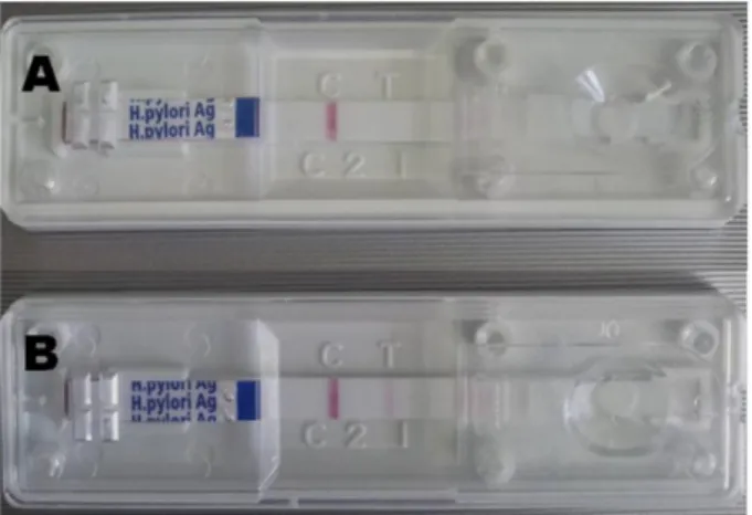
Fig. 1.The results of H. pylori stool antigen (HpSA) test stool specimens of the dogs infected with H. felis ATCC 49179. (A) One red line indicated negative, (B) Double red line indicated Helicobacter positive result.
(Mégraud et al, 2012). The 13C-urea breath test (UBT) is the most recommended non-invasive test for detecting H. pylori infection and has high sensitivity and specific- ity (Leal et al, 2011). However, the UBT cannot be ap- plied to animals due to its high cost and the require- ment for expensive analytical instruments (Nyan et al, 2004). Thus, many researchers have used polymerase chain reaction (PCR) assays to monitor the infection in stools without biopsy or sacrifice of animals (Santos et al, 2011; Neiger et al, 1998). However, PCR assay need time-consuming and high techniques and high-cost labo- ratory instrument like as Thermal Cycler (Kim et al, 2006). Furthermore, stool samples remain the most diffi- cult specimens for DNA extraction and amplification (Monteiro et al, 1997). Recently, several companies have been released H. pylori stool antigen (HpSA) test kits. HpSA tests are non-invasive diagnostic modules for H. pylori infection with human patient’s stool samples (Shimoyama, 2013; Patel et al, 2014). However, there was little information about the usefulness of HpSA test on the H. felis, which is a major Helicobacter species in dogs and cats.
The aim of the present study was to compare the non-invasive methods for the diagnosis of H. felis with HpSA kit-based detection method and H. felis-specific PCR assay with dog’s stool samples without sacrifice.
MATERIAL AND METHODS
Bacterial culture
H. felis (ATCC 49179; American Type Culture Collection, USA) was incubated in a brain-heart in- fusion broth containing 10% fetal bovine serum at 37oC overnight under a microaerophilic atmosphere and al- lowed to grow to a density of ∼2.0×10⁹ colony-forming units (CFU) per 1 ml of culture broth.
Animals and treatment
8-week-old male Beagle dogs (n=6) were obtained from the animal facility of Wonkwang University and housed in a room with constant environmental con-
ditions (22±2oC; 40∼70% relative humidity; 12-hour light-dark cycle; 150∼300 lux brightness). Pellet feed and purified water were available ad libitum. Before the inoculation, preceding PCR assays with their stool speci- mens indicated that the dogs were free of H. felis. All animal experiments were conducted according to Standard Operation Procedures and were approved by the Institutional Animal Care and Use Committee of Wonkwang University, Korea.
Bacterial inoculation
After a 24-hour fast, the dogs were orally inoculated twice at 3-day intervals by oral administration of 1.0×10⁹ CFU of H. felis suspended in 10 ml of broth.
Stool antigen kit
After H. felis inoculation, stool specimens were gath- ered in days 1, 3, 5, 7, 14, and 21. The H. felis antigen was evaluated using the commercially available SD Bioline H. pylori Ag kit according to the manufacturer’s instructions. Specimens (250 mg) were incubated with diluents solution at room temperature for 30 min and then 100 l was placed on the Helicobacter Ag exami- nation device. The test results were checked about 15 min later. One red line indicated negative, and double red line indicated Helicobacter positive result (Fig. 1).
Genomic DNA extraction and PCR
After inoculation, stool specimens were gathered in days 1, 3, 5, 7, 14, and 21. Stool samples of H. felis infected dogs were homogenized and resuspended in PBS, and DNAs were extracted using the AccuPrep Stool DNA Extraction Kit (Bioneer, Korea) according to the manufacturer's instructions. AccuPrep Stool DNA Extraction Kit is designed for the rapid, convenient ex- traction of DNA from fresh or frozen stool, or other samples containing large amounts of material that can inhibit PCR. The kit uses a glass filter, fixed in a col- umn tube that can efficiently bind DNA in the presence of chaotropic salts. Using the spin-column method, con- taminants and enzyme inhibitors (such as heparin, bilir- ubin bile salts, porphyrin) are eliminated and high-purity DNA is obtained, ready for use in a variety of applica- tions (Lee et al, 2007). After washing steps which re- move proteins and salt, high-purity DNA is finally elut- ed using a low-concentration elution buffer. It provides 2∼5 g DNA yields from 100 mg stool (Lee et al, 2007).
DNA was eluted in Tris-EDTA buffer (pH 8.0), and an aliquot was used for PCR amplification. All DNA samples were stored at −20oC until the PCR assays were performed. Previously, the primers were designed to amplify the 16S rRNA gene of H. felis (Kong et al, 1996). The sequence of H. felis primer HF-L was 5’-ATGACATGCCCTTTAGTTTGGGATAGCCA-3’, and that of primer HF-R was 5’-CGTTCACCCTCTCA GGCCGGATACC-3’. This primer set amplified a 169 bp fragment and was used for detection of H. felis in samples (Kong et al, 1996).
The template DNA (400 ng) and 20 pmol of each primer were added to a PCR mixture tube (AccuPower PCR PreMix; Bioneer) containing 2.5 U of Taq DNA polymerase, 250 M of each deoxynucleoside triphos- phate, 10 mM Tris-HCl (pH 8.3), 40 mM KCl, 1.5 mM MgCl2, and gel loading dye. The final volume was ad- justed to 20 l with distilled water. The reaction mix- ture was subjected to denaturation at 94oC for 5 min followed by 35 cycles of 94oC for 20 s, 55oC for 30 s, 72oC for 1 min, and a final extension step of 72oC for 10 min. Samples were maintained at 4oC until analysis.
Reactions were conducted using My Genie 32 Thermal Block PCR (Bioneer). Eight microliters of each sample were mixed with 2 l of loading buffer, and electro- phoretically separated on 2.0% agarose gels stained with 0.5 g/ml ethidium bromide. DNA bands were observed under ultraviolet light.
Statistical analysis
The statistical analysis was performed using analysis of variance and SPSS for Windows v.12.0 (Chicago, IL, USA). A P-value<0.05 was considered statistically significant. The positive ratio was measured using MINITAB software and 95% confidential intervals (Minitab Inc., State College, PA, USA). A positive ratio indicated a significant difference.
RESULTS
Stool Ag Kit
After the final inoculation of H. felis into the Beagle dogs, we collected stool specimens on days 1, 3, 5, 7, 14, and 21. The stools contained less water. We in- cubated the stool specimens in diluent solution for 30 min with vortexing every 10 min. Then, we placed 100
l of sample on the SD Bioline H. pylori Ag kit de- tection device. On day 1, we observed no positive results. But, the positive ratio increased on days 3 [16.7% (1/6)], days 5 [16.7% (1/6)], and days 7∼21 [50.0% (3/6)] (Table 1).
PCR of fecal samples
A PCR analysis method using 16S rRNA gene of H.
felis was employed to detect H. felis infection. The 16S rRNA gene of H. felis was specifically amplified by PCR with the 16S rRNA gene-specific primers (HF-L and HF-R). The target nucleic acid fragments were spe- cifically amplified by PCR with 16S rRNA primers (Fig. 2). As a result, five of six stool samples were pos- itive by the PCR reaction (5/6, 83.3%) (Table 2).
Table 1. Helicobacter Stool Antigen Test (HpSA) for diagnosing Helicobacter felis infected dogs
Day H. felis inoculation n Positive reactiona (positive percent)
1 Yes 6 0 (0%, CIb 0∼39.0)
3 Yes 6 1 (16.7%, CI 3.0∼56.4)
5 Yes 6 1 (16.7%, CI 3.0∼56.4)
7 Yes 6 3 (50.0%, CI 18.8∼81.2)
14 Yes 6 3 (50.0%, CI 18.8∼81.2)
21 Yes 6 3 (50.0%, CI 18.8∼81.2)
aA positive reaction revealed Helicobacter colonization, which was observed as a red colored double line.
bIncidence percentage (95% confidential interval) was calculated with MiniTab statistical software.
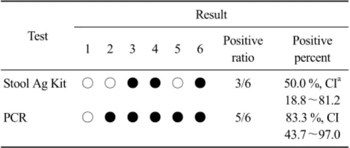
Table 2. Comparison of the test results of dogs infected with Helicobacter felis.
Test
Result
1 2 3 4 5 6 Positive
ratio
Positive percent
Stool Ag Kit ○ ○ ● ● ○ ● 3/6 50.0 %, CIa
18.8∼81.2
PCR ○ ● ● ● ● ● 5/6 83.3 %, CI
43.7∼97.0
○, negative; ●, positive.
aIncidence percentage (95% confidential interval) was calculated with MiniTab statistical software.
Fig. 2. Amplification with H. felis-specific primers in the 16S rRNA gene was identified on a 2.0% agarose gel electrophoresis. Lane 1: Stool sample of No. 1, N: distilled water, LT1: Lung tissue of No. 1 cat, LT2: Lung tissue of No. 2 cat.
DISCUSSION
Diagnosis of H. pylori infection can be made with both invasive and non-invasive tests. Invasive tests in- clude histology, culture, and the rapid urease test, which require endoscopy to obtain gastric mucosa biopsies.
Non-invasive tests for diagnosis of H. pylori infection, which are based on analyzing of samples of breath, blood, or stool, have been developed (Polk and Peek, 2010). However, serological tests are unable to dis- tinguish active from past infections (Vaira et al, 1999).
Non-invasive tests require demonstrating the micro- organism in gastric biopsy samples; therefore, an endos- copy must be performed. The current gold standard for diagnosing H. pylori infection is endoscopic biopsy of gastric tissue for the rapid urease test, histology, and culture. However, such an invasive procedure has major
disadvantages of anesthesia, discomfort, and the possi- bility for ethical problems (Hoshina et al, 1990). But, noninvasive tests are easy to perform and do not pro- duce significant discomfort and allow a patient to avoid the discomfort and risk of invasive endoscopy. These in- clude serological antibody testing for H. pylori, the urea breath test, and the stool antigen assay (HpSA) test (Hoshina et al, 1990).
Among several diagnostic tests, the HpSA test and PCR for diagnosing H. felis infection may offer a useful non-invasive method without sacrificing animals during an in vivo study. In this study, the sensitivity of the HpSA and the PCR analysis was 50.0%, 83.3%
respectively. Despite all the above observation on per- formance of antigen detection H. feslis in stool, it has certain disadvantage: antigen excretion may vary over the time period and antigen may degrade while passing through intestine. Further, use of N-acetylcysteine like mucolytic agent may decrease the accuracy of the diag- nosis (Demirtürk et al, 2003). Cut off titer, though diffi- cult to decide but crucial to reach the conclusion by us- ing antigen detection technique. However, stool antigen detection using monoclonal antibody has been recom- mended by EHSG as it gives equivalent diagnosis accu- racy to UBT (Malfertheiner et al, 2012).
In this study, the PCR analysis was more sensitive than the HpSA test. Since the target of HpSA test was H. pylori antigens, it may not detect completely whole H. felis antigens. Therefore, for non-invasive diagnosis of H. felis infection with animal stool samples, the PCR analysis was more sensitive. However, PCR analysis al- so has been reported several limitations (Kim et al,
2006). There is no single gold standard among the diag- nostic tests for Helicobacter infection but all of the tests have their pitfalls and limitations (Rautelin et al, 2003).
Among noninvasive methods, stool specimens are easy to obtain and consequently of high potential interest for the development of a direct method of Helicobacter spe- cies detection (Kim et al, 2006; Lee et al, 2007). PCR was successfully used to detect the bacterium in stools (Kim et al, 2006; Lee et al, 2007). Indeed stool samples remain the most difficult specimens for DNA extraction and amplification (Monteiro et al, 1997). The difficulty has been associated with the DNA polymerase inhibitors present in stools, identified as complex polysaccharides (Monteiro et al, 1997). In this study, we used the spin-column method and it eliminated effectively con- taminants and enzyme inhibitors (such as heparin, bilir- ubin bile salts, porphyrin) and high-purity DNA is obtained. It provides 2∼5 g DNA yields from 100 mg stool (Kim and Kim, 2005). Although HpSA test is less sensitive, it could be used for rapid, cheap and easy screening assay for H. felis infection in dog and cats. It may be confirmed the positive results using H. felis-spe- cific PCR.
We suggest that the H. pylori stool antigen kit, HpSA, is useful and effective for monitoring H. felis infection. If HpSA test would be made with H. felis an- tibodies in the future, its sensitivity could be increased.
Also, PCR assay could be successfully used to detect the H. felis in stools. Applying the H. pylori stool anti- gen kit and PCR assay may be the recommended non-invasive strategy to identify H. felis in dog and cats.
ACKNOWLEDGEMENTS
This study was supported by the research fund of Wonkwang University in 2015. We wish to appreciate Gi-Wook Oh, research assistants of Center for Animal Resources Development, Wonkwang University, for car- rying out the technical support.
REFERENCES
Demirtürk L, Yazgan Y, Tarçin O, Ozel M, Diler M, Oncül O, Yildirim S. 2003. Does N-acetyl cystein affect the sensi- tivity and specificity of Helicobacter pylori stool antigen test? Helicobacter 8: 120-123.
Eaton KA, Brooks CL, Morgan DR, Krakowka S. 1991. Essential role of urease in pathogenesis of gastritis induced by Helicobacter pylori in gnotobiotic piglets. Infect Immun 59: 2470-2475.
Fox JG, Yan LL, Dewhirst FE, Paster BJ, Shames B, Murphy JC, Hayward A, Belcher JC, Menes EN. 1995. Helicobacter bilis sp. nov., a novel Helicobacter species isolated from bile, livers, and intestines of aged, inbred mice. J Clin Microbiol 33: 445-454.
Hoshina S, Kahn SM, Jiang W, Green PH, Neu HC, Chin N, Morotomi M, LoGerfo P, Weinstein IB. 1990. Direct de- tection and amplification of Helicobacter pylori riboso- mal 16S gene segments from gastric endoscopic biopsies.
Diagn Microbiol Infect Dis 13: 473-479.
Hodzic E, McKisic M, Feng S, Barthold SW. 2001. Evaluation of diagnostic methods for Helicobacter bilis infection in laboratory mice. Comp Med 51: 406-412.
Hu LT, Mobley HL. 1990. Purification and N-terminal analysis of urease from Helicobacter pylori. Infect Immun 58:
992-998
Kim S, Cho S, Kim O. 2006. Detection and identification of se- creting Helicobacter species from cats. Lab Anim Res 22: 243-247.
Kim SH, Kim O. 2005. Genus-specific polymerase chain reaction to detect Mycoplasma species. Lab Anim Res 21: 189-192.
Kong L, Smith JG, Bramhill D, Abruzzo GK, Bonfiglio C, Cioffe C, Flattery AM, Gill CJ, Lynch L, Scott PM, Silver L, Thompson C, Kropp H, Bartizal K. 1996. A sensitive and specific PCR method to detect Helicobacter felis in a conventional mouse model. Clin Diagn Lab Immunol 3: 73-78.
Leal YA, Flores LL, Fuentes-Pananá EM, Cedillo-Rivera R, Torres J. 2011. 13C-urea breath test for the diagnosis of Helicobacter pylori infection in children: a systematic review and meta-analysis. Helicobacter 16: 327-337.
Lee A, Fox JG, Otto G, Murphy J. 1990. A small animal model of human Helicobacter pylori active chronic gastritis.
Gastroenterology 99: 1320-1323.
Lee H, Park Y, Kim O. 2007. Prevalence of Helicobacter Species in Feces of Dogs. Lab Anim Res 3: 339-344.
Malfertheiner P, Megraud F, O’Morain CA, Atherton J, Axon AT, Bazzoli F, Gensini GF, Gisbert JP, Graham DY, Rokkas T, El-Omar EM, Kuipers EJ. 2012. Management of Helicobacter pylori infection-the Maastricht IV/
Florence Consensus Report. Gut 61: 646-664.
Mégraud F. 2012. The most important diagnostic modalities for Helicobacter pylori, now and in the future. Eur J Gastroenterol Hepatol 9(Suppl 1): S13-15.
Monteiro L, Bonnemaison D, Vekris A, Petry KG. 1997.
Complex polysaccharides as PCR inhibitors in feces:
Helicobacter pylori model. J Clin Microbiol 35: 995-998.
Neiger R, Dieterich C, Burnens A, Waldvogel A, Corthesy-Theulaz I, Halter F, Lauterburg B, Schmassman A. 1998.
Detection and prevalence of Helicobater infection in pet cats. J Clin Microbiol 36: 634-637.
Nyan DC, Welch AR, Dubois A, Coleman WG Jr. 2004.
Development of a noninvasive method for detecting and monitoring the time course of Helicobacter pylori infection. Infect Immun 72: 5358-5364
Patel SK, Pratap CB, Jain AK, Gulati AK, Nath G. 2014.
Diagnosis of Helicobacter pylori: what should be the gold standard? World J Gastroenterol 20: 12847-12859.
Polk DB, Peek RM Jr. 2010. Helicobacter pylori: gastric cancer and beyond. Nat Rev Cancer. 10: 403-414.
Rautelin H, Lehours P, Megraud F. 2003. Diagnosis of Helicobacter pylori infection. Helicobacter 8 (Suppl 1):
13-20.
Santos AM, Lopes T, Oleastro M, Chaves P, Cordeiro R, Ferreira M, Pereira T, Machado J, Guerreiro AS. 2011. Role of
13C-urea breath test in experimental model of Helicobacter pylori infection in mice. Helicobacter 16: 320-326.
Shimoyama T. 2013. Stool antigen tests for the management of Helicobacter pylori infection. World J Gastroenterol 19:
8188-8191.
Vaira D1, Holton J, Menegatti M, Ricci C, Landi F, Ali' A, Gatta L, Acciardi C, Farinelli S, Crosatti M, Berardi S, Miglioli M. 1999. New immunological assays for the di- agnosis of Helicobacter pylori infection. Gut 45(Suppl 1): I23-27.