• 검색 결과가 없습니다.

Inhibition of Adenovirus 36 Replication and Lipid Accumulation by Distylium racemosum

N/A
N/A
Protected

Academic year: 2021

Share "Inhibition of Adenovirus 36 Replication and Lipid Accumulation by Distylium racemosum"

Copied!
10
0
0

로드 중.... (전체 텍스트 보기)

전체 글

(1)

http://dx.doi.org/10.12925/jkocs.2018.35.2.492

Inhibition of Adenovirus 36 Replication and Lipid Accumulation by Distylium racemosum

Hye-Ran Kim

1

․Gyu-Nam Park

1

․Bo-Kyoung Jung

1

Weon-Jong Yoon

2

․Kyung-Soo Chang

1,✝

1

Department of Clinical Laboratory Science, College of Health Sciences, Catholic University of Pusan, Busan 46252, Republic of Korea,

2

Jeju Biodiversity Research Institute,

Jeju Technopark, Jeju 63208, Republic of Korea

(Received January 1, 2018; Revised June 22, 2018; Accepted June 28, 2018)

Abstract : Obesity is a worldwide disease and one of the major risk factors. Virus among many factors can lead to obesity. Adenovirus 36 (Ad-36) is the adipogenic virus linked with human obesity.

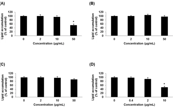
Nevertheless, there is no drug to treat both Ad-36 infection and obesity associated with virus. For the precedent study on anti-cholesterol test, Distylium racemosum ( D. racemosum ), Quercus salicina ( Q.

salicina ) and Raphiolepis indica ( R. indica ) were selected . This study was carried out to evaluate the anti-cholesterol effects, anti-lipid effects and inhibition of Ad-36 replication from three extracts. D.

racemosum (50 µg/mL) inhibited lipid accumulation on 3T3-L1 adipocyte. D. racemosum inhibited adipocyte differentiation through suppression of regulator peroxisome proliferator-activated receptor- γ (PPARγ) genes and adipocyte-specific genes such as adipocyte protein 2 (aP2). D. racemosum inhibited replication of Ad-36 at 50 µg/mL of concentration. Therefore, the extract of D. racemosum could be a candidate for development of anti-Ad-36 and anti-obesity drugs.

Keywords : Distylium racemosum, Adenovirus 36, Anti-lipid, Anti-cholesterol, Anti-obesity

1. Introduction

Obesity is defined as having an abnormal or excessive amount of body fat accumulation and is one of the major risk factors for chronic diseases including diabetes, cardiovascular disease and cancer [1]. At least 2.8 million of the world popularity are dying due to obesity and it has reached epidemic proportions worldwide [2]. Therefore, obesity



Corresponding author

(E-mail: [email protected])

is the most serious public health problem declared from the World Health Organization (WHO) [3].

Obesity is occurred by many causes. Recent studies have been made actively about correlation between virus infection and obesity.

Viruses such as canine distemper virus, Rous-associated virus 7, Borna virus and SMAM-1 cause obesity in animals, and nearly every known Adenovirus 36 (Ad-36) is associated with obesity in humans [4].

Adenoviruses are contained of 50 different

types and causes acute upper respiratory

infections. Ad-36 was first isolated from the

(2)

slurry of a six year old girl affected by diabetes and enteritis in 1978 [5]. Eleven percent of non-obese people of American adults, 30% of obese people of American adults, and 30% of obese children of South Korea were reported that they were infected with Ad-36 [6]. Fat cells infected with Ad-36 have been known for the differentiation to more fat cells and more lipid accumulation in cells. When E4orf1 gene of Ad-36 was inserted into preadipocyte, adipogenic transcription factors were stimulated [7].

Group infected with Ad-36 has shown significantly low serum cholesterol and triglyceride levels compared with the control group [8].

Obesity is generated by adipocyte differentiation and fat accumulation. Adipocyte differentiation is controlled by specific gene expression [9]. Major specific genes associated with fat accumulation are peroxisome proliferator-activated receptor-γ (PPARγ) and CCAAT/enhancer binding protein-α (C/EBPα). Especially, adipogenesis are mainly controlled by the PPARγ, belonged to nuclear receptor superfamily [10, 11]. PPARγ induces the expression fatty acid binding protein such as adipocyte protein 2 (aP2) as well as many other genes [12].

Jeju Island of the Korea has a valuable ecosystem, and is known for the variety and richness of plants and over 7800 species of them have been classified to date [13].

Therefore, natural extracts from Jeju Island were tested by anti-cholesterol screening test.

Three natural extracts among them were selected as cholesterol inhibitors. The extracts are extracted from the leaves of Distylium racemosum ( D. racemosum ) , Quercus salicina ( Q. salicina ) and Raphiolepis indica ( R. indica ) which are ones of oriental medical plants grown wildly in Jeju. D. racemosum is known for anti-oxidant effects [14]. Q. salicina is known for anti-inflammatory, anti-edemic, diuretic and litholytic activities [15]. R. indica has been a decorative form of art. However,

scientific data on biological and physiological effects of those are insufficient.

This study investigated and compared anti-cholesterol effects of extracts from D.

racemosum, Q. salicina and R. indica.

Quercetin was used as single component and positive control. Quercetin is well known as scavenge superoxide radicals and protect from lipid peroxidation [16]. The effect of extracts on the adipocyte differentiation of 3T3-L1 cells were investigated by measuring lipid accumulation and the expression major adipogenic transcriptional factor such as PPAR γ and adipocyte-specific genes such as aP2.

Moreover, Ad-36 replication in A549 cell line treated with the extracts was examined by comparing Ad-36 hexon DNA expression.

Inhibition of Ad-36 by medicinal herbs having anti-cholesterol and anti-lipid effects, might be used for drug development to prevent obesity and to treat viruses.

2. Materials and Methods

2.1. Extraction of medicinal plants

Ethanol extraction approach is employed to

extract medical plant materials which are five

hundred forty eight species of medical plants

from Jeju, Korea. The fresh leaves of them

were collected, washed with tap water, air

dried, homogenized to a fine powder and

stored in air-tight containers. The air dried

powder (100 g) was extracted with ethanol

(40-60°C) in a extractor for 18-20 hours

and solution was evaporated to dryness under

reduced pressure and controlled temperature by

using rotary evaporator. The extract was

stored in a refrigerator at 4℃ in air-tight

bottles until further use. The concentration of

extracts was prepared to be 10 mg/mL in

ethanol and used. Quercetin was used as

positive control. The extracts were tested by

anti-cholesterol screening test.

(3)

2.2. Cholesterol adsorptivity

Cholesterol adsorptivity was measured by total cholesterol kit (Asan-Pharm, Seoul, Korea). Briefly, 100 μL of different concentration of nature extracts ranging from 0.1 to 10 mg/mL were mixed with 1.8 mg/mL cholesterol (Sigma-Aldrich, St. Louis, MO, USA) in Ethanol, and reacted at 20℃ for 20 minutes. 50 μL of 0.1 M hexadecyl-trimethylammonium bromide (Sigma-Aldrich, St. Louis, MO, USA) was added and centrifuged at 4°C for 10 minutes at 15,000 rpm. After supernatant was collected, it was reacted with enzyme solution at 37°C for 5 minutes. Observance density (OD) was measured at 500 nm using Spectronic Genesys 5 (Milton Roy Company, New York, USA).

2.3. Cell viability assays

3T3-L1 preadipocyte cells (Korean Cell Line Bank, Seoul, Korea) were cultured in Dulbecco's modified Eagle's medium (DMEM) high glucose containing a 10% bovine calf serum (BCS), 1% penicillin and streptomycin (PS) for proliferation at 37°C and 5% CO

2.

Human epithelial (A549) cells (Korean Cell Line Bank, Seoul, Korea) were cultured in DMEM high glucose a 10% fetal bovine serum (FBS), 1% non essential amino acids, 1% PS. After cell was counted, it was diluted to 1☓10

5

cells/mL. Then, it was pipetted to 96 well plate each 100 μL. It was incubated for one night. The extracts from natural materials were diluted 2, 10, 50, 100 and 200 μg/mL concentrations, it was pipetted each 100 μL.

It was shaked at 150 rpm for 5 minutes and incubated at 37°C and 5% CO

2

for 3 days.

3-[4,5-dimethylthiazol-2-yl]-2,5-diphenyltetra zoliumbromide (MTT) solution (Ambresco, Ohio, USA) concentrated on 5 mg/mL in phosphate-buffered saline (PBS) was pipetted each 20 μL. After shaked at 150 rpm for 5 minutes, it was incubated for 2 hours.

Medium were deleted completely. dimethyl sulfoxide (Ambresco, Ohio, USA) were pipetted

each 200 μL and it was shaked at 150 rpm for 5 minutes. OD was measured at 560 nm using ELISA reader (Amersham Life Science, Buckinghamshire, UK).

2.4. Differentiation of 3T3-L1 preadipocytes

3T3-L1 preadipocytes were grown in DMEM high glucose with 10% BCS and 1%

PS. At 2 day post confluence, cell differentiation was induced with 10% FBS, 10 μg/mL insulin (INS), 1 μM dexamethasone (DEX), 0.5 μM isobutylmethylxanthine (IBMX) and 1% PS. After 3 days, cells were then maintained in DMEM high glucose with 10% FBS, 10 μg/mL INS and 1% PS for 4 additional days. This medium was changed every 2 days. The adipocytes were treated into the culture medium containing the extracts at 2 day. Cells were treated with 0, 2, 10 and 50 μg/mL of extracts. After treatment with extracts for 7 days, the 3T3-L1 adipocytes were staining and RT-PCR.

2.5. Oil Red O staining of 3T3-L1 adipocytes

3T3-L1 adipocytes were incubated in culture media containing with various concentration of the extracts, and then fixed in 10%

formaldehyde prepared for 1 hour. The cells were washed with distilled water (DW) 3 times, and were stained with the Oil red O (Sigma-Aldrich, St. Louis, MO, USA) solution.

The cells were then washed in DW three times for 1 hour each time. 3T3-L1 adipocytes were eluted into isopropanol.

2.6. Reverse transcription-PCR (RT-PCR) RNA was isolated by accuzol RNA extraction solution (Bioneer, Daejeon, Korea).

Isolated RNA was reverse transcribed to

cDNA for 60 minutes at 42°C and then 10

minutes at 72°C by RT-PCR. The primers

were designed following as: GAPDH forward

- GCT AGG ACT GGA TAA GCA GGG,

GAPDH reverse - GAT GGG CTT CCC GTT

(4)

GAT GA, PPARγ forward-GTG AGA CCA ACA GCC TGA CG, PPARγ reverse - ACA GAC TCG GCA CTC AAT GG, aP2 forward - GAA ATC ACC GCA GAC GAC AG, aP2 reverse - AAC TCT TGT GGA AGT CAC GCC. The amplification cycles were 94°C for 1 minute, 56°C for 1 minute and 72°C for 1 minute. After 35 cycles, PCR products were separated by electrophoresis on 1.5% agarose gel for 25 minutes at 100 V. Gels were stained with ethidium bromide visualized by Alpha Innotech imaging system (San Leandro, CA, USA).

2.7. Inhibition of Adenovirus 36 replication A549 cells were infected of adenovirus 36 for 2 hours. After infection, the cells were cultured in complete DMEM containing 10%

FBS and 1% PS in an incubator with the condition of 37°C and 5% CO

2

. After 48 hours, the infected cells were collected for further analyses. Genomic DNA was extracted using an AccuPrep Genomic DNA extraction Kit (Bioneer, Daejeon, Korea). The primers were designed following as: GAPDH forward primer– CCC ACC ACA CTG AAT CTC CC, GAPDH reverse primer – CTC ACC TTG ACA CAA GCC CA , Ad-36 hexon forward primer - ATG GCC AGC TAC TTT GA, Ad-36 hexon reverse primer – TGA GGT TCT GGC TGG AAA GT. The amplification cycles were 94°C for 1 minute, 56°C for 1 minute and 72°C for 1 minute. After 35 cycles, PCR products were separated by electrophoresis on 1.5% agarose gel for 25 minutes at 100 V. Gels were stained with ethidium bromide visualized by Alpha Innotech imaging system (San Leandro, CA, USA).

2.8. Statistical analysis

Statistical analysis of data was performed with ANOVA followed by P -values less than 0.05 were considered significant.

3. Results and Discussion

3.1. Cholesterol reduction effects of the natural extracts

The medical plants are D. racemosum, Q.

salicina and R. indica which are ones of oriental medical plants grown wildly in Jeju.

Cholesterol adsoptivity of the extracts (10 mg/mL) from D. racemosum , Q. salicina, R.

indica and quercetin were 69%, 39%, 32%

and 84%, respectively. Cholesterol adsorptivity of the extracts from D. racemosum , Q.

salicina and R. indica were not very high compared to that of quercetin, the single component. Cholesterol adsoptivities by the all extracts were gradually increased depending on concentration. Cholesterol adsorptivity of the extracts from D. racemosum and quercetin was increased compared to those of Q.

salicina and R. indica through dose-dependent increase. Cholesterol adsoptivity of the extract from D. racemosum was highest among the natural extracts (Fig. 1).

Fig. 1. Cholesterol adsorptivity of extracts from D. racemosum, Q. salicina, R. indica and quercetin. Cholesterol adsoptivity of D. racemosum , Q. salicina, R.

indica and quercetin were 69, 39, 32

and 84%, respectively, at 10 mg/mL of

concentration. quercetin as material and

positive control material was physiolo-

gically active. D. racemosum among

the natural extracts showed the highest

cholesterol reduction effects (p<0.05).

(5)

Fig. 2. Cell viability of natural extracts from D. racemosum, Q. salicina, R. indica and quercetin in 3T3-L1 cells. (A) D. racemosum , (B) Q. salicina , (C) R. indica, (D) quercetin. 3T3-L1 cells were treated with various concentrations of quercetin, D. racemosum, Q. salicina and R. indica. Cell viability was measured by the MTT assay after 72 hours. The concentration of the three natural extracts and quercetin to not affect cell viability is 50 μg/mL and 10 μg/mL, respectively. Therefore, the concentration is determined as maximum concentration in every experiment (p<0.05).

3.2. Cell viability of the natural extracts on 3T3-L1 and A549 cells

Cell viability of the extracts was determined by MTT assay on 3T3-L1 cells and A549 cells. The cells were treated with various concentrations of D. racemosum, Q. salicina , R. indica and quercetin . The extracts of D.

racemosum and Q. salicina showed cell toxicity over 100 μg/mL concentration on 3T3-L1 cells. The extracts of R. indica showed cell toxicity over 200 μg/mL concentration on 3T3-L1 cells. The concentration of quercetin to not affect cell viability is under 10 μg/mL concentration on 3T3-L1 cells (Fig. 2). Extracts of D.

racemosum and quercetin showed cell toxicity

over 100 μg/mL concentration on A549 cells.

Extracts of Q. salicina and R. indica showed cell toxicity over 200 μg/mL concentration on A549 cells (Fig. 3).

3.3. Inhibitory effect on adipogenesis and lipid accumulation by the natural extracts

To examine the anti-lipid effects of D.

racemosum , Q salicina, R. indica and

quercetin, 3T3-L1 preadipocytes were treated

with the extracts for 7 days, and stained with

Oil red O. Adipogenesis and lipid

accumulation in the cells was inhibited over

50% at 10 μg/mL quercetin and 50 μg/mL

D. racemosum. Adipogenesis in the cells were

(6)

Fig. 3. Cell viability of natural extracts from D. racemosum, Q. salicina, R. indica and quercetin in A549 cells. (A) D. racemosum , (B) Q. salicina , (C) R. indica, (D) quercetin. A549 cells were treated with various concentrations of D. racemosum, Q. salicina, R. indica and quercetin. Cell viability was measured by MTT assay after 72 hours. The concentration of the three natural extracts and quercetin to not affect cell viability is 50 μg/mL. Therefore, the concentration is determined as maximum concentration in every experiment (p<0.05).

not inhibited by the extracts of both Q.

salicina and R. indica. These results indicate that the extract from D. racemosum might be used effectively for development of anti-lipid drugs. The extracts from Q. salicina and R.

indica showed no anti-lipid effects (Fig. 4).

3.4. Inhibition of adipogenic specific genes expression by the natural extracts

To examine whether the expression of adipogenic transcription factors is inhibited by the extracts from D. racemosum , Q salicina, R. indica and quercetin, 3T3-L1 preadipocytes were treated with various concentration of the extracts and incubated for 7 days. The expression of PPARγ gene was inhibited from

10 μg/mL quercetin and 50 μg/mL D.

racemosum. The expression of aP2 gene was inhibited from 2 μg/mL quercetin and 50 μ g/mL D. racemosum. PPARγ and aP2 genes were inhibited by the extract of D. racemosum and quercetin in dose-dependent manner.

However, PPARγ and aP2 gene were not

inhibited by the extracts of both Q. salicina

and R. indica. D. racemosum inhibited

adipocyte differentiation through suppression of

regulator PPARγ genes and adipocyte-specific

genes such as aP2 (Fig. 5). PPARγ was

constitutes a expression of adipocyte-specific

genes through a master regulator of adipocyte

differentiation such as aP2 [17]. Kim et al .

[18] showed that Citrus aurantium flavonoid

suppressed adipogenesis by down regulation of

(7)

Fig. 4. Inhibitory effects of lipid accumulation by the natural extracts in 3T3-L1 cells. (A) D.

racemosum , (B) Q. salicina , (C) R. indica, (D) quercetin. Isopropyl alcohol was used to elute the Oil-red O. Lipid accumulation was decreased in the cell treated with D.

racemosum and quercetin, significantly (p<0.05).

PPARγ and C/EBPα that are relevant to lipid accumulation and lipid metabolism. Song et al . [9] showed that the anti-obesity effects of the extracts of blueberry peel on 3T3-L1.

3.5. Inhibitory effects of Ad-36 replication by the natural extracts To examine whether Ad-36 replication is inhibited by the natural extracts, A549 cells were infected with Adenovirus 36 for 2 hours and were cultured for 48 hours in the cell culture media containing the natural extracts.

After 48 hours, hexon gene of Adenovirus 36 and GAPDH as cell control gene was analyzed by RT-PCR. Hexon gene of Adenovirus 36 was inhibited in the cell treated with 10 μ g/mL quercetin and 50 μg/mL D. racemosum, significantly. Hexon gene of Adenovirus 36 was inhibited by the extract of D. racemosum

and quercetin in dose-dependent manner.

However, Hexon gene of Adenovirus 36 was not inhibited by the extracts of both Q.

salicina and R. indica (Fig. 6). Many factors

and contributions lead to obesity, but

researchers have been pondering the infection

obesity for years. Human Ad-36 belonging to

Adenovirus subgroup D was first isolated by

fecal sample of a girl suffering from enteritis

[19]. Dhurandhar et al . [20] reported that

Ad-36 increased visceral fat, total fat and

body weight. Ad-36 enhanced lipid

accumulation in 3T3-L1 cells in the presence

of adipogenic-inducer such as dexamethasone

and insulin [21]. Na et al . [22] reported that

the anti-obesity and anti-inflammation effects

using mulberry extract on Ad-36 were

evaluated in vivo.

(8)

Fig. 5. Inhibitory effects of transcriptional factors and adipocyte-specific genes by the natural extracts. (A) D. racemosum , (B) Q. salicina , (C) R. indica, (D) quercetin. Confluent 3T3-L1 preadipocytes were differentiated into adipocytes in medium containing various concentrations of D. racemosum , Q. salicina and R. indica and quercetin for 7 days. On day 7, PPARγ and aP2 genes which are transcriptional factors and adipocyte- specific genes, and GAPDH as cell control gene was analyzed by RT-PCR. PPARγ and aP2 gene transcriptions were inhibited in the cell treated with D. racemosum and quercetin, significantly.

Fig. 6. Inhibitory effects of Adenovirus 36 replication by the natural extracts. (A) D. racemosum ,

(B) Q. salicina , (C) R. indica, (D) quercetin. A549 cells were infected with adenovirus 36

for 2 hours. After infection, the cells were cultured for 48 hours in the cell culture media

containing the natural extracts. After 48 hours, hexon gene of Adenovirus 36 and GAPDH

as cell control gene was analyzed by PCR. Hexon gene of Adenovirus 36 was inhibited in

the cell treated with D. racemosum and quercetin, significantly.

(9)

4. Conclusion

This results revealed that the suppression of adipogenesis by D. racemosum was caused by expression inhibition of PPARγ and aP2 gene and replication inhibition of Ad-36. Therefore, the extract of D. racemosum might be one candidate for development of anti-Ad-36 and anti-obesity drugs.

Acknowledgements

This work was supported by grants from Brain Busan 21 program. This research was supported by the Basic Science Research Program through the National Research Foundation of Korea (NRF) funded by the ministry of Education, Science and Technology (2012007586).

References

1. World Health Organization, Health topic, http://www.who.int/topics/obesity, (accessed May., 2014)

2. World Health Organization, 10 facts on obesity, http://www.who.int/features/

factfiles/obesity, (accessed May., 2014).

3. Y. Baek, K. Park, S. Lee, E. Jang, “The prevalence of general and abdominal obesity according to sasang constitution in Korea”, BMC Complement Altern Med , Vol.14, pp. 298, (2014).

4. S. D. Vangipuram, J. Sheele, R.L. Atkinson, T. C. Holland, N. V. Dhurandhar, “A human adenovirus enhances preadipocyte differentiation”, Obes. Res , Vol.12, No.5 pp. 770-777, (2004).

5. N. V. Dhurandhar, “Infectobesity: obesity of infectious origin”, J Nutr , Vol.131, No.10 pp. 2794S-2797S, (2001).

6. R. L. Atkinson, “Prevalence of infection with adenovirus-36 in Belgium and Holland and association with obesity”,

Obesity , Vol.19, No.1 pp. 2, (2011).

7. L. Suplicy Hde, A. Bornschein, “Infeccions as the etiology for obesity”, Arq Bras Endocrinol Metabol , Vol.53, No.2 pp.

159-164, (2009).

8. M. Pasarica, A. C. Shin, M. Yu, H. M.

Ou Yang, M. Rathod, K. L. Jen, S.

MohanKumar, P. S. MohanKumar, N.

Markward, N. V. Dhurandhar, “Human adenovirus 36 induces adiposity, increases insulin sensitivity, and alters hypothalamic monoamines in rats”, Obesity , Vol.14, No.11 pp. 1905-1913, (2006).

9. Y. Song, H. J. Park, S. N. Kang, S. H.

Jang, S. J. Lee, Y. G. Ko, G. S. Kim, J.

H. Cho, “Blueberry peel extracts inhibit adipogenesis in 3T3-L1 cells and reduce high-fat diet-induced obesity”, PLoS One , Vol.8, No.7 pp. e69925, (2013).

10. Q. Zhang, M. K. Ramlee, R. Brunmeir, C.

J. Villanueva, D. Halperin, F. Xu,

“Dynamic and distinct histone modifications modulate the expression of key adipogenesis regulatory genes”, Cell Cycle , Vol.11, No.23 pp. 4310-4322, (2012).

11. V. García-Alonso, C. López-Vicario, E.

Titos, E. Morán-Salvador, A.

González-Périz, B. Rius, M. árrizas, O.

Werz, V. Arroyo, J. Clària, “Coordinate functional regulation between microsomal prostaglandin E synthase-1 (mPGES-1) and peroxisome proliferator-activated receptor γ (PPARγ) in the conversion of white-to-brown adipocytes”, J Biol Chem , Vol.288, No.39 pp. 28230-28242, (2013).

12. H. J. Park, B. Y. Chung, M.K. Lee, Y.

Song, S. S. Lee, G. M. Chu, S. N. Kang, Y. M. Song, G. S. Kim, J. H. Cho,

“Centipede grass exerts anti-adipogenic activity through inhibition of C/EBPβ, C/EBPα, and PPARγ expression and the AKT signaling pathway in 3T3-L1 adipocytes”, BMC Complement Altern Med , Vol.12, pp. 230, (2012).

13. J. Y. Moon, E. Y. Yim, G. SONG, N. H.

(10)

Lee, C. G. Hyun, “Screening of elastase and tyrosinase inhibitory activity from Jeju Island plants”, EurAsia J BioSci , Vol.4, pp.

41-53, (2010).

14. R. K. Ko, G. O. Kim, C. G. Hyun, D. S.

Jung, N. H. Lee, “Compounds with tyrosinase inhibition, elastase inhibition and DPPH radical scavenging activities from the branches of Distylium racemosum Sieb.

et Zucc”, Phytother Res , Vol.25, No.10 pp. 1451-1456, (2011).

15. J. L. Song, X. Zhao, Q. Wang, “Protective effects of Quercus salicina on alloxan-induced oxidative stress in HIT-T15 pancreatic β cells”, Exp Ther Med , Vol.5, No.3 pp. 947-951, (2013).

16. T. A. Raju, A. N. Lakshmi, T. Anand, L.

V. Rao, G. Sharma, “Protective effects of quercetin during influenza virus-induced oxidative stress”, Asia Pac J Clin Nutr , Vol.9, No.4 pp. 314-317, (2000).

17. H. J. Park, J. Yun, S. H. Jang, S. N.

Kang, B.S. Jeon, Y. G. Ko, H. D. Kim, C.

K. Won, G. S. Kim, J. H. Cho, “ Coprinus comatus cap inhibits adipocyte differentiation via regulation of PPARγ and Akt signaling pathway”, PLoS One , Vol.9, No.9 pp. e105809, (2014).

18. G. S. Kim, H. J. Park, J. H. Woo, M. K.

Kim, P. O. Koh, W. Min, Y. G. Ko, C.

H. Kim, C. K. Won, J. H. Cho, “ Citrus aurantium flavonoids inhibit adipogenesis through the Akt signaling pathway in 3T3-L1 cells”, BMC Complement Altern Med , Vol.12, pp. 31, (2012).

19. N. V. Dhurandhar, “Countering the Effects of Chronic Inflammation in Obesity”, Korean J Obes , Vol.23, No.2 pp. 73-78, (2014).

20. N. V. Dhurandhar, B. A. Israel, J. M.

Kolesar, G. F. Mayhew, M. E. Cook, R.

L. Atkinson, “Increased adiposity in animals due to a human virus”, Int J Obes Relat Metab Disord , Vol.24, No.8 pp. 989-996, (2000).

21. P. M. Rogers, K. A. Fusinski, M. A.

Rathod, S. A. Loiler, M. Pasarica, M. K.

Shaw, G. Kilroy, G. M. Sutton, E. J.

McAllister, N. Mashtalir, J. M. Gimble, T.

C. Holland, N. V. Dhurandhar, “Human adenovirus Ad-36 induces adipogenesis via its E4 orf-1 gene”, Int J Obes , Vol.32, No.3 pp. 397-406, (2008).

22. H. N. Na, S. Park, H. J. Jeon, H. B.

Kim, J. H. Nam, “Reduction of adenovirus

36-induced obesity and inflammation by

mulberry extract”, Microbiol Immunol ,

Vol.58, No.5 pp. 303-306, (2014).

수치

Fig.  1.  Cholesterol  adsorptivity  of  extracts  from D.  racemosum,  Q.  salicina,  R
Fig.  2.  Cell  viability  of  natural  extracts  from   D.  racemosum,  Q.  salicina,  R
Fig.  3.  Cell  viability  of  natural  extracts  from   D.  racemosum,  Q.  salicina,  R
Fig.  4.  Inhibitory  effects  of  lipid  accumulation  by  the  natural  extracts  in  3T3-L1  cells
+2

참조

관련 문서

ATE also decreased the lipid accumulation in 3T3-L1 adipocytes without cell cytotoxicity (Fig. Expression levels of adipogenic key transcriptional factors, such as

Effects of AY on adipogenic transcription factors and adipocyte expressed genes expression of differentiated 3T3-L1.. and significantly different between

We report that tanshinone I strongly inhibits lipid accumulation in 3T3-L1 cells and zebrafish, and its anti-adi- pogenic/lipogenic effect on 3T3-L1 cells is associated with

Here we demonstrate that among the 12 natural compounds tested, cryptotanshinone most strongly inhibits lipid accumulation in differentiating 3T3-L1

Here we demonstrate firstly that tanshinone IIA inhibits lipid accumulation in 3T3-L1 preadipocytes and zebrafish, and its anti-adipogenic effect on 3T3-L1 cells is attributable

In the present study, the effects and mechanisms of water extracts of raw garlic (WERG) and aged black garlic (WEABG) on adipocyte differentiation and adipogenesis in

We examined the inhibitory effects of Eriobotrya japonica leaf and seed extracts on lipid absorption in vitro and fat accumulation during the differentiation of 3T3-L1

Induction of G1 cell cycle arrest on clonal expansion phase of adipocyte differentiation process in Rosae laevigatae fruc- tus 30% EtOH extracts (RLE) treated 3T3-L1