288
서 론
위암은 우리나라에서 가장 흔한 악성종양이며, 조기진단 과 치료방법의 발달에 따라 생존율은 꾸준히 향상되고 있 지만 여전히 암으로 인한 사망원인 중 폐암에 이어 2위를 차지하는 종양이다.(1) 위암의 발생에는 식생활과 관련된 환경적 요인이 가장 중요하게 작용하는 것으로 알려져 있 으나, 개인의 유전적 소인도 위암의 발생인자로 작용한다 는 것이 밝혀졌다. 즉, 종양의 발생과정에서 일어나는 체세 포 변이(somatic mutation)가 아닌, 개인의 고유한 유전 형질 에 의하여 종양의 발생 위험도가 증가될 수 있으며, 위암의 발생과 관련된 유전적 소인으로 interleukin-1β (IL-1β), IL1RN, mucin, HLA, tumor necrosis factor-α (TNF-α), methyle- netetrahydrofolate reductase (MTHFR), cytochrome P450 2E1, glutathione S-transferase (GST), N-acetyl transferase (NAT), L- myc, XRCC1 등의 유전자 다형성이 제시되어 있다.(2) TNF-α는 대표적인 염증성 사이토카인 (cytokine)이며, 감
위암의 악성도와 Tumor Necrosis Factor-αPromoter 다형성의 연관성
1인제대학교 의과대학 동래백병원 외과학교실, 2부산백병원 외과학교실, 3생화학교실, 4백인제기념임상의학연구소
세포유전자치료연구부, 5병리학교실
김운원1․홍관희2․장원희3,4․김형인4․서지연4․양영일4,5
Tumor Necrosis Factor-α-308G/A Promoter Polymorphism is Associated with the Severity of Gastric Carcinomas
Woon Won Kim, M.D, Ph.D1, Kwan Hee Hong, M.D, Ph.D2, Won Hee Jang, Ph.D.3,4, Hyeong-In Kim, M.S.4, Ji Yeon Seo, M.S.4 and Young-Il Yang, M.D., Ph.D.4,5
Purpose: The pro-inflammatory cytokine, tumor necrosis factor-α (TNF-α), is a central mediator of the immune re- sponse involved in a wide range of immuno-inflammatory and infectious diseases. There is increasing evidence that TNF-α may promote the development and spread of the cancer. Polymorphisms in the TNF-α promoter have been related to TNF-α production. Therefore, we investigated the potential association of TNF-α genotypes with gastric cancer in the Korean population.
Methods: The study included 66 patients with gastric ade- noma, 75 patients with gastric carcinoma, and 551 healthy controls. The -308 and -238 polymorphisms in the TNF-α promoter were analyzed by PCR- restriction fragment length polymorphism (RFLP). Distributions of TNF-α promoter pol- ymorphisms were compared between groups byχ2 test. P values smaller than 0.05 were considered to be significant.
Results: The proportion of individuals carrying the TNF-α -308A allele was higher in the carcinoma group compared to controls and adenomas, but the differences were not significant (P=0.124). However, the TNF-α -308A allele was significantly associated with advanced gastric carcinoma (P=0.026), serosa invasion (P=0.004), neural invasion (P=
0.021), and lymph node metastasis (P=0.005). On the other hand, the TNF-α -238G/A polymorphism was not associ- ated with the development of gastric adenoma and carcino
ma and the severity of gastric carcinoma.
Conclusion: These results suggest that the TNF-α -308A allele is associated with the severity of gastric carcinoma in terms of invasion and metastasis in the Korean population.
Therefore, TNF-α promoter polymorphism could be used as a predictive marker of the severity of gastric carcinoma. (J Korean Surg Soc 2005;68:288-295)
Key Words: Gastric cancer, Progression, TNF-α, Promoter polymorphism
중심 단어: 진행성 위암, TNF-α, Promoter 다형성 ꠏꠏꠏꠏꠏꠏꠏꠏꠏꠏꠏꠏꠏꠏꠏꠏꠏꠏꠏꠏꠏꠏꠏꠏꠏꠏꠏꠏꠏꠏꠏꠏꠏꠏꠏꠏꠏꠏꠏꠏꠏꠏꠏꠏꠏꠏꠏꠏꠏꠏ
1Department of General Surgery, Dongrae Paik Hospital, Inje University, 2Department of Surgery, Busan Paik Hospital, Inje University, 3Department of Biochemistry, College of Medicine, Inje University, 4Department of Cell and Gene Therapy, Paik Institute for Clinical Research, College of Medicine, Inje University, 5Department of Pathology, College of Medicine, Inje University, Busan, Korea
책임저자:양영일, 부산광역시 부산진구 개금2동 633-165 ꂕ 614-735, 인제대학교 의과대학 병리학교실 Tel: 051-890-6885, Fax: 051-892-1733 E-mail: [email protected]
접수일:2004년 10월 29일, 게재승인일:2005년 1월 27일
ꠏꠏꠏꠏꠏꠏꠏꠏꠏꠏꠏꠏꠏꠏꠏꠏꠏꠏꠏꠏꠏꠏꠏꠏꠏꠏꠏꠏꠏꠏꠏꠏꠏꠏꠏꠏꠏꠏꠏꠏꠏꠏꠏꠏꠏꠏꠏꠏꠏꠏꠏꠏꠏꠏꠏꠏꠏꠏꠏꠏꠏꠏꠏꠏꠏꠏꠏꠏꠏꠏꠏꠏꠏꠏꠏꠏꠏꠏꠏꠏꠏꠏꠏꠏꠏꠏꠏꠏꠏꠏꠏꠏꠏꠏꠏꠏꠏꠏꠏꠏꠏꠏꠏꠏꠏꠏꠏꠏꠏꠏꠏꠏꠏꠏꠏ 염성 질환, 염증성 질환, 면역성 질환 등의 다양한 질환에서
중추적인 매개체로 작용한다. TNF-α는 숙주의 면역반응과 염증반응을 활성화하여 질환의 발생과 진행을 제어하는 방 어인자로서 작용하지만, 조절되지 않은 TNF-α의 과다한 발현은 오히려 질환을 야기하고 악화시킨다.(3) 생쥐에 종 양을 이식하였을 때 이식된 종양이 괴사되는 현상이 관찰 되었고, 이 때 TNF-α가 종양의 괴사를 유발하는 핵심적인 매개체로 작용한다는 것이 밝혀짐으로써 항암성 인자로서 TNF-α의 기능이 제시되었으며, 또한 임상연구에서도 TNF- α의 항암성 기능이 입증되었다.(4) 반면에, TNF-α가 오히 려 종양의 발생을 유발하고 종양의 진행을 촉진시킨다는 상반된 증거들도 보고되었다. 즉, 정상인에 비하여 암환자 에서 TNF-α의 혈청 수치가 증가되어 있었고, TNF-α 혈청 수치는 불량한 임상적 예후와 비례적인 연관성을 보였 다.(5) 또한, TNF-α는 in vitro 실험에서 종양 생성을 유발하 는 인자로 작용하였고, sarcophytol A, canventol, epigalloca- techin gallate, tamoxifen 등과 같은 다양한 암 억제제들은 공 통적으로 발암물질에 의한 TNF-α의 발현 유도를 억제하였 다.(6) 게다가, TNF-α유전자가 결핍된(TNF-α-/-) 생쥐는 발 암물질에 의한 피부 종양 발생에 저항성을 보이는 것이 밝혀 졌다.(7) 그러므로, TNF-α가 종양의 발생과 진행에 중요한 매개체로 작용한다는 것을 생각할 수 있다.
TNF-α의 발현은 주로 전사단계에서 조절되며, TNF-α promoter의 다형성은 TNF-α의 발현에 영향을 미치는 것으 로 알려졌다.(8-10) 따라서, TNF-α가 매개체로 작용하는 감 염성, 염증성, 그리고 종양성 질환을 대상으로 질환의 발생 과 진행에 미치는 TNF-α promoter 다형성의 영향에 대한 연구가 활발히 진행되었다. 특히, adenine (A) 유전자형 (TNF-α-308A)을 갖는 경우에 guanine (G) 유전자형 (TNF-α -308G)의 경우보다 TNF-α 발현이 증가된다고 보고된,(8) TNF-α promoter의 -308 위치의 다형성이 많이 연구되었 고, 실제로 여러 질환과의 연관성이 밝혀졌다.(11) 한편, TNF-α promoter의 또 다른 대표적인 다형성인 -238 위치 의 G/A 다형성에서는 -238 염기가 A인 경우 TNF-α 유전 자의 전사가 감소되는 것으로 알려졌고,(12) -238 염기의 다형성도 일부 자가면역질환과 감염성 질환과 연관성이 있 다고 제시되었다.(13)
위선종(gastric adenoma)은 위암의 대표적인 전암성 병변 (precancerous lesion)으로 추정되고 있다. 하지만, 위에서 선 종이 암으로 진행하는 빈도는 3.5∼30.8%로 다양하게 보고 되고 있고, 따라서 위선종이 위암의 전암성 병변으로서 갖 는 생물학적 의의는 불분명하다.(14) 위선종의 발생에 관련 되어 많은 생물학적 발생기전이 제시되었으나 현재까지 확 실히 정립되지 않았으며, 또한 선종의 생성에 관련된 유전 적 소인에 대한 자료는 찾기가 어렵다.
본 연구에서는 우리나라의 정상인과 위선종 및 위암 환 자에서 TNF-α promoter 다형성의 분포를 조사하여, TNF-α
promoter 다형성이 위선종과 위암의 발생위험도 및 악성도 와 연관되어 있는 지를 연구함으로써, TNF-α promoter 다 형성이 위선종과 위암의 유전적 인자로서 갖는 의의를 규 명하고자 하였다.
방 법
1) 연구대상
1998년부터 2000년 사이에 인제대학교 부산백병원에서 조직학적 검사를 통하여 위선종으로 진단된 66명과 위암으 로 진단된 75명을 무작위로 추출하였다. 선종군은 위내시 경과 적출된 위의 육안적 검사 및 조직학적 검사를 통하여 위암의 동반이 없는 경우만 연구에 포함하였으며, 위암군 은 조직학으로 선암종(adenocarcinoma)인 경우만을 포함하 였다. 한편, 본원 건강검진센터에 내원한 일반인들 중 혈청 학적, 이학적, 방사선과적 검사에서 당뇨병, 고혈압, 자가면 역질환, 만성간염 등의 만성질환 및 종양성 질환이 없다고 판정된 551명을 정상 대조군으로 하였다. 환자군 및 대조군 모두는 부산, 경남지역에 거주하고 있었다. 본 연구는 부산 백병원 임상시험심사위원회의 승인을 받았으며, 모든 연구 대상으로부터 시료 제공에 대한 동의를 받아 수행하였다.
2) 임상적, 병리학적 특성 분석
대상 환자들의 임상기록지와 병리결과보고서를 검토하 여 성별, 연령, 종양의 크기를 조사하였다. 적출된 위암 및 위선종 조직을 10% 중성 포르말린에 고정한 후 통상적인 방법으로 파라핀 블록을 제작하였고, 블록을 4∼5μm의 두 께로 박절한 후 Hematoxylin-Eosin (H&E) 염색을 시행하여 병리학적 특성은 분석하였다. 위선종은 대한 병리학회 소 화기 병리 연구회의 진단 기준에 의거하여 저등급과 고등 급으로 나누었다.(14) 위암의 육안적 유형은 Borrmann 분류 에 따라 나누었고, Lauren 분류법에 따라 장형(intestinal ty- pe)과 미만형(diffuse type)으로 나누었으며, WHO 기준에 따 라 암종의 조직학적 분화도를 조사하였다.(15) 또한 종양의 침윤 깊이, 림프절 전이, 신경관 침윤 등을 조사하였다.
3) DNA 분리
위선종과 위암 환자군의 정상 위점막 조직과 정상 대조 군의 말초혈액에서 DNA를 분리하였다. 위점막 조직은 파 라핀 블록 혹은 신선한 조직의 상태였다. 파라핀 블록의 경 우 7μm 두께로 15개 이상의 절편을 박절하여 1.5 ml micro- tube에 옮겨 담고 60oC에서 1시간 동안 가열한 후 xylene으 로 10분간 2회 처리하여 파라핀을 제거하였고, 이어서 100%
에탄올로 2회 세척하여 xylene을 제거한 다음 공기 중에서 건조하였다. 건조된 조직을 lysis buffer (10 mM Tris-HCl, pH 8.0, 400 mM NaCl, 2 mM EDTA) 500μl에 풀어준 후 200μg 의 Proteinase K (Boehringer Mannheim, Mannheim, Germany)
ꠏꠏꠏꠏꠏꠏꠏꠏꠏꠏꠏꠏꠏꠏꠏꠏꠏꠏꠏꠏꠏꠏꠏꠏꠏꠏꠏꠏꠏꠏꠏꠏꠏꠏꠏꠏꠏꠏꠏꠏꠏꠏꠏꠏꠏꠏꠏꠏꠏꠏꠏꠏꠏꠏꠏꠏꠏꠏꠏꠏꠏꠏꠏꠏꠏꠏꠏꠏꠏꠏꠏꠏꠏꠏꠏꠏꠏꠏꠏꠏꠏꠏꠏꠏꠏꠏꠏꠏꠏꠏꠏꠏꠏꠏꠏꠏꠏꠏꠏꠏꠏꠏꠏꠏꠏꠏꠏꠏꠏꠏꠏꠏꠏꠏꠏ
를 첨가하여 55oC에서 12시간 동안 흔들면서 반응시켰다.
반응액을 95oC에서 10분간 가열하여 Proteinase K를 불활성 화한 후 phenol-chloroform 추출과 에탄올 침전을 통하여 DNA를 최종 분리하였다.
신선한 위점막 조직은 먼저 phosphate-buffered saline (PBS) 용액으로 세척한 후 micropipette을 이용하여 분쇄하 고 PBS로 2회 세척하여 점액질과 적혈구를 가능한 한 제거 하였다. 분쇄된 위점막 조직에 lysis buffer와 Proteinase K를 첨가한 후 위와 동일한 방법으로 DNA를 분리하였다.
정상 대조군에서 얻어진 말초혈액은 EDTA로 처리한 후 PBS로 2배 희석하였다. Ficoll-Paque PLUS (Amersham Bio- sciences, Piscataway, NJ, USA) 450μl를 1.5 ml microtube에 넣은 후 그 위에 희석된 말초혈액 600μl를 서서히 가하여 올려 놓고 400×g에서 30분간 실온에서 원심분리하였다.
적혈구와 분리되어 별도의 층을 형성하는 단핵구 세포만을 회수하여 새 microtube로 옮기고 PBS로 2회 세척하였다. 분 리된 단핵구 세포에 lysis buffer와 Proteinase K를 첨가한 후 위와 동일한 방법으로 DNA를 분리하였다.
4) PCR-RFLP를 통한 TNF-α promoter 다형성 분석 TNF-α promoter의 다형성 부위를 증폭하기 위하여, 분리 한 genomic DNA를 대상으로 nested PCR을 수행하였다. 먼 저, TNFP11 primer (5'-TAGAA GGAAA CAGAC CACAG ACC-3')와 TNFP22 primer (5'-CTCAT CTGGA GGAAG CGGTA GTG-3')를 사용하여 1차 PCR을 수행한 후 1차 PCR 산물 2μl를 template로 하고 TNF-Nco3 primer (5'- AGGCA ATAGG TTTTG AGGTC CATG-3')와 TNF-Bg primer (5'-CACAC TCCCC ATCCT CCCAG ATC-3')를 사용 하여 nested PCR을 수행하였다. PCR 반응액의 부피는 20μl 로 하였고, DNA를 제외한 반응액의 최종 조성은 10 mM
Tris- HCl (pH 9.0), 50 mM KCl, 0.1% TritonⓇ X-100, 1.5 mM MgCl2, 각 0.5 M primer, 200 nM dNTP, 0.5 U Taq DNA poly- merase (Promega, Madison, WI, USA) 이었다. 일차 PCR에서 는 94oC에서 5분간 pre-denaturation한 후 94oC에서 30초, 57oC에서 30초, 72oC에서 30초의 반응을 35회 반복하였고, 최종적으로 72oC에서 5분간 post-extension하였다. Nested PCR의 반응 조건은 annealing 온도가 55oC인 것을 제외하고 1차 PCR과 동일하였다.
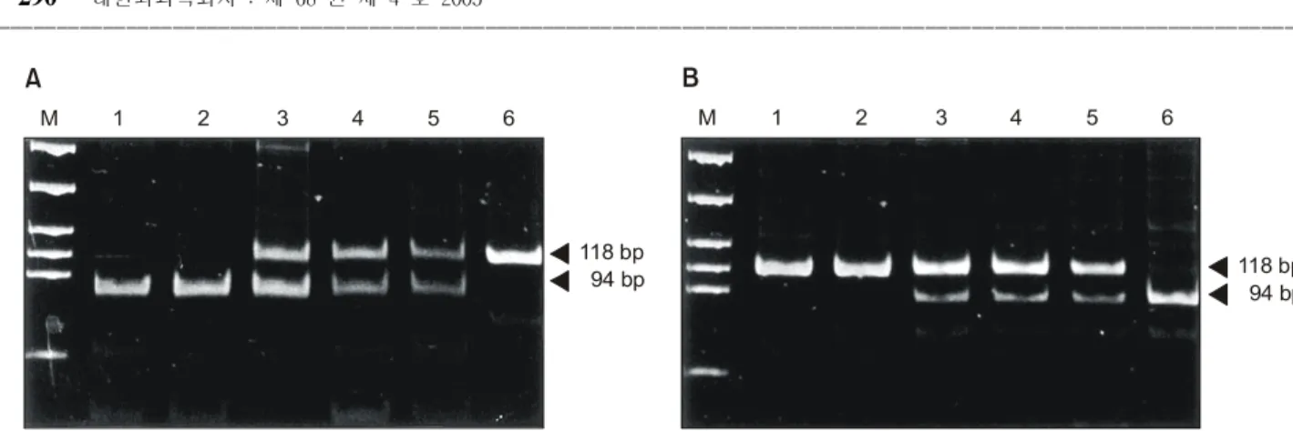
TNF-α promoter의 -308과 -238 위치의 다형성을 조사 하기 위하여 PCR 산물을 각각 NcoI (New England Biolabs, Beverly, MA, USA)과 BglII (Amersham Biosciences, Piscata- way, NJ, USA) 제한효소로 절단하였다. 본 연구에서는 -308 위치의 염기가 G인 경우 NcoI 효소에 의해 절단되나 A인 경우는 절단되지 않고, -238 위치가 G이면 BglII 효소에 의해 절단되지 않으나 A이면 절단되도록 primer를 디자인 하였다. PCR 산물 4μl에 2.5 U의 효소를 가하고 최종 부피 10μl로 맞춘 후 37oC에서 4시간 이상 반응시켰다. 반응산물 을 15% polyacrylamide gel electrophoresis를 통해 분석하였 다. PCR 산물의 길이는 118 bp이며 NcoI과 BglII 효소에 의해 절단될 경우 각각 94와 95 bp의 산물이 만들어진다(Fig. 1).
5) 통계학적 분석
위선종 및 위암 환자와 정상 대조군 사이에 TNF-α pro- moter 다형성의 분포를 비교하기 위하여 Mantel-Haenszelχ2 방법을 이용하여 통계적 의의를 조사하였다. P 값이 0.05 미만인 경우를 통계적으로 유의성이 있는 것으로 정의하였 다. 모든 통계학적 분석은 SAS system (SAS Institute Inc., Cary, NC, USA, Version 6.12)을 이용하여 수행하였다.
Fig. 1. Genotyping of TNF-α promoter by PCR-RFLP. A 118-bp fragment was amplified by the nested PCR using primers, TNF-Nco3 (from -332 to -309) and TNF-Bg (from -237 to -215). The PCR products were digested with: (A), NcoI; or (B), BglII, and subjected to a 15% polyacrylamide gel electrophoresis. (A) Polymorphism at position -308. Lanes: M, molecular weight marker; 1 and 2, G homozygotes; 3-5, G/A heterozgotes; 6, A homozygote. (B) Polymorphism at position -238. Lanes: M, molecular weight marker; 1 and 2, G homozygotes; 3-5, G/A heterozygotes; 6, A homozygote.
M 1 2 3 4 5 6 M 1 2 3 4 5 6
118 bp
118 bp 94 bp
94 bp
A B
ꠏꠏꠏꠏꠏꠏꠏꠏꠏꠏꠏꠏꠏꠏꠏꠏꠏꠏꠏꠏꠏꠏꠏꠏꠏꠏꠏꠏꠏꠏꠏꠏꠏꠏꠏꠏꠏꠏꠏꠏꠏꠏꠏꠏꠏꠏꠏꠏꠏꠏꠏꠏꠏꠏꠏꠏꠏꠏꠏꠏꠏꠏꠏꠏꠏꠏꠏꠏꠏꠏꠏꠏꠏꠏꠏꠏꠏꠏꠏꠏꠏꠏꠏꠏꠏꠏꠏꠏꠏꠏꠏꠏꠏꠏꠏꠏꠏꠏꠏꠏꠏꠏꠏꠏꠏꠏꠏꠏꠏꠏꠏꠏꠏꠏꠏ
결 과
1) TNF-α promoter 다형성의 분포
정상 대조군 551예와 위선종 66예 그리고 위암 75예에 대 한 TNF-α -308 promoter 다형성의 분포 및 allele 빈도는 Table 1과 같다. TNF-α -308A allele의 빈도는 위암 환자군 에서 0.100로 정상 대조군의 0.064 및 위선종 환자군의
0.053보다 높았으나 통계적 유의성은 없었다(P=0.124). 한 편, TNF-α -238A 다형성은 정상 대조군 보다 위선종과 위암 환자군에서 낮게 나타났으나 통계적 유의성은 없었다 (Table 2)(P=0.558).
2) 선종의 등급에 따른 TNF-α promoter 다형성의 분포 위선종의 등급에 따른 TNF- promoter 다형성의 분포 분석 에서 TNF-α -308A 다형성의 빈도는 고등급 선종군보다 저등급 선종군에서 높았고, 반면에 TNF-α -238A 다형성 의 빈도는 저등급 선종군보다 고등급 선종군에서 높았으나 통계적 유의성은 관찰되지 않았다(Table 3)(P>0.05).
3) 위암의 병리학적 특성에 따른 TNF-α promoter 다 형성의 분포
TNF-α -308 promoter 다형성의 분포는 종양의 크기, Lauren 분류, 분화도와 같은 병리학적 특성과는 연관성이 없었다(Table 4)(P>0.05). 그런데, 조기 위암보다 진행성 위 암에서, 종양의 장막 침범, 신경관 침범, 그리고 림프절 전 이가 각각 없는 경우보다 동반된 경우에 TNF-α -308A 다 형성이 유의하게 높은 빈도로 나타나므로, TNF-α -308A 다형성은 위암의 생물학적 악성도와 유의한 연관성이 있었 다(Table 4)(P<0.05). 반면에, TNF-α -238A 다형성의 분포 는 종양의 크기, Lauren 분류, 분화도와 같은 병리학적 특성 Table 1. Genotype distribution of TNF-α promoter polymorphism at position -308 in controls and patients with gastric adenoma or
carcinoma
ꠚꠚꠚꠚꠚꠚꠚꠚꠚꠚꠚꠚꠚꠚꠚꠚꠚꠚꠚꠚꠚꠚꠚꠚꠚꠚꠚꠚꠚꠚꠚꠚꠚꠚꠚꠚꠚꠚꠚꠚꠚꠚꠚꠚꠚꠚꠚꠚꠚꠚꠚꠚꠚꠚꠚꠚꠚꠚꠚꠚꠚꠚꠚꠚꠚꠚꠚꠚꠚꠚꠚꠚꠚꠚꠚꠚꠚꠚꠚꠚꠚꠚꠚꠚꠚꠚꠚꠚꠚꠚꠚꠚꠚꠚꠚꠚꠚꠚꠚꠚꠚꠚꠚꠚꠚꠚꠚꠚꠚꠚꠚꠚꠚꠚꠚ
TNF-α-308 genotype Allele frequency
Groups* ꠏꠏꠏꠏꠏꠏꠏꠏꠏꠏꠏꠏꠏꠏꠏꠏꠏꠏꠏꠏꠏꠏꠏꠏꠏꠏꠏꠏꠏꠏꠏꠏꠏꠏꠏꠏꠏꠏꠏꠏꠏꠏꠏꠏꠏꠏꠏꠏ ꠏꠏꠏꠏꠏꠏꠏꠏꠏꠏꠏꠏꠏꠏꠏꠏꠏꠏꠏꠏꠏ P value†
G/G G/A A/A A G
ꠏꠏꠏꠏꠏꠏꠏꠏꠏꠏꠏꠏꠏꠏꠏꠏꠏꠏꠏꠏꠏꠏꠏꠏꠏꠏꠏꠏꠏꠏꠏꠏꠏꠏꠏꠏꠏꠏꠏꠏꠏꠏꠏꠏꠏꠏꠏꠏꠏꠏꠏꠏꠏꠏꠏꠏꠏꠏꠏꠏꠏꠏꠏꠏꠏꠏꠏꠏꠏꠏꠏꠏꠏꠏꠏꠏꠏꠏꠏꠏꠏꠏꠏꠏꠏꠏꠏꠏꠏꠏꠏꠏꠏꠏꠏꠏꠏꠏꠏꠏꠏꠏꠏꠏꠏꠏꠏꠏꠏꠏꠏꠏꠏꠏꠏ
Control (n=551) 484 (87.8%) 63 (11.4%) 4 (0.7%) 0.0644 0.9356
GA (n=66) 60 (90.9%) 5 (7.6%) 1 (1.5%) 0.0530 0.9470 0.124
GC (n=75) 63 (84.0%) 9 (12.0%) 3 (4.0%) 0.1000 0.9000
ꠏꠏꠏꠏꠏꠏꠏꠏꠏꠏꠏꠏꠏꠏꠏꠏꠏꠏꠏꠏꠏꠏꠏꠏꠏꠏꠏꠏꠏꠏꠏꠏꠏꠏꠏꠏꠏꠏꠏꠏꠏꠏꠏꠏꠏꠏꠏꠏꠏꠏꠏꠏꠏꠏꠏꠏꠏꠏꠏꠏꠏꠏꠏꠏꠏꠏꠏꠏꠏꠏꠏꠏꠏꠏꠏꠏꠏꠏꠏꠏꠏꠏꠏꠏꠏꠏꠏꠏꠏꠏꠏꠏꠏꠏꠏꠏꠏꠏꠏꠏꠏꠏꠏꠏꠏꠏꠏꠏꠏꠏꠏꠏꠏꠏꠏ
*GA = gastric adenoma; GC = gastric carcinoma. †The probability from the χ2 test comparing the genotype distribution for controls and patients with gastric adenoma or gastric carcinoma.
Table 2. Genotype distribution of TNF-α promoter polymorphism at position -238 in controls and patients with gastric adenoma or carcinoma
ꠚꠚꠚꠚꠚꠚꠚꠚꠚꠚꠚꠚꠚꠚꠚꠚꠚꠚꠚꠚꠚꠚꠚꠚꠚꠚꠚꠚꠚꠚꠚꠚꠚꠚꠚꠚꠚꠚꠚꠚꠚꠚꠚꠚꠚꠚꠚꠚꠚꠚꠚꠚꠚꠚꠚꠚꠚꠚꠚꠚꠚꠚꠚꠚꠚꠚꠚꠚꠚꠚꠚꠚꠚꠚꠚꠚꠚꠚꠚꠚꠚꠚꠚꠚꠚꠚꠚꠚꠚꠚꠚꠚꠚꠚꠚꠚꠚꠚꠚꠚꠚꠚꠚꠚꠚꠚꠚꠚꠚꠚꠚꠚꠚꠚꠚ
TNF-α -238 genotype Allele frequency
Groups* ꠏꠏꠏꠏꠏꠏꠏꠏꠏꠏꠏꠏꠏꠏꠏꠏꠏꠏꠏꠏꠏꠏꠏꠏꠏꠏꠏꠏꠏꠏꠏꠏꠏꠏꠏꠏꠏꠏꠏꠏꠏꠏꠏꠏꠏꠏ ꠏꠏꠏꠏꠏꠏꠏꠏꠏꠏꠏꠏꠏꠏꠏꠏꠏꠏꠏꠏꠏꠏꠏꠏ P value†
G/G G/A A/A A G
ꠏꠏꠏꠏꠏꠏꠏꠏꠏꠏꠏꠏꠏꠏꠏꠏꠏꠏꠏꠏꠏꠏꠏꠏꠏꠏꠏꠏꠏꠏꠏꠏꠏꠏꠏꠏꠏꠏꠏꠏꠏꠏꠏꠏꠏꠏꠏꠏꠏꠏꠏꠏꠏꠏꠏꠏꠏꠏꠏꠏꠏꠏꠏꠏꠏꠏꠏꠏꠏꠏꠏꠏꠏꠏꠏꠏꠏꠏꠏꠏꠏꠏꠏꠏꠏꠏꠏꠏꠏꠏꠏꠏꠏꠏꠏꠏꠏꠏꠏꠏꠏꠏꠏꠏꠏꠏꠏꠏꠏꠏꠏꠏꠏꠏꠏ
Control (n=551) 499 (90.6%) 51 (9.3%) 1 (0.2%) 0.0481 0.9519
GA (n=66) 63 (95.5%) 3 (4.5%) 0 (0.0%) 0.0227 0.9773 0.558
GC (n=75) 71 (94.7%) 4 (5.3%) 0 (0.0%) 0.0267 0.9733
ꠏꠏꠏꠏꠏꠏꠏꠏꠏꠏꠏꠏꠏꠏꠏꠏꠏꠏꠏꠏꠏꠏꠏꠏꠏꠏꠏꠏꠏꠏꠏꠏꠏꠏꠏꠏꠏꠏꠏꠏꠏꠏꠏꠏꠏꠏꠏꠏꠏꠏꠏꠏꠏꠏꠏꠏꠏꠏꠏꠏꠏꠏꠏꠏꠏꠏꠏꠏꠏꠏꠏꠏꠏꠏꠏꠏꠏꠏꠏꠏꠏꠏꠏꠏꠏꠏꠏꠏꠏꠏꠏꠏꠏꠏꠏꠏꠏꠏꠏꠏꠏꠏꠏꠏꠏꠏꠏꠏꠏꠏꠏꠏꠏꠏꠏ
*GA = gastric adenoma; GC = gastric carcinoma. †The probability from the χ2 test comparing the genotype distribution for controls and patients with gastric adenoma or gastric carcinoma.
Table 3. Genotype distribution of TNF-α promoter polymorphism in gastric adenomas according to grade of dysplasia ꠚꠚꠚꠚꠚꠚꠚꠚꠚꠚꠚꠚꠚꠚꠚꠚꠚꠚꠚꠚꠚꠚꠚꠚꠚꠚꠚꠚꠚꠚꠚꠚꠚꠚꠚꠚꠚꠚꠚꠚꠚꠚꠚꠚꠚꠚꠚꠚꠚꠚꠚꠚꠚꠚꠚ
TNF-α Low grade High grade
Position P value
genotype dysplasia (n=42) dysplasia (n=24) ꠏꠏꠏꠏꠏꠏꠏꠏꠏꠏꠏꠏꠏꠏꠏꠏꠏꠏꠏꠏꠏꠏꠏꠏꠏꠏꠏꠏꠏꠏꠏꠏꠏꠏꠏꠏꠏꠏꠏꠏꠏꠏꠏꠏꠏꠏꠏꠏꠏꠏꠏꠏꠏꠏꠏ
-308 G/G 36 (85.7%) 24 (100.0%)
G/A 5 (11.9%) 0 (0.0%)
0.152 A/A 1 (2.4%) 0 (0.0%)
A allele frequency 0.0833 0.0000
-238 G/G 41 (97.6%) 22 (91.7%)
G/A 1 (2.4%) 2 (8.3%)
0.264
A/A 0 (0.0%) 0 (0.0%) A allele frequency 0.0119 0.0417
ꠏꠏꠏꠏꠏꠏꠏꠏꠏꠏꠏꠏꠏꠏꠏꠏꠏꠏꠏꠏꠏꠏꠏꠏꠏꠏꠏꠏꠏꠏꠏꠏꠏꠏꠏꠏꠏꠏꠏꠏꠏꠏꠏꠏꠏꠏꠏꠏꠏꠏꠏꠏꠏꠏꠏ
ꠏꠏꠏꠏꠏꠏꠏꠏꠏꠏꠏꠏꠏꠏꠏꠏꠏꠏꠏꠏꠏꠏꠏꠏꠏꠏꠏꠏꠏꠏꠏꠏꠏꠏꠏꠏꠏꠏꠏꠏꠏꠏꠏꠏꠏꠏꠏꠏꠏꠏꠏꠏꠏꠏꠏꠏꠏꠏꠏꠏꠏꠏꠏꠏꠏꠏꠏꠏꠏꠏꠏꠏꠏꠏꠏꠏꠏꠏꠏꠏꠏꠏꠏꠏꠏꠏꠏꠏꠏꠏꠏꠏꠏꠏꠏꠏꠏꠏꠏꠏꠏꠏꠏꠏꠏꠏꠏꠏꠏꠏꠏꠏꠏꠏꠏ
Table 5. Genotype distribution of TNF-α promoter polymorphism at position -238 in gastric carcinomas according to grade of pathologic parameters
ꠚꠚꠚꠚꠚꠚꠚꠚꠚꠚꠚꠚꠚꠚꠚꠚꠚꠚꠚꠚꠚꠚꠚꠚꠚꠚꠚꠚꠚꠚꠚꠚꠚꠚꠚꠚꠚꠚꠚꠚꠚꠚꠚꠚꠚꠚꠚꠚꠚꠚꠚꠚꠚꠚꠚꠚꠚꠚꠚꠚꠚꠚꠚꠚꠚꠚꠚꠚꠚꠚꠚꠚꠚꠚꠚꠚꠚꠚꠚꠚꠚꠚꠚꠚꠚꠚꠚꠚꠚꠚꠚꠚꠚꠚꠚꠚꠚꠚꠚꠚꠚꠚꠚꠚꠚꠚꠚꠚꠚꠚꠚꠚꠚꠚꠚ
TNF-α genotype Allele frequency
Variables* ꠏꠏꠏꠏꠏꠏꠏꠏꠏꠏꠏꠏꠏꠏꠏꠏꠏꠏꠏꠏꠏꠏꠏꠏꠏꠏꠏꠏꠏꠏꠏꠏꠏꠏꠏꠏꠏꠏꠏꠏꠏꠏꠏ ꠏꠏꠏꠏꠏꠏꠏꠏꠏꠏꠏꠏꠏꠏꠏꠏꠏꠏ P value†
G/G G/A A/A A G
ꠏꠏꠏꠏꠏꠏꠏꠏꠏꠏꠏꠏꠏꠏꠏꠏꠏꠏꠏꠏꠏꠏꠏꠏꠏꠏꠏꠏꠏꠏꠏꠏꠏꠏꠏꠏꠏꠏꠏꠏꠏꠏꠏꠏꠏꠏꠏꠏꠏꠏꠏꠏꠏꠏꠏꠏꠏꠏꠏꠏꠏꠏꠏꠏꠏꠏꠏꠏꠏꠏꠏꠏꠏꠏꠏꠏꠏꠏꠏꠏꠏꠏꠏꠏꠏꠏꠏꠏꠏꠏꠏꠏꠏꠏꠏꠏꠏꠏꠏꠏꠏꠏꠏꠏꠏꠏꠏꠏꠏꠏꠏꠏꠏꠏꠏ
EGC 27 (93.1%) 2 (6.9%) 0 (0.0%) 0.0345 0.9655
JRSGC 0.447
AGC 34 (97.1%) 1 (2.9%) 0 (0.0%) 0.0143 0.9857
<5 cm 37 (94.9%) 2 (5.1%) 0 (0.0%) 0.0256 0.9744
Tumor size 0.890
≥5 cm 22 (95.7%) 1 (4.3%) 0 (0.0%) 0.0217 0.9783
Intestinal 55 (94.8%) 3 (5.2%) 0 (0.0%) 0.0259 0.9741
Lauren classification 0.909
Diffuse 16 (94.1%) 1 (5.9%) 0 (0.0%) 0.0294 0.9706
W 7 (100.0%) 0 (0.0%) 0 (0.0%) 0.0000 1.0000
WHO classification M 25 (92.6%) 2 (7.4%) 0 (0.0%) 0.0370 0.9630 0.873
P 23 (95.8%) 1 (4.2%) 0 (0.0%) 0.0208 0.9792
- 34 (94.4%) 2 (5.6%) 0 (0.0%) 0.0278 0.9722
Serosa invasion 0.709
+ 27 (96.4%) 1 (3.6%) 0 (0.0%) 0.0179 0.9821
- 46 (97.9%) 1 (2.1%) 0 (0.0%) 0.0106 0.9894
Neural invasion 0.733
+ 15 (93.8%) 1 (6.3%) 0 (0.0%) 0.0313 0.9688
- 38 (95.0%) 2 (0.0%) 0 (0.0%) 0.0250 0.9750
LN metastasis 0.880
+ 23 (95.8%) 1 (4.2%) 0 (0.0%) 0.0208 0.9792
ꠏꠏꠏꠏꠏꠏꠏꠏꠏꠏꠏꠏꠏꠏꠏꠏꠏꠏꠏꠏꠏꠏꠏꠏꠏꠏꠏꠏꠏꠏꠏꠏꠏꠏꠏꠏꠏꠏꠏꠏꠏꠏꠏꠏꠏꠏꠏꠏꠏꠏꠏꠏꠏꠏꠏꠏꠏꠏꠏꠏꠏꠏꠏꠏꠏꠏꠏꠏꠏꠏꠏꠏꠏꠏꠏꠏꠏꠏꠏꠏꠏꠏꠏꠏꠏꠏꠏꠏꠏꠏꠏꠏꠏꠏꠏꠏꠏꠏꠏꠏꠏꠏꠏꠏꠏꠏꠏꠏꠏꠏꠏꠏꠏꠏꠏ
*JSRGC = Japanese Reserach Society of Gastric Cancer; EGC = early gastric carcinoma; Advanced = advanced carcinoma; W = well- differentiated; M = moderate-differentiated; P = poorly-differentiated.
Table 4. Genotype distribution of TNF-α promoter polymorphism at position -308 in gastric carcinomas according to grade of pathologic parameters
ꠚꠚꠚꠚꠚꠚꠚꠚꠚꠚꠚꠚꠚꠚꠚꠚꠚꠚꠚꠚꠚꠚꠚꠚꠚꠚꠚꠚꠚꠚꠚꠚꠚꠚꠚꠚꠚꠚꠚꠚꠚꠚꠚꠚꠚꠚꠚꠚꠚꠚꠚꠚꠚꠚꠚꠚꠚꠚꠚꠚꠚꠚꠚꠚꠚꠚꠚꠚꠚꠚꠚꠚꠚꠚꠚꠚꠚꠚꠚꠚꠚꠚꠚꠚꠚꠚꠚꠚꠚꠚꠚꠚꠚꠚꠚꠚꠚꠚꠚꠚꠚꠚꠚꠚꠚꠚꠚꠚꠚꠚꠚꠚꠚꠚꠚ
TNF-α genotype Allele frequency
Variables* ꠏꠏꠏꠏꠏꠏꠏꠏꠏꠏꠏꠏꠏꠏꠏꠏꠏꠏꠏꠏꠏꠏꠏꠏꠏꠏꠏꠏꠏꠏꠏꠏꠏꠏꠏꠏꠏꠏꠏꠏꠏ ꠏꠏꠏꠏꠏꠏꠏꠏꠏꠏꠏꠏꠏꠏꠏꠏꠏꠏ P value
G/G G/A A/A A G
ꠏꠏꠏꠏꠏꠏꠏꠏꠏꠏꠏꠏꠏꠏꠏꠏꠏꠏꠏꠏꠏꠏꠏꠏꠏꠏꠏꠏꠏꠏꠏꠏꠏꠏꠏꠏꠏꠏꠏꠏꠏꠏꠏꠏꠏꠏꠏꠏꠏꠏꠏꠏꠏꠏꠏꠏꠏꠏꠏꠏꠏꠏꠏꠏꠏꠏꠏꠏꠏꠏꠏꠏꠏꠏꠏꠏꠏꠏꠏꠏꠏꠏꠏꠏꠏꠏꠏꠏꠏꠏꠏꠏꠏꠏꠏꠏꠏꠏꠏꠏꠏꠏꠏꠏꠏꠏꠏꠏꠏꠏꠏꠏꠏꠏꠏ
EGC 28 (96.6%) 1 (3.4%) 0 (0.0%) 0.0172 0.9828
JRSGC 0.026
AGC 27 (77.1%) 5 (14.3%) 3 (8.6%) 0.1571 0.8429
<5 cm 33 (84.6%) 5 (12.8%) 1 (2.6%) 0.0897 0.9103
Tumor size 0.332
≥5 cm 20 (87.0%) 1 (4.3%) 2 (8.7%) 0.1087 0.8913
Intestinal 47 (81.0%) 8 (13.8%) 3 (5.2%) 0.1207 0.8793
Lauren classification 0.180
Diffuse 16 (94.1%) 1 (5.9%) 0 (0.0%) 0.0294 0.9706
W 5 (71.4%) 2 (28.6%) 0 (0.0%) 0.1429 0.8571
WHO classification M 22 (81.5%) 3 (11.1%) 2 (7.4%) 0.1296 0.8704 0.360
P 20 (83.3%) 3 (12.5%) 1 (4.2%) 0.1042 0.8958
- 35 (97.2%) 1 (2.8%) 0 (0.0%) 0.0139 0.9861
Serosa invasion 0.004
+ 20 (71.4%) 5 (17.9%) 3 (10.7%) 0.1964 0.8036
- 44 (91.7%) 3 (6.3%) 1 (2.1%) 0.0521 0.9479
Neural invasion 0.021
+ 11 (68.8%) 3 (18.8%) 2 (12.5%) 0.2188 0.7812
- 38 (95.0%) 2 (5.0%) 0 (0.0%) 0.0250 0.9750
LN metastasis 0.005
+ 17 (70.8%) 4 (16.7%) 3 (12.5%) 0.2083 0.7917
ꠏꠏꠏꠏꠏꠏꠏꠏꠏꠏꠏꠏꠏꠏꠏꠏꠏꠏꠏꠏꠏꠏꠏꠏꠏꠏꠏꠏꠏꠏꠏꠏꠏꠏꠏꠏꠏꠏꠏꠏꠏꠏꠏꠏꠏꠏꠏꠏꠏꠏꠏꠏꠏꠏꠏꠏꠏꠏꠏꠏꠏꠏꠏꠏꠏꠏꠏꠏꠏꠏꠏꠏꠏꠏꠏꠏꠏꠏꠏꠏꠏꠏꠏꠏꠏꠏꠏꠏꠏꠏꠏꠏꠏꠏꠏꠏꠏꠏꠏꠏꠏꠏꠏꠏꠏꠏꠏꠏꠏꠏꠏꠏꠏꠏꠏ
*JSRGC = Japanese Reserach Society of Gastric Cancer; EGC = early gastric carcinoma; AGC, advanced gastric carcinoma; W = well- differentiated; M = moderate-differentiated; P = poorly-differentiated.
ꠏꠏꠏꠏꠏꠏꠏꠏꠏꠏꠏꠏꠏꠏꠏꠏꠏꠏꠏꠏꠏꠏꠏꠏꠏꠏꠏꠏꠏꠏꠏꠏꠏꠏꠏꠏꠏꠏꠏꠏꠏꠏꠏꠏꠏꠏꠏꠏꠏꠏꠏꠏꠏꠏꠏꠏꠏꠏꠏꠏꠏꠏꠏꠏꠏꠏꠏꠏꠏꠏꠏꠏꠏꠏꠏꠏꠏꠏꠏꠏꠏꠏꠏꠏꠏꠏꠏꠏꠏꠏꠏꠏꠏꠏꠏꠏꠏꠏꠏꠏꠏꠏꠏꠏꠏꠏꠏꠏꠏꠏꠏꠏꠏꠏꠏ 과 연관성이 없었으며, 또한 생물학적 악성도를 의미하는
암의 진행 정도, 종양의 장막 침범, 신경관 침범, 그리고 림 프절 전이와도 연관성을 보이지 않았다(Table 5)(P>0.05).
고 찰
단일 염기 다형성(single nucleotide polymorphism, 이하 SNP로 명함)과 같은 개개인의 유전적 특성은 질환이나 약 물대사와 관련된 특정 유전자의 발현과 발현된 단백질의 기능을 변화시킬 수 있다. 결과적으로 개개인의 유전적 특 성이 질환의 발생과 진행, 그리고 약물학적 반응 등에 영향 을 미칠 수 있는 고유한 인자로서 작용할 것으로 생각되었 고, 따라서 특정 질환의 발생과 진행에 관련되는 유전자를 규명하려는 연구가 활발히 진행되고 있다.
TNF-α는 염증성 반응의 초기 단계에서 증가하여, IL-1, interleukin-6, NF-κB 등의 발현을 증가시킴으로써 다양한 세포와 조직에서 광범위한 생물학적 영향을 미치는 염증성 사이토카인이다.(16) 염증성 질환과 더불어 TNF-α가 종양 의 발생과 진행에 관계하는 기전이 밝혀져,(6,7) 종양의 발 생과 진행에 영향을 미치는 유전인자로서의 TNF-α promo- ter 유전자형의 역할에 대한 연구가 시도되고 있다. 연구 결 과 TNF-α promoter 유전자형이 비호치킨 림프종, 유방암, 자궁내막암, 전립선암 등에서 발암 위험인자로 작용하는 것으로 보고되었지만,(17-19) 호치킨씨병, 만성림프구성 백 혈병, 췌장암, 다발성 골수종 등의 암과는 연관성이 없는 것 으로 나타나,(20-22) 종양의 발생 위험인자로서 TNF-α pro- moter 유전자형의 의의는 아직 확실히 정립되지 않았다.
위암은 한국에서 가장 높은 빈도를 보이는 악성종양이며, 위암이 빈발하는 지역에서는 장형 위암이 많으며, 또한 장 형 위암의 발생에는 환경적 요인이 중요하게 작용할 것으 로 생각된다. 장형 위암의 전암성 병변으로는 장형화생, 위 축성 위염, 선종, 그리고 이형성증(dysplasia) 등이 알려져 있 다.(14) 이 중에서 특히 선종과 이형성증이 위암의 전암성 병변으로 간주되고 있으나, 위선종이 어느 정도의 빈도로 어떤 단계를 거쳐 암종으로 진행하는지는 아직도 분명하지 않다. 본 연구에서는 TNF-α promoter 유전자형이 위선종과 위암의 유전적 인자로서 갖는 의의를 규명하고자 하였다.
현재까지 TNF-α 유전자의 promoter 영역에서 10가지 이 상의 SNP가 밝혀져 있으며, 특히 -308과 -238 위치의 SNP 는 TNF-α 발현에 영향을 주는 것으로 보고되었다.(8,12) TNF-α promoter의 -308과 -238 위치의 염기는 guanine (G) 혹은 adenine (A)으로 구성되어 있고, 다수가 G 염기를 가지고 있다. 본 연구에서 조사된 정상 한국인의 TNF-α -238A allele 빈도는 일본이나 스웨덴과 비교할 때 1.5배 이상 높았으나 남아프리카 흑인 집단이나 이탈리아보다는 1.5배 가량 낮게 조사되어 인종간 차이를 확인할 수 있었 다.(9,10) 마찬가지로 본 연구에서 정상 한국인의 TNF-α
-308A 유전자형 빈도는 대만, 잠비아, 튀니지, 이탈리아, 스웨덴, 미국 백인 집단 등과 비교할 때 2배에서 3배 정도 낮은 반면에, 일본보다는 4배 가까이 높게 나타났다.(9,10,17) 그런데, 기존에 조사된 한국인 집단의 -308A allele의 빈도 는 0.060∼0.122로 다양하게 보고되었다(23- 26). 기존의 연 구들과 본 연구에서 모두 PCR-RFLP 기법을 사용했기 때문 에 이러한 차이가 서로 다른 기법을 사용함에 따른 연구의 오류에 의한 것은 아닐 것이며 아마도 지역적 차이에 따른 표본 모집단의 차이에 기인한 것으로 생각된다. 한편, 본 연 구에서 551예를 대상으로 조사하였는데 이는 기존의 연구 들 중에서 610예를 조사한 엄 등(26)의 연구와 더불어 가장 많은 예를 대상으로 조사한 것으로서 결과의 신뢰도는 상 대적으로 높다고 할 수 있다.
위선종과 위암 환자를 대상으로 TNF-α promoter 유전자 형의 분포를 조사한 결과, TNF-α의 발현을 높이는 것으로 알려진 TNF-α-308A 유전자형의 빈도는 정상인에 비하여 위선종 환자에서 오히려 낮게 나타났고, 저등급 이형성증 보다 고등급 이형성증에서 낮은 분포를 보였다. 또한, TNF-α 의 발현을 낮추는 TNF-α -238A 유전자형의 빈도는 정상 인에 비해 위선종 환자에서 낮게 나타났으나 통계적인 유 의성은 없었고, 저등급 이형성증보다 고등급 이형성증에서 높게 나타났다. 따라서, TNF-α promoter 유전자형은 위선 종의 생성과는 연관성이 없는 것을 확인할 수 있었다. 위암 의 경우, TNF-α -308A 유전자형의 빈도는 정상인에 비하 여 위암 환자에서 높게 나타났고, TNF-α -238A 유전자형 의 빈도는 위암 환자에서 낮게 나타났다. 이러한 경향은 종 양의 발생과 관련된 TNF-α의 역할과 부합하는 것이기는 하지만 분석 결과 통계적 유의성은 보이지 않았다. 그런데, 병리학적 악성도가 높은 진행성 위암, 장막 침범, 신경관 침 범, 림프절 전이가 동반된 위암에서 TNF-α -308A 유전자 형의 빈도가 통계적으로 유의하게 증가되었고, 이는 TNF- 의 발현을 높이는 TNF-α -308A 유전자형이 위암의 악성 도와 연관되어 있음을 의미한다.
종양성 질환에서 TNF-α의 발현은 종양세포의 자체 분비 에 의하여 증가되며, TNF-α는 이차적으로 NF-κB 발현을 증가시키고, NF-κB는 다시 종양세포의 TNF-α 발현을 증 가시키는 자가 증폭과정을 통하여 발현이 증가되는 것으로 알려졌다.(27) TNF-α는 종양으로 인한 만성염증반응을 증 가시켜 악액질(cachexia), 체중감소, 저알부민혈증 등과 같 은 전신증상을 유발함으로써 종양에 대한 환자의 저항력을 저하시킨다.(28) 또한, 과다한 TNF-α의 발현은 수지상 세 포의 항원 제시(antigenic presentation) 기능을 저하시키며, 성숙 T 세포와 자연살해세포의 세포자멸사를 유도하여 세 포성 면역반응을 억제하는 현상이 밝혀졌다.(29) 즉, 과다한 TNF-α 발현은 환자의 전신상태를 악화시키고 항 종양성 면역반응을 저하시켜 종양의 진행과 불량한 임상경과를 초 래할 수 있다. 이와 관련하여 림프종 환자에서 TNF-α의 발
ꠏꠏꠏꠏꠏꠏꠏꠏꠏꠏꠏꠏꠏꠏꠏꠏꠏꠏꠏꠏꠏꠏꠏꠏꠏꠏꠏꠏꠏꠏꠏꠏꠏꠏꠏꠏꠏꠏꠏꠏꠏꠏꠏꠏꠏꠏꠏꠏꠏꠏꠏꠏꠏꠏꠏꠏꠏꠏꠏꠏꠏꠏꠏꠏꠏꠏꠏꠏꠏꠏꠏꠏꠏꠏꠏꠏꠏꠏꠏꠏꠏꠏꠏꠏꠏꠏꠏꠏꠏꠏꠏꠏꠏꠏꠏꠏꠏꠏꠏꠏꠏꠏꠏꠏꠏꠏꠏꠏꠏꠏꠏꠏꠏꠏꠏ 현이 높을 수록 종양이 크고, β2-microglobulin 및 LDH의
혈청수치가 높았으며, TNF-α의 발현 정도가 진행성 병기, 높은 재발률, 그리고 낮은 생존율과 연관성이 있다는 것이 보고되었다.(27) 한편, TNF-α는 vascular endothelial growth factor 및 A1의 발현을 증가시켜 혈관생성을 촉진하는 기능 을 하는 것으로 밝혀졌다.(30) 또한, TNF-α가 종양세포의 침윤과 전이에 관여하는 metalloproteinase의 발현을 증가시 킨다는 것이 보고되었다.(31) 결과적으로 TNF-α의 과다발 현은 신생혈관의 생성, 침윤과 전이와 같은 종양의 생물학 적 악성도에 영향을 미칠 수가 있으며, 동물실험에서 TNF- α가 난소암의 복강 내 전파를 촉진하고,(32) 임상적으로도 전립선암의 골전이에 작용하는 것이 보고되었다.(33) 그러 므로, TNF-α의 발현을 높이는 TNF-α -308A 유전자형이 진행성 위암, 장막 침범, 신경관 침범, 림프절 전이가 동반 된 위암에서 높은 분포를 보이는 본 연구의 결과는, 종양의 생물학적 악성도에 작용하는 TNF-α의 기능과 잘 부합하는 것이다.
결론적으로, TNF-α promoter 다형성은 위선종과 위암의 발생과는 연관성이 없었으나, TNF-α -308A 유전자형은 진행성, 악성 위암과 연관성이 있었다. 따라서, TNF-α -308A 유전자형은 위암의 생물학적 악성도를 나타내는 유전인자로서 사용될 수 있을 것으로 생각된다.
감사의 글
본 논문은 2003년도 인제대학교 석사 후 연수과정 지원 연구비 보조에 의한 것입니다.
REFERENCES
1) The Cause of Death Statistics in 2003. Korea National Statis- tical Office. 2004.
2) Gonzalez CA, Sala N, Capella G. Genetic susceptibility and gastric cancer risk. Int J Cancer 2002;100:249-60.
3) Strieter RM, Kunkel SL, Bone RC. Role of tumor necrosis factor-alpha in disease states and inflammation. Crit Care Med 1993;21S:447-63.
4) Gutman M, Inbar M, Lev-Shlush D, Abu-Abid S, Mozes M, Chaitchik S, et al. High dose tumor necrosis factor-alpha and melphalan administered via isolated limb perfusion for ad- vanced limb soft tissue sarcoma results in a >90% response rate and limb preservation. Cancer 1997;79:1129-37.
5) Nakashima J, Tachibana M, Ueno M, Miyajima A, Baba S, Murai M. Association between tumor necrosis factor in serum and cachexia in patients with prostate cancer. Clin Cancer Res 1998;4:1743-8.
6) Suganuma M, Okabe S, Sueoka E, Iida N, Komori A, Kim SJ, et al. A new process of cancer prevention mediated
through inhibition of tumor necrosis factor alpha expression.
Cancer Res 1996;56:3711-5.
7) Moore RJ, Owens DM, Stamp G, Arnott C, Burke F, East N, et al. Mice deficient in tumor necrosis factor-alpha are resistant to skin carcinogenesis. Nature Medicine 1999;5:828-31.
8) Kroeger KM, Carville KS, Abraham LJ, The -308 tumor ne- crosis factor-alpha promoter polymorphism effects transcrip- tion. Mol Immunol 1997;34:391-9.
9) Higuchi T, Seki N, Kamizono S, Yamada A, Kimura A, Kato H, et al. Polymorphism of the 5'-flanking region of the human tumor necrosis factor (TNF)-alpha gene in Japanese. Tissue Antigens 1998;51:605-12.
10) Skoog T, van't Hooft FM, Kallin B, Jovinge S, Boquist S, Nilsson J, et al. A common functional polymorphism (C→A substitution at position -863) in the promoter region of the tumour necrosis factor-alpha (TNF-alpha) gene associated with reduced circulating levels of TNF-alpha. Hum Mol Genet 1999;8:1443-9.
11) Abraham LJ, Kroeger KM. Impact of the -308 TNF promoter polymorphism on the transcriptional regulation of the TNF gene: relevance to disease. J Leukoc Biol 1999;66:562-6.
12) Kaluza W, Reuss E, Grossmann S, Hug R, Schopf RE, Galle PR, et al. Different transcriptional activity and in vitro TNF-alpha production in psoriasis patients carrying the TNF- alpha 238A promoter polymorphism. J Invest Dermatol 2000;
114:1180-3.
13) Brinkman BM, Huizinga TW, Kurban SS, van der Velde EA, Schreuder GM, Hazes JM, et al. Tumour necrosis factor alpha gene polymorphisms in rheumatoid arthritis: association with susceptibility to, or severity of, disease? Br J Rheumatol 1997;
36:516-21.
14) Kim H, Jin SY, Jang JJ, Kim WH, Song SY, Kim KR, et al. Grading system for gastric epithelial proliferative diseases.
Korean J Pathol 1997;31:389-400.
15) Watanabe H, Jass JR, Sabin LH. Histological typing of e- sophageal and gastric tumours. WHO international histological classification of tumours. 2nd ed. Berlin: Springer-Verlag;
1990. p.1-26.
16) Bazzoni F, Beutler B. The tumor necrosis factor ligand and receptor families. N Engl J Med 1996;334:1717-25.
17) Chouchane L, Ahmed SB, Baccouche S. Remadi BS.
Polymorphism in the tumor necrosis factor-alpha promoter region and in the heat shock protein 70 genes associated with malignant tumors. Cancer 1997;80:1489-96.
18) Sasaki M, Nakajima K, Perinchery G, Fujino T, Oh BR, Fujimoto S, et al. Frequent genotype changes at -308 of the human tumor necrosis factor-alpha promoter region in human uterine endometrial cancer. Oncol Rep 2000;7:369-73.
19) Oh BR, Sasaki M, Perinchery G, Ryu SB, Park YI, Carroll P, et al. Frequent genotype changes at -308, and 488 regions of the tumor necrosis factor-alpha (TNF-alpha) gene in pa- tients with prostate cancer. J Urol 2000;163:1584-7.
ꠏꠏꠏꠏꠏꠏꠏꠏꠏꠏꠏꠏꠏꠏꠏꠏꠏꠏꠏꠏꠏꠏꠏꠏꠏꠏꠏꠏꠏꠏꠏꠏꠏꠏꠏꠏꠏꠏꠏꠏꠏꠏꠏꠏꠏꠏꠏꠏꠏꠏꠏꠏꠏꠏꠏꠏꠏꠏꠏꠏꠏꠏꠏꠏꠏꠏꠏꠏꠏꠏꠏꠏꠏꠏꠏꠏꠏꠏꠏꠏꠏꠏꠏꠏꠏꠏꠏꠏꠏꠏꠏꠏꠏꠏꠏꠏꠏꠏꠏꠏꠏꠏꠏꠏꠏꠏꠏꠏꠏꠏꠏꠏꠏꠏꠏ 20) Wihlborg C, Sjoberg J, Intaglietta M, Axdorph U, Pisa EK,
Pisa P. Tumour necrosis factor-alpha cytokine promoter gene polymorphism in Hodgkin's disease and chronic lymphocytic leukaemia. Br J Haematol 1999;104:346-9.
21) Barber MD, Powell JJ, Lynch SF, Gough NJ, Fearon KC, Ross JA. Two polymorphisms of the tumour necrosis factor gene do not influence survival in pancreatic cancer. Clin Exp Immunol 1999;117:425-9.
22) Zheng C, Huang DR, Bergenbrant S, Sundblad A, Osterborg A, Bjorkholm M, et al. Interleukin 6, tumour necrosis factor alpha, interleukin 1beta and interleukin 1 receptor antagonist promoter or coding gene polymorphisms in multiple myeloma.
Br J Haematol 2000;109:39-45.
23) Park KS, Kim MY, Mok JW. NcoI restriction fragment length polymorphism at -308 of the tumor necrosis factor alpha (TNFA) promoter region in Korean. Jpn J Hum Genet 1997;
42:241-7.
24) Park YJ, Park H, Park MH. TNF-alpha promoter polymor- phisms and extended HLA and TNF-alpha haplotypes in Ko- reans based on 100 families. Tissue Antigens 2004;63:75-9.
25) Lee BC, Ahn SY, Doo HK, Yim SV, Lee HJ, Jin SY, et al.
Susceptibility for ischemic stroke in Korean population is as- sociated with polymorphisms of the interleukin-1 receptor an- tagonist and tumor necrosis factor-alpha genes, but not the interleukin-1beta gene. Neurosci Lett 2004;357:33-6.
26) Um JY, Kim HM. Tumor necrosis factor alpha gene polymor- phism is associated with cerebral infarction. Brain Res Mol
Brain Res 2004;122:99-102.
27) Warzocha K, Ribeiro P, Bienvenu J, Roy P, Charlot C, Rigal D, et al. Genetic polymorphisms in the tumor necrosis factor locus influence non-Hodgkin's lymphoma outcome. Blood 1998;91:3574-81.
28) Tracey KJ, Wei H, Manogue KR, Fong Y, Hesse DG, Nguyen HT, et al. Cachectin/tumor necrosis factor induces cachexia, a- nemia, and inflammation. J Exp Med 1988;167:1211-27.
29) Ross ME, Caligiuri MA. Cytokine-induced apoptosis of human natural killer cells identifies a novel mechanism to regulate the innate immune response. Blood 1997;89:910-8.
30) Ferrer FA, Miller LJ, Andrawis RI, Kurtzman SH, Albertsen PC, Laudone VP, et al. Angiogenesis and prostate cancer: in vivo and in vitro expression of angiogenesis factors by prostate cancer cells. Urology 1998;51:161-7.
31) Mann EA, Hibbs MS, Spiro JD, Bowik C, Wang XZ, Clawson M, et al. Cytokine regulation of gelatinase production by head and neck squamous cell carcinoma: the role of tumor necrosis factor-alpha. Ann Otol Rhinol Laryngol 1995;104:203-9.
32) Malik ST, Griffin DB, Fiers W, Balkwill FR. Paradoxical ef- fects of tumour necrosis factor in experimental ovarian cancer.
Int J Cancer 1989;44:918-25.
33) Sugihara A, Maeda O, Tsuji M, Tsujimura T, Nakata Y, Akedo H, et al. Expression of cytokines enhancing the osteoclast ac- tivity, and parathyroid hormone-related protein in prostatic can- cers before and after endocrine therapy: an immunohistoche- mical study. Oncol Rep 1998;5:1389-94.