482
Korean Circulation J 2006;36:482-489 ⓒ 2006, The Korean Society of Circulation
Curcumin Attenuates Nuclear Factor-κB, c-Jun N-Terminal Kinase and p38 in Tumor Necrosis Factor-α-Stimulated Endothelial Cells
Yong Sook Kim, PhD, Youngkeun Ahn, MD, Moon Hwa Hong, BS, Soo Yeon Joo, BS, Kye Hun Kim, MD, Il Suk Sohn, MD, Hyung Wook Park, MD, Young Joon Hong, MD, Ju Han Kim, MD, Weon Kim, MD, Myung Ho Jeong, MD,
Jeong Gwan Cho, MD, Jong Chun Park, MD and Jung Chaee Kang, MD The Heart Center of Chonnam National University Hospital, Gwangju, Korea ABSTRACT
Background and Objectives:Curcumin, a yellow pigment of turmeric in curry, has been reported to interfere with nuclear factor (NF)-κB. This study was designed to investigate the underlying pathway of the anti-inflam- mation effect of curcumin on endothelial cells. Materials and Methods:Human umbilical vein endothelial cells (HUVECs) were stimulated with tumor necrosis factor (TNF)-α (10 ng/mL). The levels of intracellular reactive oxygen species (ROS) were examined using a fluorescent dye DCFH-DA, and the adhesion of U-937 monocytes to the HUVECs was then examined. Nuclear factor kappa B (NF-κB) activation was determined by the NF-κB p65 translocation to the nucleus via immunocytochemistry. The expression of the NF-κB dependent pro-inflam- matory molecules were measured by RT-PCR and ELISA. The phosphorylations of c-Jun N-terminal protein kinase (JNK), p38 and STAT-3 (signal transducer and activator of transcription-3) were measured by Western blotting. Results:Curcumin blocked the activation of NF-κB by TNF-α, and it also reduced the ROS, monocyte adhesion and the phosphorylation of JNK, p38 and STAT-3 in the TNF-α-stimulated HUVECs. The expression of intracellular cell adhesion molecule (ICAM)-1, monocyte chemoattractant protein (MCP)-1, and interleukin (IL)-8 were attenuated by curcumin at both the transcription and translation levels. Conclusion:We suggest that curcumin could contribute to protection against the adverse vascular effects of the pro-inflammatory response through the modulation of NF-κB, JNK, p38 and STAT-3, and this is in addition to its antioxidant effect in endothelial cells. (Korean Circulation J 2006;36:482-489)
KEY WORDS:Curcumin;Nuclear factor-κB;Tumor necrosis factor-α;Inflammation;Endothelial cell.
Introduction
Curcumin [1, 7-bis-(4-hydroxy-3-methoxyphenyl)- 1, 6-heptadiene-3, 5-dione] is the yellow pigment of turmeric in curry. It is derived from the rhizome of the plant Curcuma longa, a widely used spice and food coloring agent that has anti-inflammatory and anti- cancer properties. In addition, curcumin has been re- ported to inhibit ROS production, nitric oxide synthase activity and the lipoxygenase and cyclooxygenase acti- vities that are involved in the inflammatory pathways.1)2)
Accumulated evidence suggests that curcumin is a po- tential chemopreventive agent that could suppress tumor initiation, promotion and metastasis.3) Consumption of turmeric and curcumin has been associated with beneficial effects on human health with the most pro- minent among them being anti-inflammatory and cancer chemopreventive activities. Some recent studies have shown that curcumin is a potent inhibitor of pro- tein kinase C and I kappa B(IκB) kinase.4)
Much evidence has demonstrated that inflammation and endothelial dysfunction are key initiating events in atherosclerosis.5)6) Atherosclerotic lesions that are prone to rupture are characterized by the increased expression of pro-inflammatory cytokines like tumor necrosis factor(TNF)-α, and so this promotes an acti- vated immune response.5) TNF-α is one of the major inflammatory cytokines that mediates a wide range of biological responses including inflammation, infection,
Received: February 2, 2006 Accepted:March 14, 2006
Correspondence:Youngkeun Ahn, MD, PhD, FACC, FSCAI, The Heart Center of Chonnam National University Hospital, 8 Hak-dong, Dong-gu, Gwangju 501-757, Korea
Tel: 82-62-220-4764, Fax: 82-62-223-3105 E-mail: [email protected]
injury and apoptosis.7)8) The effects of TNF-α are ini- tiated by this cytokine binding to its receptors, which causes activation of two major transcription factors, AP-1 and nuclear factor kappa B(NF-κB); these in turn induce genes that are involved in inflammatory responses and apoptosis.9) NF-κB is a pivotal tran- scription factor that’s been implicated in the regulation of many genes, and particularly those genes of the in- flammatory and immune responses and the genes of both the cytoprotective and cell death pathways.10-12)
Cytokine-derived signaling is amplified by several pathways, including mitogen-activated protein kinase (MAPK). MAPKs that contain extracellular signal- regulated kinase(ERK), c-Jun N-terminal kinase(JNK), and p38 are well known stress-activated kinases. ERK is involved in cell survival and proliferation, while JNK and p38 are activated by various extracellular stimu- lations such as reactive oxygen species(ROS), ultraviolet irradiation and cytotoxins.13) The Janus kinase(JAK)- signal transducer and activator of transcription(STAT) pathway have recently been shown to be activated in the remodeling of post-myocardial infarctions.14) It could be proposed that STAT proteins play an important role in the maintenance of cardiac function; however, their molecular mechanisms remain to be fully elucid-ated and especially under pathological conditions in vivo.
Thus, gaining control of the cytokine signaling mole- cules may play a critical role in protecting against in- flammatory conditions.
The purpose of this study was to demonstrate the protective effects of curcumin on TNF-α-stimulated human endothelial cells. The data from this study pro- vides a mechanism for how curcumin is involved in TNF-α-induced inflammatory pathways, as well as providing a possible strategy for preventing cytokine- induced endothelial dysfunction.
Materials and Methods Materials
Curcumin, dichlorofluorescein diacetate(DCFH- DA), 2’, 7’-Bis-(2-carboxyethyl)-5(6)-carboxyfluo- rescein, acetoxymethyl ester(BCECF-AM), and bovine serum albumin(BSA) were purchased from Sigma (MO, USA). TNF-α and the Duoset ELISA kit were obtained from R & D Systems(Minneapolis, MS).
Fetal bovine serum(FBS) and the antibiotics/antimy- cotics were obtained from Gibco-BRL(Grand Island, NY). Endothelial basal medium(EBM)-2 and Single Quot were obtained from Cambrex(East Rutherford, NJ). Vascular cell adhesion molecule(VCAM)-1, β- actin, mouse-horse radish peroxidase conjugate, rabbi- thorse radish peroxidase conjugate and WesternBreeze were obtained from Santa Cruz. The images were ob-
tained from a LAS-3000 analyzer(Fuji, Japan). 4’, 6- Diamidino-2-phenylindole(DAPI) and Alexa Fluor 488 goat anti-rabbit antibody were purchased from Molecular Probes(CA, USA), and the NF-κB p65 antibody was obtained from SantaCruz(CA, USA).
MMLV reverse transcriptase and SuperScript reverse transcriptase were purchased from Invitrogen(Carls- bad, CA).
Cell culture
The human umbilical vein endothelial cells(HU- VECs) were purchased from Modern Tissue Techno- logies(Seoul, Korea) and they were cultured in EBM-2 supplemented with a Single Quot kit. The HUVECs were grown to 70-80% confluence and they were used at the 7th passage.
Determination of the intracellular reactive oxygen species (ROS)
The cells were cultured on a 96-well plate and they were pretreated with curcumin. After 1 hour, cells were preloaded with 10 μM DCFH-DA for 30 minutes at 37℃; this was followed by incubation with TNF-α (10 ng/mL). The fluorescence intensity was analyzed by a fluorescence reader(Fluoroscan Ascent FL, Lab- systmes, Finland) with using a 485 nm excitation filter and a 538 nm emission filter.
U937 Adhesion assay
The U937 cells were purchased from the Korean Cell Line Bank(Seoul, Korea) and they were maintained in RPMI-1640 supplemented with 10% FBS. The U937 cells were labeled with BCECF-AM(10 μg/mL) for 30 minutes at 37℃, and then they were washed and resuspended in serum-free media. The HUVECs were cultured and incubated with reagents on a 24-well cul- ture plate; they were then co-cultured with the BCECF- AM-labeled U937 cells(106 cells/well) for 30 minutes at 37℃. The non-adhering U937 cells were removed by gentle aspiration and the wells were washed with PBS.
The cells were lysed in 0.1% Triton X-100 in 0.1 M Tris- HCl, pH 7.4 to evaluate the U937 cells’ adhesion to the HUVECs. The fluorescence was measured with a mi- croplate fluorescence reader using an excitation wave- length of 510 nm and an emission wavelength of 531 nm. Each adhesion assay was performed 12 hours after treating the HUVECs with TNF-α. The effect of cur- cumin on U937 cellular adhesion was assessed by pre- incubating curcumin with the HUVECs for 1 hour before adding the TNF-α.
RNA isolation and reverse transcription polymerase chain reaction(RT-PCR)
The total RNA of the HUVECs was extracted with
using Trizol reagent(Invitrogen) according to the ma- nufacturer’s instruction. 1 μg of the total RNA was used as a template for cDNA synthesis under the con- ditions of 65℃ for 15 minutes, 25℃ for 10 minutes, 42℃ for 60 minutes and 95℃ for 10 minutes with using SuperScript reverse transcriptase; the products were then stored at 4℃. The first-strand cDNA was amplified by PCR with using specific primers. The amplified PCR products were fractionated by 1.5%
agarose gel electrophoresis, and the amplified products were then visualized by ultraviolet fluorescence after being stained with ethidium bromide. The primer se- quences used for the RT-PCR were as follows: human intercellular adhesion molecule(ICAM)-1(forward- aatgcccagacatctgtgtccc, reverse-ggcagcgtagggtaaggttctt), monocyte chemoattractant protein(MCP)-1 (forward- cagccagatgcaatcaatgc, reverse-gtggtccatggaatcctgaa), in- terleukin(IL)-8(forward-atgacttccaagctggccgtggct, re- verse-tctcagccctcttcatcaaaaacttctc), and β-actin(forward- gactacctcatgaagatc, reverse-gatccacatctgctggaa).
Western blot analysis
The HUVECs were washed with ice-cold PBS, resu- spended in lysis buffer(20 mM Tris-HCl, pH 7.4, 0.1 mM EDTA, 150 mM NaCl, 1 mM phenylmethylsulph- onyl fluoride, 1(g/mL leupeptin and 1 mM Na3VO4), and then briefly sonicated. After centrifugation, the supernatant was prepared as a protein extract. The whole cell extracts(30 μg/well) were fractionated by electrophoresis on 8% or 10% acrylamide gels and the proteins were transferred onto a PVDF membrane; this was followed by blotting with the indicated antibodies.
The protein levels were determined using Western Breeze reagents and an Image Reader(LAS-3000 Imaging System, Fuji Photo Film).
Cytokine analysis via enzyme-linked immunosorbent assay(ELISA)
The HUVECs were seeded onto a 96-well plate at 3×10⁴ cells/well, and they were pretreated with cur- cumin for 1 hour; this was followed by the addition of TNF-α. After stimulation with TNF-α for 12 hours, the cell culture medium was collected for measurement.
The concentrations of ICAM-1, MCP-1 and IL-8 were quantified using a commercially available ELISA de- velopment system according to the manufacturer’s protocols.
Immunocytochemistry
The HUVECs were pretreated with curcumin for 1 hour and then they were stimulated with TNF-α for 1 hour. The cells were fixed for 10 minutes with 2% pa- raformaldehyde at room temperature and next washed three times with PBS. They were then permeabilized for 10 minutes with 0.5% Triton X-100 in PBS, washed three times with PBS and incubated for 10 minutes in 1% BSA in PBS to block the non-specific binding sites before labeling with the NF-κB p65 antibody. The pri- mary antibodies were applied for 1 hour at room tem- perature, and this was followed by incubation with Alexa Fluor 488 goat anti-rabbit antibody. The HUVECs’
nuclei were counterstained with DAPI. Recording and analysis of the fluorescence signals were performed using ImagePro software 5.0(Media Cybernetics, Inc. MD, USA).
Statistical analysis
Each experiment was performed at least three times.
The data are presented as means±SDs. Differences were analyzed by ANOVA testing. p<0.05 were deemed sta- tistically significant.
0 3 10 30 50 100 Cureumin (μm) 120
100 80 60 40 20 0
Cell viability (%) *
- - 3 10 30 +TNF-α
2
1.5
1
0.5
DCF fluorescence (fold) 0
Cureumin (μm)
*
*
- - 3 10 30 +TNF-α
3
2
1
DCF fluorescence (fold) 0
Cureumin (μm)
*
*
*
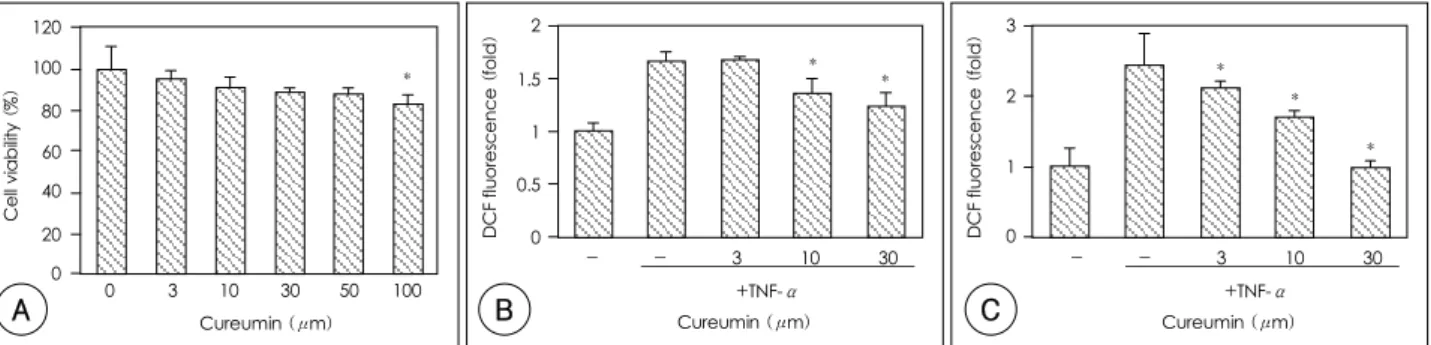
Fig. 1. The effects of curcumin on cell viability, the intracellular ROS level and U937 monocyte adhesion in the TNF-α-stimulated HUVECs. A:
cells were incubated with curcumin at the indicated dosage for 24 hours, and the cell viability was assayed by MTT assay as described in Materials and Methods. *: p<0.05 compared with control (the non-treated cells). B: the ROS level was increased by TNF-α and reduced by the addition of curcumin. The cells were pretreated with curcumin 1 hour prior to the addition of TNF-α (10 ng/mL). The cells were then preloaded with DCFH- DA (10 μM) for 30 minutes, and this was followed by incubation with curcumin at the indicated dosage. After 30 minutes, the fluorescence intensity was measured as described in Materials and Methods. *: p<0.05 compared with the TNF-α-treated cells. C: curcumin inhibited U937 cell adhesion to the basal level. The cells were pretreated with curcumin for 1 hour, and this was followed by the treatment with TNF-α (10 ng/mL) for 12 hours. BCECF-labeled U937 cells were added to be co-cultured for 30 minutes. The non-adherent U937 cells were removed and the fluore- scence intensity was measured as described in Materials and Methods. *: p<0.05 compared with the TNF-α-treated cells. ROS: reactive oxygen species, HUVECs: human umbilical vein endothelial cells, TNF-α: tumor necrosis factor-α, MTT: 3-(4, 5-dimethylthiazol-z-yl)-2, 5-diphenylte- trazoliumbromide, BCECF: 2’, 7’-bis-(2-carboxyethyl)-5(6)-carboxyfluorescein.
A B C
Results
Curcumin reduced the ROS production and U937 monocyte adhesion induced by TNF-α
The cytotoxicity of curcumin on the HUVECs was examined by performing MTT assay. The cell viability was not different from the control cells with treatment of curcumin at concentrations under 50 μM for 24 hours(p>0.05)(Fig. 1A). The intracellular ROS level was increased 1.7±0.1-fold by the TNF-α. Curcumin at concentrations of 3 μM, 10 μM and 30 μM re- duced the intracellular ROS level by 3.0±0.3%, 47.8±
5.3% and 62.7±6.0%, respectively(Fig. 1B). U937 mo- nocyte adhesion to the HUVECs was increased 2.4±
0.5-fold by stimulation with TNF-α treatment(10 ng/mL) for 12 hours. Pretreatment with curcumin at 3 μM, 10 μM and 30 μM showed inhibitory effects of 21.8±1.1%, 50.7±2.7% and 98.9±8.7%, respectively (Fig. 1C).
Curcumin inhibited the NF-κB activation induced by TNF-α
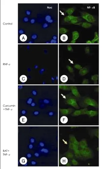
As shown in Fig. 2, unstimulated HUVECs have NF- κB(green) only in the cytoplasm(red), and not the nuclear area(blue). TNF-α(10 ng/mL) activated the NF-κB, leading to its translocation to the nucleus in 30 minutes. Pretreatment of curcumin(10 μM) signi- ficantly attenuated NF-κB translocation and it re- mained in the cytosol to a significant degree. BAY 11- 7082(10 μM), a commercially available inhibitor of NF-κB, impaired this nuclear translocation.
Curcumin suppressed the TNF-α induced expressions of CAM, MCP-1 and IL-8
The effects of curcumin on the TNF-α induced ex- pressions of adhesion molecules and cytokines at the transcription level in the HUVECs were assessed by RT-PCR. After being incubated with curcumin for 1 hour, the cells were treated for 3 hours with TNF-α.
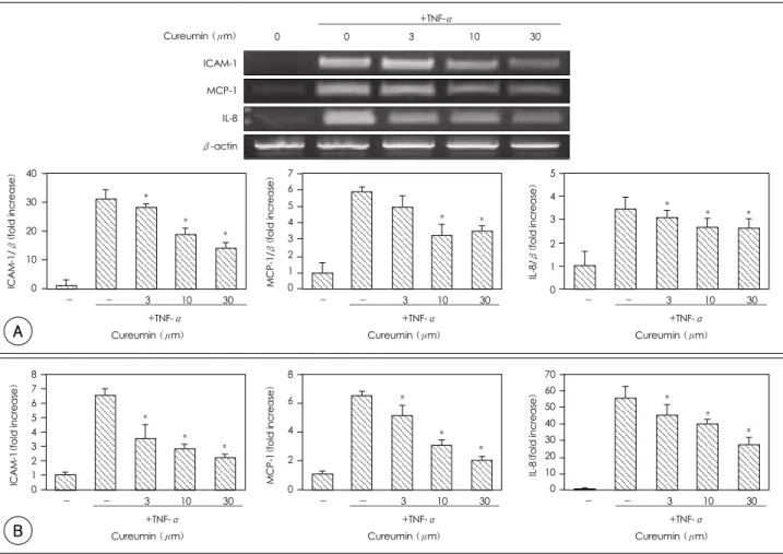
ICAM-1, MCP-1 and IL-8 mRNAs were expressed at very low levels in the HUVECs and their expressions were drastically induced by TNF-α. Pretreatment with curcumin decreased the TNF-induced expression of ICAM-1, MCP-1 and IL-8 mRNAs(Fig. 3A). In addi- tion to transcriptional analysis, the effect of curcumin at the translational level was accessed by ELISA. The ICAM-1, MCP-1 and IL-8 proteins were also dramati- cally increased by TNF to 6.6 units(0.4-fold), 6.5 units(0.3-fold), and 55.0 units(7.0-fold), respectively, compared to the control. After treatment with curcu- min at 10M, the ICAM-1, MCP-1 and IL-8 were de- creased by 68.0%, 62.8% and 27.8%, respectively(Fig.
3B).
Curcumin suppressed the TNF-α induced phosphorylation of JNK, p38 and STAT-3
The signaling molecules JNK, p38 and STAT-3 that were activated by TNF-α were examined by Western blotting. The HUVECs were pretreated with curcumin (10 μM) for 1 hour, and this was followed by incuba- tion with TNF-α for 30 minutes, 1 hour, or 2 hours.
TNF-α increased the phosphorylation of JNK and p38 in 1 hour, and STAT-3 was increased in 2 hours;
however, pretreatment of curcumin significantly blocked the phosphorylations of JNK, p38 and STAT-3(Fig.
4A). To investigate which phosphorylation residue was inhibited by curcumin, western blotting was performed with using two different antibodies; anti-Ser phosph-
Nuc NF-κB
Control
RNF-α
Curcumin +TNF-α
BAT+
TNF-α
A B
C D
E F
G H
Fig. 2. Immunocytochemical analysis of NF-κB p65 localization.
HUVECs were incubated with TNF-α (10 ng/mL) for 30 minutes in the absence (C, D) or presence of curcumin (10 μM, E, F) and BAY 11-7082 (10 μM, G, H), and the cells were subjected to im- munocytochemistry as described in Materials and Methods. NF-κB p65 was stained green and the nuclei were counter-stained blue. Nuc:
nucleus, Cyto: cytoplasm, NF-κB: nuclear factor-kappa B, HUV- ECs: human umbilical vein endothelial cells, TNF-α: tumor necrosis factor-α.
orylated STAT-3 and anti-Tyr phosphorylated STAT-3.
As seen in Fig. 4B, the Ser residue stayed phosphory- lated to some extent and was not induced by TNF-α.
On the other hand, phosphorylation of the Tyr residue was induced by TNF-α and this was inhibited by cur- cumin.
The effects of AG490, BAY 11-7082 and NAC on the TNF-α in the HUVECs
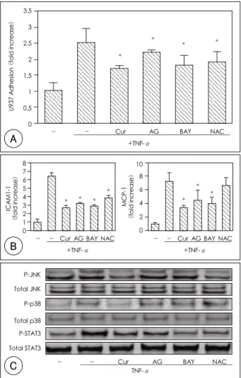
Curcumin attenuated the intracellular ROS level, NF-κB activation and STAT-3 activation during the stimulation with TNF-α. To examine the effects of NF- κB, STAT-3 and oxidative stress on the TNF-α-stim- ulated HUVECs, AG490(50 μM), BAY 11-7082(10 μM), and N-acetylcysteine(NAC, 10 mM) were used as a NF-κB inhibitor, a STAT-3 inhibitor, and an anti- oxidant, respectively. The U937 adhesion was signifi- cantly inhibited by curcumin(53.3%), AG490(20.0%),
BAY11-7082(46.7%), and NAC(40.0%) significantly (Fig. 5A). The protein expressions of ICAM-1 was also significantly inhibited by curcumin(69.4%), AG490 (60.5%), BAY11-7082(65.8%), and NAC(47.4%) significantly(Fig. 5B). The release of MCP-1 protein release was significantly reduced by pretreatment withof curcumin(66.7%), AG490(44.6%), BAY11-7082(53.
2%), with statistical significance but not by NAC(10.
6%, p>65.8%), and NAC(47.4%)(Fig. 5B). The re- lease of MCP-1 protein was significantly reduced by pretreatment with curcumin(66.7%), AG490(44.6%), BAY11-7082(53.2%), but not by NAC(10.6%, p>
0.05)(Fig. 5B). In addition, the phosphorylation of JNK, p38, and STAT-3 was examined by immunoblot- ting(Fig. 5C). The phosphorylation of JNK was inhi- bited by curcumin and NAC, and the phosphorylation of p38 was only inhibited by curcumin. On the other hand, STAT-3 phosphorylation was inhibited by cur-
- - 3 10 30 +TNF-α
8 7 6 5 4 3 2 1 ICAM-1(fold increase) 0
Cureumin (μm)
*
*
*
- - 3 10 30 +TNF-α
8 6
4
2
MCP-1(fold increase) 0
Cureumin (μm)
*
*
*
- - 3 10 30 +TNF-α
70 60 50 40 30 20 10 0
IL-8(fold increase)
Cureumin (μm)
*
*
*
Fig. 3. The effects of curcumin on the ICAM-1, MCP-1 and IL-8 levels in the HUVECs stimulated with TNF-α. A: the levels of mRNA were determined by RT-PCR. Curcumin was pretreated and then TNF-α (10 ng/mL) was added. After 3 hour incubation, the mRNA was extracted and subjected to RT-PCR. The cells were incubated with TNF-α for 3 hours before treatment of curcumin at the indicated dosage for 1 hour. The representative RT-PCR is presented. In the graph, the expressions of ICAM-1, MCP-1 and IL-8 were normalized to that of β-actin and they were expressed as fold increases; the control ratio was defined as 1.0. B: the protein levels were quantitated by ELISA. Curcumin was pretreated onto the cells and TNF-α was then added. After 12 hours, the cell culture medium was collected to determine the protein level. *: p<0.05 compared with the TNF-α-treated cells. ICAM-1: intracellular adhesion molecule, MCP-1: monocyte chemoattractant protein, IL-8: interleukin, HUVECs:
human umbilical vein endothelial cells, TNF-α: tumor necrosis factor-α, mRNA: messeuger ribonucleic acid, RT-PCR: reverse transcriptase- polymerase chain reaction, ELISA: enzyme-linked immunosorbent assay.
Cureumin (μm)
- - 3 10 30 +TNF-α
40
30
20
10
ICAM-1/β(fold increase) 0
*
*
*
- - 3 10 30 +TNF-α
5 4 3 2 1 0
IL-8/β(fold increase)
Cureumin (μm)
*
* *
- - 3 10 30 +TNF-α
7 6 5 4 3 2 1 MCP-1/β(fold increase) 0
Cureumin (μm)
* *
+ +TNF-α 0 0 3 10 30 ICAM-1
MCP-1
IL-8
β-actin Cureumin (μm)
A
B
cumin, AG490, BAY11-7082 and NAC.
Discussion
The goal of the present study was to examine the mechanism by which curcumin exert its inhibitory effects on the proinflammatory response of human endothelial cells.
In the previous studies, curcumin has been reported to have a ROS scavenging property in addition to its anti-inflammatory and anti-cancer effects.3) Curcumin inhibited the NF-κB activation that may participate in the reaction to various stimuli such as oxidative stress, cytokines and hypoxic injury. Extensive research has recently shown that curcumin can prevent inflam- mation and cellular injury via NF-κB inhibition in steatohepatitis mice, intestine epithelial cells and mic- roglial cells.15) However, the molecular mechanisms of curcumin for reducing endothelial inflammation have not been clearly elucidated.
We examined the effect of curcumin on HUVECs viability to exclude the possible cytotoxic effects. Cur- cumin was noted to relatively preserve the HUVECs’
viability and so it is regarded as safe when it is used at a level less than 30 μM as compared to when it is used at 10 μM, which inhibited the ROS elevation and U937 adhesion induced by TNF-α within a non-toxic range.
NF-κB is a well-known transcription factor that is
critical for the pro-inflammatory gene regulation re- lated to cancer, atherosclerosis, myocardial infarction, diabetes, arthritis and etc.11) We confirmed the acti- vation of NF-κB by immunocytochemistry(ICC) and the representative ICC pictures are illustrated in Fig. 2.
NF-κB activation was significantly increased in the HUVECs subjected to TNF-α treatment, which in- dicated a rapid activation process, while curcumin and BAY11-7082, an inhibitor of NF-κB, inhibited NF-κ B p65 nuclear translocation.
NF-κB has been known to induce cytokines and adhesive molecules, and we examined whether cur- cumin inhibits the NF-κB-dependent pro-inflamma- tory molecules. In the TNF-α-stimulated HUVECs, the ICAM-1, MCP-1 and IL-8 expressions were atte-
Fig. 4. The effects of curcumin on JNK, p38 and STAT-3 activation.
A: the HUVECs were treated with TNF-α for the indicated time in the presence or absence of curcumin pretreatment. Cells were har- vested and then subjected to Western blotting against phos-phory- lated JNK, p38 and STAT-3. B: the phosphorylation residue that was inhibited by curcumin was confirmed by immunoblotting with using phospho-serine-STAT-3 antibody or phospho-tyrosine-STAT-3 anti- body. JNK: c-Jun N-terminal kinase, STAT-3: signal transducer and activator of transcription-3, HUVECs: human umbilical vein end- othelial cells, TNF-α: tumor necrosis factor-α.
TNF-a (hr) P-JNK JNK p-p38 p38 p-STAT3 STAT3
+ +Curcumin 0 0.5 1 2 0.5 1 2
A
30 min 2 hr Total STAT3
p-Ser-STAT3 p-Tyr-STAT3 Con TNF Cur+TNF Con TNF Cur+TNF
B Fig. 5. The effects of curcumin, AG490, BAY11-7082 and NAC on
TNF-α-stimulated HUVECs. A: the cells were treated with curcumin (10 μM), AG490 (10 μM), BAY11-7082 (10 μM), and NAC (10 mM) 1 hour prior to the challenge with TNF-α. After 12 hours, U937 adhesion to the HUVECs was assayed. The results are ex- pressed as fold increases, in which the control value is defined as 1.0.
*: p<0.05 compared with e ICAM-1 and MCP-1 protein levels were then determined. *: p<0.05 compared with the TNF-α-treated cells.
TNF-α: tumor necrosis factor-α, STAT-3: signal transducer and acti- vator of transcription-3, NAC: N-acetylcysteine, ICAM-1: intra- cellular adhesion molecule, MCP-1: monocyte chemoattractant protein.
- - Cur AG BAY NAC +TNF-α
3.5 3 2.5 2 1.5 1 0.5 0
U937 Adhesion (fold increase)
*
*
* *
- - Cur AG BAY NAC +TNF-α 8
7 6 5 4 3 2 1 0 ICAM1-1 (fold increase)
*
* * *
- - Cur AG BAY NAC +TNF-α 10
8 6 4 2 0 MCP-1 (fold increase)
*
*
*
P-JNK Total JNK P-p38 Total p38 P-STAT3 Total STAT3
- - Cur AG BAY NAC TNF-α
A
B
C
nuated by curcumin at both the transcriptional and translational levels. The up-regulation of adhesion mo- lecules, including ICAM-1, on the surface of the endo- thelium is required for the firm adhesion of rolling monocytes. Curcumin inhibited the TNF-α-induced ICAM-1 and MCP-1 expressions, which indicated that the mechanism of anti-adhesion, at least in part, was related to the down-regulation of these proteins. These results suggest that suppressing the surface proteins and their mRNA expression was one of the pathways of curcumin inhibiting the adhesion of U937 to TNF- α-stimulated HUVECs.
Of all the signaling molecules involved in TNF-α- mediated pathways, we detected the activation of JNK, p38 and STAT-3 by Western blotting. JNK and p38 are members of the MAPK family, which is a family of proteins that are known to be involved in cellular da- mage or cell death.13)16) They are activated by extra- cellular stresses such as ROS, UV and cytokines, and their activation leads to cellular death. STAT factors are a family of cytoplasmic transcription factors that mediate the intracellular signaling that’s initiated at the cytokine cell surface receptors and is then tran- smitted to the nucleus. STAT-3 is a key molecule down- stream of gp130, and is activated under various stressful conditions such as pressure-overload and myocardial infarction.17)18) Previous research has demonstrated that STAT-3 may be a survival factor in the heart that is able to protect the myocardium following ischemic injury. In malignant cells, however, curcumin suppressed JAK- STAT signaling and so it suppressed tumor cell growth in brain microglia,19) multiple myeloma cells20) and T lymphocytes.21) Thus, STAT-3 seems to mediate either a pro-apoptotic or anti-apoptotic effect depending on the cell or tissue type.
It is known that there are two phosphorylation sites in Stat-3: tyrosine 705 and serine 727. These two pho- sphorylation sites appear to be induced by distinct signaling.22)23) Phosphorylation of both sites is neces- sary for the maximal activation of transcription tyrosine 705 phosphorylation, which is required for STAT-3 dimerization, nuclear translocation and gene activation.
Serine 727 phosphorylation is necessary for maximal transcription efficiency, although it was not known why.
In our study, curcumin attenuated the tyrosine phosph- orylation of STAT-3 as well as the phosphorylation of JNK and p38 in the TNF-α-stimulated HUVECs. Fur- ther, curcumin inhibited the phosphorylation of JNK, p38 and STAT-3.
Curcumin inhibited the TNF-α-induced intracellular ROS production(Fig. 1B), and we thought that curcu- min’s antioxidative activity could partly contribute to its inhibitory effects. Several research groups have re- ported that oxidative stress might contribute to the NF-κB-dependent inflammatory responses. Rocksen
et al.24) have reported that dexamethasone treatment resulted in reduced ROS production and the reduced expression of TNF-α, IL-1α, IL-1α, IL-6 and IL-12 in lipopolysaccharide(LPS)-stimulated mice. Hayashi et al.25) have reported that the anti-oxidant NAC sup- pressed vascular NF-κB activation and this inhibition reduced the pathological thickening of the arterial wall.
NF-κB has been shown to be ROS-sensitive. Therefore, we think that the anti-oxidant property of curcumin inhibits the activation of NF-κB. In our work, TNF-α produced a significant increase in ROS after 1 hour of exposure. Therefore, we examined whether treatment with an anti-oxidant will inhibit the TNF-α induced phosphorylation of JNK and p38.
To determine what property of curcumin contributes to its anti-inflammatory effects, we used the chemicals AG490, BAY11-7082 and NAC. AG490 is an inhibitor of Jak2, an upstream kinase of STAT-3; BAY11-7082 is a NF-κB inhibitor and NAC is a well-known an- tioxidant.
AG490 and BAY 11-7082 inhibited the expression of ICAM-1 and MCP-1 in the TNF-α-stimulated HUVECs. It also suppressed U937 adhesion and STAT- 3 activation, and these events were considered to be dependent on NF-κB. On the other hand, the activa- tions of JNK, p38 and intracellular ROS(unpublished data) were inhibited by curcumin, but not by BAY 11- 7082. These data revealed that the activation of JNK and p38 by TNF-α was an independent event on NF- κB activation. NAC, an anti-oxidative reagent, blocked the activation of JNK and STAT-3 that was induced by TNF-α, but NAC did not block the activation of p38.
Although AG490, BAY 11-7082 and NAC all showed inhibitory effects on TNF-α, the extent of their in- hibitory effect was less than that of curcumin.
In this work, we studied whether the vascular in- flammation could be influenced by curcumin, and we checked the possible molecules that had important roles. STAT-3 has been reported to be a survival factor and it is constitutively activated in some type of cancers.
We did not address the physiological meaning of STAT-3 activation by TNF-α as well as its inhibition by curcumin, but we presumed STAT-3 activation was a compensatory response, and inhibition by curcumin counteracted the TNF-α.
The MAPK and STAT3 pathways have been im- plicated in the responses to pro-inflammatory stress in endothelial cells, and curcumin may block these res- ponses by counteraction. Our data indicate that cur- cumin suppresses the expression of gene products involved in inflammation via NF-κB-dependent and independent pathways. Other pathways are possibly involved in the capacities of curcumin to block the MAPK and STAT-3 pathways and to scavenging ROS.
In TNF-α-stimulated endothelial cells, curcumin ap-
pears to function as a repressor in this model of acute inflammation.
We propose that curcumin could be a potential the- rapeutic agent for achieving endothelial protection against the pro-inflammatory cytokine-induced cyto- toxicity that’s been observed in several pathological conditions.
This study was supported by funds(CUHRI-U-200523) of Chonnam National University Hospital Research Institute of Clinical Medicine.
REFERENCES
1) Brouet I, Ohshima H. Curcumin, an anti-tumour promoter and anti-inflammatory agent, inhibits induction of nitric oxide syn- thase in activated macrophages. Biochem Biophys Res Commun 1995;206:533-40.
2) Kang G, Kong PJ, Yuh YJ, et al. Curcumin suppresses lipopoly- saccharide-induced cyclooxygenase-2 expression by inhibiting activator protein 1 and nuclear factor kappab bindings in BV2 microglial cells. J Pharmacol Sci 2004;94:325-8.
3) Lin JK. Suppression of protein kinase C and nuclear oncogene expression as possible action mechanisms of cancer chemopre- vention by Curcumin. Arch Pharm Res 2004;27:683-92.
4) Wessler S, Muenzner P, Meyer TF, Naumann M. The anti-in- flammatory compound curcumin inhibits Neisseria gonorrhoeae- induced NF-kappaB signaling, release of pro-inflammatory cy- tokines/chemokines and attenuates adhesion in late infection. Biol Chem 2005;386:481-90.
5) Lusis AJ. Atherosclerosis. Nature 2000;407:233-41.
6) Ross R. Atheroclerosis: an inflammatory disease. N Engl J Med 1999;340:115-26.
7) Baud V, Karin M. Signal transduction by tumor necrosis factor and its relatives. Trends Cell Biol 2001;11:372-7.
8) Kwak MH, Kim MK, Kim SH, Lee WH, Ryoo UH, Park JE.
Expression of tumor necrosis factor receptor superfamily in car- otid atheroma. Korean Circ J 2000;30:1563-73.
9) Barnes PJ, Karin M. Nuclear factor-kappaB: a pivotal transcrip- tion factor in chronic inflammatory diseases. N Engl J Med 1997;336:1066-71.
10) Ahn YK, Kim YS, Park HW, et al. In vivo cardiac gene transfer of dominant negative IKK-β reduces myocardial inflammation, apoptosis, and infarction after ischemia-reperfusion injury. Korean Circ J 2005;35:206-14.
11) Kim YS, Ahn Y, Hong MH, et al. Carvedilol inhibits expressions of vascular cell adhesion molecule-1, intercellular adhesion mo- lecule-1, monocyte chemoattractant-1, and interleukin-8 via NF- κB inhibition in human endothelial cells. Korean Circ J 2005;
35:576-82.
12) Baines CP, Molkentin JD. Stress signaling pathways that modulate cardiac myocyte apoptosis. J Mol Cell Cardiol 2005;38:47-62.
13) Matsuzawa A, Ichijo H. Stress-responsive protein kinases in re- dox-regulated apoptosis signaling. Antioxid Redox Signal 2005;
7:472-81.
14) el-Adawi H, Deng L, Tramontano A, et al. The functional role of the JAK-STAT pathway in post-infarction remodeling. Cardiovasc Res 2003;57:129-38.
15) Leclercq IA, Farrell GC, Sempoux C, dela Pena A, Horsmans Y.
Curcumin inhibits NF-kappaB activation and reduces the se- verity of experimental steatohepatitis in mice. J Hepatol 2004;41:
926-34.
16) Ventura JJ, Cogswell P, Flavell RA, Baldwin AS Jr, Davis RJ.
JNK potentiates TNF-stimulated necrosis by increasing the pro- duction of cytotoxic reactive oxygen species. Genes Dev 2004;18:
2905-15.
17) Negoro S, Kunisada K, Tone E, et al. Activation of JAK/STAT pathway transduces cytoprotective signal in rat acute myocar- dial infarction, Cardiovasc Res 2000;47:797-805.
18) Alonzi T, Middleton G, Wyatt S, et al. Role of STAT3 and PI 3- kinase/Akt in mediating the survival actions of cytokines on sen- sory neurons. Mol Cell Neurosci 2001;18:270-82.
19) Kim HY, Park EJ, Joe EU, Jou I. Curcumin suppresses Janus kinase-STAT inflammatory signaling through activation of Src homology 2 domain-containing tyrosine phosphatase 2 in brain microglia. J Immunol 2003;171:6072-9.
20) Bharti AC, Donato N, Aggarwal BB. Curcumin (diferuloylme- thane) inhibits constitutive and IL-6-inducible STAT3 phosphory- lation in human multiple myeloma cells. J Immunol 2003;171:
3863-71.
21) Natarajan C, Bright JJ. Curcumin inhibits experimental allergic encephalomyelitis by blocking IL-12 signaling through Janus kinase-STAT pathway in T lymphocytes. J Immunol 2002;168:
6506-13.
22) Heinrich PC, Behrmann I, Haan S, Hermanns HM, Muller-Newen G, Schaper F. Principles of interleukin (IL)-6-type cytokine signalling and its regulation. Biochem J 2003;374:1-20.
23) Stark GR, Kerr IM, Williams BR, Silverman RH, Schreiber RD.
How cells respond to interferons. Annu Rev Biochem 1998;67:
227-64.
24) Rocksen D, Lilliehook B, Larsson R, Johansson T, Bucht A. Diffe- rential anti-inflammatory and anti-oxidative effects of dexame- thasone and N-acetylcysteine in endotoxin-induced lung inflam- mation. Clin Exp Immunol 2000;122:249-56.
25) Hayashi K, Takahata H, Kitagawa N, Kitange G, Kaminogo M, Shibata S. N-acetylcysteine inhibited nuclear factor-kappaB expression and the intimal hyperplasia in rat carotid arterial injury.
Neurol Res 2001;23:731-8.