I. Introduction
Current bio-imaging devices are able to display and re- construct three-dimensional (3D) anatomical details, and diagnostics are becoming increasingly vital to the quality of patient treatment planning and clinical treatment [1,2].
It is important to know that radiographic images obtained by techniques, such as computed tomography (CT) and magnetic resonance imaging, can be converted to 3D files and produce a 3D solid model with a 3D printer [3]. Radio- graphic images and image processing tools are being used for the segmentation and reconstruction of tissue images in 3D to determine bone and muscle characteristics [4]. However,
Clinical Application of Solid Model Based on Trabecular Tibia Bone CT Images Created by 3D Printer
Jaemo Cho, BEng, Chan-Soo Park, MS, Yeoun-Jae Kim, PhD, Kwang Gi Kim, PhD
Biomedical Engineering Branch, Division of Convergence Technology, National Cancer Center, Goyang, Korea
Objectives: The aim of this work is to use a 3D solid model to predict the mechanical loads of human bone fracture risk as- sociated with bone disease conditions according to biomechanical engineering parameters. Methods: We used special image processing tools for image segmentation and three-dimensional (3D) reconstruction to generate meshes, which are necessary for the production of a solid model with a 3D printer from computed tomography (CT) images of the human tibia’s trabecu- lar and cortical bones. We examined the defects of the mechanism for the tibia’s trabecular bones. Results: Image process- ing tools and segmentation techniques were used to analyze bone structures and produce a solid model with a 3D printer.
Conclusions: These days, bio-imaging (CT and magnetic resonance imaging) devices are able to display and reconstruct 3D anatomical details, and diagnostics are becoming increasingly vital to the quality of patient treatment planning and clinical treatment. Furthermore, radiographic images are being used to study biomechanical systems with several aims, namely, to describe and simulate the mechanical behavior of certain anatomical systems, to analyze pathological bone conditions, to study tissues structure and properties, and to create a solid model using a 3D printer to support surgical planning and reduce experimental costs. These days, research using image processing tools and segmentation techniques to analyze bone struc- tures to produce a solid model with a 3D printer is rapidly becoming very important.
Keywords: 3D Printer, Computed Tomography, Computer-Aided Design, Computer-Assisted, Image Processing, Prototype
Healthc Inform Res. 2015 July;21(3):201-205.
http://dx.doi.org/10.4258/hir.2015.21.3.201 pISSN 2093-3681 • eISSN 2093-369X
Submitted: May 26, 2015 Revised: June 30, 2015 Accepted: June 30, 2015 Corresponding Author Kwang Gi Kim, PhD
Biomedical Engineering Branch, Division of Convergence Technology, National Cancer Center, 323 Ilsan-ro, Ilsandong-gu, Goyang 410- 769, Korea. Tel: +82-31-920-2241, Fax: +82-31-920-2242, E-mail:
This is an Open Access article distributed under the terms of the Creative Com- mons Attribution Non-Commercial License (http://creativecommons.org/licenses/by- nc/4.0/) which permits unrestricted non-commercial use, distribution, and reproduc- tion in any medium, provided the original work is properly cited.
ⓒ 2015 The Korean Society of Medical Informatics
the results are not satisfactory when the focus is on the stress and strain distributions near boundaries [5]. Generally, the finite-element method is used to compensate the defects [6].
Today, radiographic images are being used to study bio- mechanical systems with several aims, namely, to describe and simulate the mechanical behavior of certain anatomical systems, to analyze pathological bone conditions, to study tissues structure and tissues properties, and to create a solid model using a 3D printer to support surgical planning and reduce experimental costs [7].
The rapid prototyping process known as 3D printing is ex- panding rapidly in clinical application; it provides novel pos- sibilities and potentials for modeling, simulation and analysis of the human body [8]. There have already been attempts to use this novel technology in various fields of clinical applica- tions. For example, in the fields of orthopedics and oral and maxillofacial surgery, it has been used in designing implant prosthetics and tissues. Moreover, it can be used in planning surgeries, to investigate possibilities, and improve accuracy before surgery [9]. Especially in the dental field, 3D printing is actively used to design implants and crowns. In addition, 3D printing can be used in a special way to facilitate facial reconstruction [10].
The use of 3D-printed solid models has many benefits in clinical fields, including determining whether a specific surgical procedure is too risky to perform for an individual patient, implant prosthetics designing, and medical research (too costly for in vivo experiments), thus improve surgical accuracy. It can also be useful for educational programs, such as medical student education and training to aid under- standing of complex anatomical structures [11,12].
Various 3D printing methods have been developed to build 3D structural models, such as stereolithography, fused- deposition modeling, selective-laser sintering, selective-laser melting, electronic-beam melting, and laminated-object manufacturing [13]. The 3D printer (ProJet 3510 HD; 3D Systems Inc., Rock Hill, SC, USA) is owned by the National Cancer Center using the stereolithography printing method.
This method is the oldest and has a wide range of applica- tion. The whole process consists of the sequential printing of layers, which is similar to the functioning of the traditional inkjet printer. However, the printer differs from the conven- tional printer in that it produces 3D solid objects layer-by- layer [14]. The 3D printer first follows the instructions of computer-aided design (CAD) to build the foundation of an object, while the printer-head moves along the X, Y-planes and along the Z-plane to build the object’s vertical layers [15].
It can build almost any shape defined in a CAD file and can use various types of materials and forms, such as plastics,
rubber, gypsum, metals powders, liquids and solid which can be selected depending on the object’s intended use [16,17].
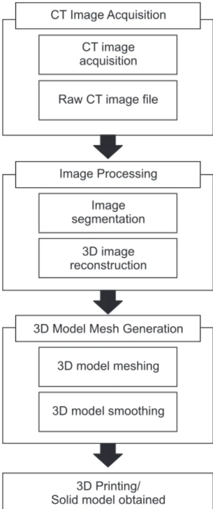
In this study, we used special image processing tools for image segmentation and 3D reconstruction to generate meshes necessary for production of solid objects with a 3D printer from CT images of the human tibia’s trabecular and cortical bones (Figure 1).
II. Case Description
1. CT Images Acquisition
To acquire cadaver tibia CT images, the CT conditions were the following: 293 × 293 × 100 μm voxel size (virtually de- graded resolution), temporal bone protocol, 120 kVp, 300 mAs, 150 mm field of view, 12 mm away from distal (Figure 2A).
2. Image Segmentation and 3D Image Reconstruction Image segmentation was applied to the acquired cadaver tibia CT images using Analyze ver. 11.0 (Biomedical Imag- ing Resource at Mayo Clinic, Rochester, MN, USA). Various
CT Image Acquisition
CT image acquisition
Raw CT image file
Image segmentation
3D image reconstruction
3D model meshing
3D model smoothing
3D Printing/
Solid model obtained 3D Model Mesh Generation
Image Processing
Figure 1. Process from image acquisition to the production of a 3D solid object. CT: computed tomography.
regions window levels/ widths (contrast) were applied to establish a clear separation between the cortical bone and the trabecular bone. The set up image segmentation was per- formed with a contrast region on 300/2000 for visualization, which was shown by yellow coloring on the trabecula bone region for clear separation during 3D reconstruction. Also, automatic detection of each region was performed, and then manual segmentation was performed as needed. The seg- mented images were accumulated layer-by-layer following the Z-plane and reconstructed as a 3D image (Figure 2B and C).
3. 3D Model Mesh Generation
The 3D reconstruct file formats were transformed to surface tessellation language (STL) to composed 3D meshes. The STL file format is accepted in 3D printer software and is widely and commonly used for many 3D processes. The 3D mesh model format constructed only surfaces; the surface format was composed of only 3D images and it was rough and irregular. In this case, an additional mesh method was
required to overcome inside defect errors. Another mesh method was required for mesh repair because mesh defects had occurred on the irregular surface in the process of con- necting between the complex 3D model and meshes. Fur- thermore, an additional smoothing method is required if the model has excessive meshes that are detected as defect elements in 3D printing. In this case, due to the character- istics of ICEM ver. 14.0 software (ANSYS Inc., Canonsburg, PA, USA), the in and out surfaces of the meshes consisted of curves and points. The meshes were composed of hexagonal and triangular surface meshes, and homogeneous meshes could only transfer a limit quantity (less than 40 million of meshes) of the whole model’s meshes. Furthermore, addi- tional composited model smoothing was performed with the Taubin mesh smoothing method (Figure 2D).
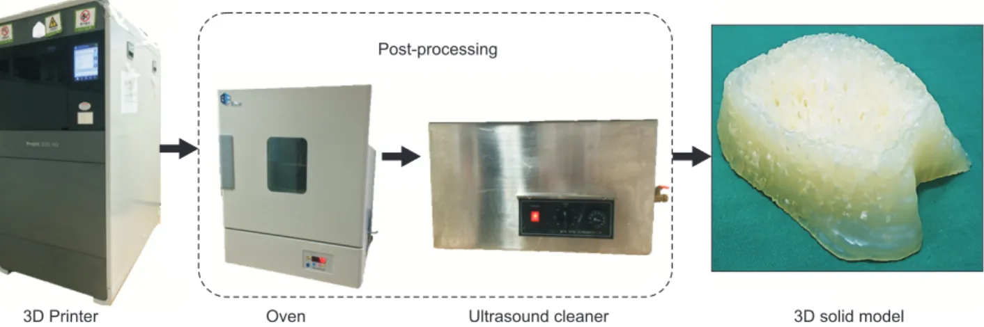
4. 3D Printing and 3D Solid Object Post-processing The 3D printing method is used to produce a 3D solid ob- ject with 3D CAD format files (STL). The STL format file is transferred to the 3D printer. Before printing a 3D model
3D Printer Oven Ultrasound cleaner 3D solid model
Post-processing
Figure 3. Process to produce a 3D solid model.
A B C D
Figure 2. Process from computed tomography (CT) image acquisition to the production of a 3D file, comprising four major steps: image is acquired from CT and raw CT data image is achieved, image segmentation is performed in image post-processing and a 3D file is reconstructed, a 3D structural computer-aided design (CAD) mesh file is constructed, smoothing method is applied to the irregular surface. (A) Raw CT image, (B) image segmentation, (C) 3D model meshing, and (D) 3D model smoothing.
from an STL file, the file must be examined for manifold er- rors, sue to such errors, surfaces do not connect each other or there are gaps in the model. It takes varying amounts of time depending on the size of the object, its resolution, and the material used. In this study, the 3D printer (ProJet 3510 HD, 3D Systems Inc.) produced the object layer-by-layer with part and support materials. The part material used the VisiJet M3 Crystal (3D Systems Inc.), which is composed of urethane acrylate oligomers and tough plastic and translu- cent materials. The material was designed for durability and reliability for functional testing and rapid tooling applica- tions. The support material, VisiJet S300 (3D Systems Inc.) is based on hydroxylated wax and is used for support during the processing.
Post-processing procedure of 3D printed object: the sup- port was removed from the 3D solid object using an oven and ultrasound cleaner. The support material was made from wax, which was easily removed by the application of heat (60oC) without affecting the object, and to remove the support from the more delicate surfaces, manual hand work is sometimes necessary (Figure 3).
III. Discussion
These days, research regarding the use of image processing tools and segmentation techniques to analyze bone struc- tures to produce a solid model with a 3D printer is rapidly becoming increasingly important. The aim of this work was to use a 3D solid model to predict the mechanical loads of human bone fracture risk for bone disease conditions us- ing biomechanical engineering parameters. The 3D printing technique has potential for use in a variety of clinical ap- plications, such as analyzing human anatomical structure, implant design, medical research, as well as surgical plan- ning and training. However, 3D printing is not used often in clinical practice yet because of its limitations.
The limited choice of materials is an issue in the clinical field. It is imperfect represented by using polymers, rubber, gypsum, and metal for human body tissues. To build an ob- ject, two kinds of materials are required for both the support and part materials. A support material is needed to build the object which uses the part material. However, the materials are expensive to build objects for only research purposes so, user should be sure to check the file before produce a 3D solid model. The software is difficult to use by people who are not experts; it is difficult to learn, and it takes time to be- come an expert user. Furthermore, the process of creating a 3D file has not been fully automated before 3D printing, and they the software is still expensive. Even expensive software
does not include a fully automated program from start to end to obtain a 3D file. The limitations are producing a small version of large structure or dividing the whole model into smaller parts which can be combined after printing. Nor- mally, a 3D printer only prints models of certain sizes with certain levels of complexity, and it usually takes a long time to obtain a solid model, depending on its size. Commonly, 3D printers use polymer or a polymer mix with rubber for the material, and these are not strong enough to build joint implant prosthetics or dental implants. They could break or crack because they are too weak.
The potential and benefits of 3D printing have already has been demonstrated in several studies in various fields. Clini- cal research has conducted on the development of artificial organs, prosthetics, and tissues using a 3D printer. Models can be produced by 3D printing that are suitable to use for the education of medical student, surgical planning, and to help patients understand surgical procedures. The additional cost of 3D printing might be compensated by reducing op- erating time and achieving higher success rates for surgical procedure. However, it is still has not been approved to use 3D printers to produce clinical support devices, and applica- tions of artificial medical tissues are limited and still in an early stage of research. Furthermore, 3D printers can only print model of certain sizes and certain levels of complex- ity. Also, the use of part and support materials depends on a model’s size and the structures which lie within time and cost spent as 3D solid object. The clinical application of 3D printing is technically in an early stage. In the near future, it will have enormous potential for expanded utilization and the development of new applications in the fields of individ- ual patient care as well as academic and research activities.
The study here is different in terms of aims and impacts on medical applications. However, it is common in the method- ological approach that is use of 3D printer based on medi- cal image processing techniques with the goal of improving clinical processes.
Conflict of Interest
No potential conflict of interest relevant to this article was reported.
Acknowledgments
This study was supported by a grant of the Korea Health technology R&D Project, Ministry of Health & Welfare, Re- public of Korea (HI13C12830100).
References
1. Mao K, Wang Y, Xiao S, Liu Z, Zhang Y, Zhang X, et al.
Clinical application of computer-designed polystyrene models in complex severe spinal deformities: a pilot study. Eur Spine J 2010;19(5):797-802.
2. Esses SJ, Berman P, Bloom AI, Sosna J. Clinical applica- tions of physical 3D models derived from MDCT data and created by rapid prototyping. AJR Am J Roentgenol 2011;196(6):W683-8.
3. Butscher A, Bohner M, Hofmann S, Gauckler L, Muller R. Structural and material approaches to bone tissue en- gineering in powder-based three-dimensional printing.
Acta Biomater 2011;7(3):907-20.
4. Janssen I, Heymsfield SB, Ross R. Low relative skeletal muscle mass (sarcopenia) in older persons is associated with functional impairment and physical disability. J Am Geriatr Soc 2002;50(5):889-96.
5. Scifert CF, Brown TD, Lipman JD. Finite element analy- sis of a novel design approach to resisting total hip dis- location. Clin Biomech (Bristol, Avon) 1999;14(10):697- 703.
6. Ventola CL. Medical applications for 3D printing: cur- rent and projected uses. P T 2014;39(10):704-11.
7. Niebur GL, Feldstein MJ, Yuen JC, Chen TJ, Keaveny TM. High-resolution finite element models with tissue strength asymmetry accurately predict failure of tra- becular bone. J Biomech 2000;33(12):1575-83.
8. Peltola SM, Melchels FP, Grijpma DW, Kellomaki M. A review of rapid prototyping techniques for tissue engi- neering purposes. Ann Med 2008;40(4):268-80.
9. Mertz L. Dream it, design it, print it in 3-D: what can 3-D printing do for you? IEEE Pulse 2013;4(6):15-21.
10. Rengier F, Mehndiratta A, von Tengg-Kobligk H, Zech- mann CM, Unterhinninghofen R, Kauczor HU, Giesel FL. 3D printing based on imaging data: review of medi- cal applications. Int J Comput Assist Radiol Surg 2010;
5(4):335-41.
11. Lee MY, Chang CC, Ku YC. New layer-based imaging and rapid prototyping techniques for computer-aided design and manufacture of custom dental restoration. J Med Eng Technol 2008;32(1):83-90.
12. Harrysson OL, Hosni YA, Nayfeh JF. Custom-designed orthopedic implants evaluated using finite element analysis of patient-specific computed tomography data:
femoral-component case study. BMC Musculoskelet Disord 2007;8:91.
13. Gross BC, Erkal JL, Lockwood SY, Chen C, Spence DM.
Evaluation of 3D printing and its potential impact on biotechnology and the chemical sciences. Anal Chem 2014;86(7):3240-53.
14. Patel P. Micro 3-D printer creates tiny structures in sec- onds [Internet]. Cambridge (MA): MIT Technology Re- view; c2013 [cited at 2015 Jul 15]. Available from: http://
www.technologyreview.com/news/511856/micro-3-d- printer-creates-tiny- structures-in-seconds/.
15. Moskvitch K. Artificial blood vessels created on a 3D printer [Internet]. London, UK: BBC News; 2011 [cited at 2015 Jul 15]. Available from: http://www.bbc.com/
news/technology-14946808.
16. Ragaert K, De Baere I, Moerman M, Cardon L, Degrieck J. Design and thermoregulation of a new microextru- sion dispense head for 3D‐plotting of thermally sensi- tive thermoplastics. Polym Eng Sci 2013;53(2):273-82.
17. Lee SJ. 3D printer technology changes future of the health industry. KHIDI Brief 2014;111:1-16.