Blood Res2014;49:196-207. bloodresearch.or.kr
198 Letters to the Editor
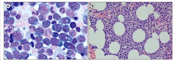
Fig. 1. Bone marrow (BM) aspiration and biopsy findings in the first case. BM aspiration showed the presence of neoplastic cells (27.8%) with oval to round nuclei, prominent nucleoli, and abundant amount of bluish cytoplasm (A, Wright staining, ×1,000). The BM clot section also showed interstitial infiltration of neoplastic cells (B, hematoxylin and eosin staining, ×400) positive for CD4, CD56, and CD123 on immunohistochemical staining.
Department of Pathology and Laboratory Medicine, Medanta-The Medicity, Gurgaon, India
Correspondence to: Ritesh Sachdev Department of Pathology and Laboratory Medicine,
Medanta-The Medicity, Sector - 38, Gurgaon, Haryana 122 001, India E-mail: sachdev05@gmail.com
Received on Mar. 26, 2014; Revised on Mar. 29, 2014; Accepted on Jun. 11, 2014 http://dx.doi.org/10.5045/br.2014.49.3.196
AuthorsÊ Disclosures of Potential Conflicts of Interest No potential conflicts of interest relevant to this article were reported.
REFERENCES
1. Coustan-Smith E, Mullighan CG, Onciu M, et al. Early T-cell pre- cursor leukaemia: a subtype of very high-risk acute lympho- blastic leukaemia. Lancet Oncol 2009;10:147-56.
2. Neumann M, Coskun E, Fransecky L, et al. FLT3 mutations in ear- ly T-cell precursor ALL characterize a stem cell like leukemia and imply the clinical use of tyrosine kinase inhibitors. PLoS One 2013;8:e53190.
3. Weir EG, Ali Ansari-Lari M, Batista DA, et al. Acute bilineal leu- kemia: a rare disease with poor outcome. Leukemia 2007;21:
2264-70.
4. Borowitz MJ, Bene MC, Harris NL, Porwit A, Matutes E. Acute leukaemias of ambiguous lineage. In: Swerdlow SH, Campo E, Harris NL, et al, eds. WHO classification of tumours of haemato- poietic and lymphoid tissues, 4th ed. Lyon, France: IARC Press, 2008:150-5.
5. Chen W. Case study interpretation-New Orleans: case 2. Mixed phenotype acute leukemia, T/myeloid. Cytometry B Clin Cytom 2013;84:342-5.
6. Borowitz MJ. Mixed phenotype acute leukemia. Cytometry B
Clin Cytom 2014;86:152-3.
Leukemic manifestation of blastic plasmacytoid dendritic cell neoplasm:
laboratory approaches in 2 cases
TO THE EDITOR: The blastic plasmacytoid dendritic cell neoplasm (BPDCN) is a rare subtype of the myeloid neo- plasm, with an incidence of 0.44% among all hematologic malignancies and characterized by clonal plasmacytoid den- dritic cell presence [1, 2]. Patients with BPDCN typically display skin lesions and aggressive clinical features with rapid systemic dissemination, exhibiting bone marrow (BM) involvement in 60–90% of cases [3, 4]. However, leukemic presentation of BPDCN is rare, with a reported incidence of less than 1% among those with acute leukemia [5].
Moreover, BPDCN with leptomeningeal involvement has been reported to be more infrequent compared with lymph node or liver/spleen involvement, as only 4 of 43 BPDCN patients experienced cerebrospinal fluid (CSF) involvement in a large-scale multicenter study [1]. Herein, we report 2 patients with BPDCN who exhibited leukemic manifes- tation (1 with simultaneous leptomeningeal involvement) diagnosed by using immunohistochemical staining and flow cytometry analysis for CD4, CD56, and CD123.
CASES
The first case of BPDCN was observed in a 36-year-old woman who was admitted to the authors’ institution for workup of a left cheek skin lesion that had developed 6 months previously. An incisional biopsy of the skin lesion
bloodresearch.or.kr Blood Res 2014;49:196-207.
Letters to the Editor 199
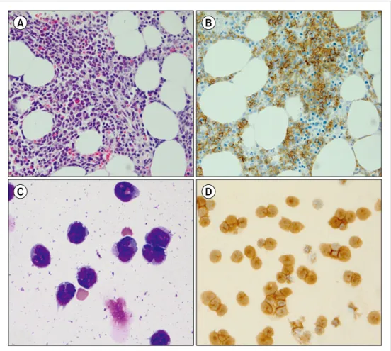
Fig. 2. Findings obtained with bone marrow biopsy and cytospin analysis of the cerebrospinal fluid in the second case. The bone marrow biopsy demonstrated an infiltration of neoplastic cells (A, hematoxylin and eosin staining,
×400) with weak positivity for CD4 and strong positivity for CD56 (B, ×400) on immuno- histochemical staining. Cytospin analysis of the cerebrospinal fluid showed neoplastic cells with at frequency of 92.0% (C, Wright staining, ×400); immunocyto- chemical staining for both CD56 and CD123 (D, ×400) demon- strated positive neoplastic cells.
demonstrated BPDCN with CD4, CD56, and CD123 positivity. Her hemogram results at admission were as fol- lows: WBC count, 7.6×10⁹/L; hemoglobin, 12.0 g/dL; and platelet count, 199.0×10⁹/L. A peripheral blood smear showed no evidence of blasts, but BM aspiration revealed infiltration of neoplastic cells with oval to round nuclei, prominent nucleoli, and abundant bluish cytoplasm at a frequency of 27.8% among BM-nucleated cells (Fig. 1A).
The BM clot section also showed neoplastic cell infiltration in an interstitial pattern (Fig. 1B) and positivity for CD4, CD56, and CD123 on immunohistochemical staining. Flow cytometry analysis also revealed positivity for both CD4 (22.7%) and CD56 (79.5%). Karyotype analysis results from the BM aspiration specimen demonstrated 46,XX, add(1) (p13),+del(1)(q21),del(6)(q21q23),add(8)(q24),-9,add(13)(q 34)[6]/46,XX[14], indicating a complex karyotype including monosomy 9. Based on these findings, the patient was diag- nosed with a leukemic manifestation of BPDCN. She re- ceived chemotherapy based on a vincristine, prednisolone, dexamethasone, and L-asparagine (VPDL) regimen for 2 months and underwent stem cell transplantation from a sibling donor 4 months previously. She has been in complete remission with no evidence of relapse.
The second case of BPDCN was observed in a 49-year-old man who was admitted to the authors’ institution because of a right inguinal lymph node mass that developed 3 years
previously. The lymph node mass was reported as BPDCN with CD4, CD56, and CD123 positivity via an excisional biopsy. The patient received chemotherapy based on a VPDL regimen and achieved complete remission for 2 years.
However, he experienced his first relapse 1 year previously and received autologous stem cell transplantation (ASCT) after 6 months; 2 months after ASCT, he developed a seiz- ure-like movement for which he was readmitted. His hemo- gram results at readmission were as follows: WBC count, 6.4×10⁹/L; hemoglobin, 14.7 g/dL; and platelet count, 78.0×10⁹/L. A peripheral blood smear showed no evidence of blasts. However, BM aspiration showed neoplastic cell infiltration, with a frequency of 20.8%. BM biopsy revealed neoplastic cells in an interstitial pattern (Fig. 2A), with weak positivity for CD4 and strong positivity for CD56 on immunohistochemical staining (Fig. 2B). Flow cytometry analysis results also demonstrated positivity for both CD4 (49.4%) and CD56 (99.0%) antigens. His karyotype was 44,XY,del(4)(p16),del(5)(q31q35),del(6)(q23q27),der(8)add (8)(p11.2)add(8)(q24),-9,der(10)t(8;10)(q22;p13),der(12;14) (q10;q10),add(19)(p13.3),der(19)t(9;19)(q13;p13.3)[18]/44, XY,t(3;15)(q21;q24),del(4),add(5)(q35),del(6),der(8)add(8)a dd(8),-9,der(10)t(8;10)der(12;14),add(19)[4]/46,XY[3], indi- cating a complex chromosomal rearrangement. Based on these results, he was diagnosed with a leukemic manifes- tation of BPDCN. Simultaneously performed cytospin analy-
Blood Res2014;49:196-207. bloodresearch.or.kr
200 Letters to the Editor
sis of the CSF showed neoplastic cells with a frequency of 92.0% (Fig. 2C) and positivity for both CD56 and CD123 on immunocytochemical staining (Fig. 2D). He was diag- nosed with leptomeningeal involvement of BPDCN. He was treated with intrathecal injections of high-dose cytarabine (40 mg) and methotrexate (15 mg) for 3 months, and he has been in complete remission for 4 months without evi- dence of residual disease.
DISCUSSION
Because markers are useful for confirming BPDCN, a recently performed multicenter study recommends im- munohistochemical staining using 6 antigens, such as CD4 (positive in 94.6% of cases), CD56 (positive in 96.5% of cases), CD123 (positive in 95.3% of cases), TCL1 (positive in 89.3% of cases), CD2AP (positive in 80.5% of cases), and BDCA2/CD303 (positive in 75.0% of cases) [1]. As a single antigen does not exhibit positivity in all cases of BPDCN, immunohistochemical staining using at least the 3 antigens mentioned previously (CD4, CD56, and CD123), all of which possess higher positivity rates compared with other antigens, should be performed to confirm BPDCN to provide maximum sensitivity. Our 2 cases demonstrated positivity for all these 3 antigens, which is adequate evidence of BPDCN involvement. Given that leptomeningeal involve- ment in BPDCN is a rare phenomenon, our second patient, who developed simultaneous leukemic manifestation and leptomeningeal involvement of BPDCN, is a very rare case and worth being reported.
In conclusion, we report 2 cases of BPDCN exhibiting leukemic manifestation (1 with simultaneous lepto- meningeal involvement) confirmed with positive CD4, CD56, and CD123 expression on immunohistochemical staining and flow cytometry. Immunohistochemical staining for CD4, CD56, and CD123 can confirm the BPDCN diag- nosis, and it should be performed in all cases suspected of having BM involvement in BPDCN.
Sang Hyuk Park1,2, Hyun-Sook Chi3, Young-Uk Cho3, Seongsoo Jang3, Chan-Jeoung Park3
1Department of Laboratory Medicine, Pusan National University School of Medicine, 2Biomedical Research Institute, Pusan National University Hospital, Busan,
3Department of Laboratory Medicine, University of Ulsan College of Medicine and Asan Medical Center, Seoul, Korea
Correspondence to: Hyun-Sook Chi Department of Laboratory Medicine, University of Ulsan
College of Medicine and Asan Medical Center, 88 Olympic-ro, 43-gil, Songpa-gu, Seoul 138-736, Korea
E-mail: hschi@amc.seoul.kr
Received on Apr. 15, 2014; Revised on May 25, 2014; Accepted on Aug. 25, 2014 http://dx.doi.org/10.5045/br.2014.49.3.198
AuthorsÊ Disclosures of Potential Conflicts of Interest No potential conflicts of interest relevant to this article were reported.
REFERENCES
1. Pagano L, Valentini CG, Pulsoni A, et al. Blastic plasmacytoid den- dritic cell neoplasm with leukemic presentation: an Italian multi- center study. Haematologica 2013;98:239-46.
2. Ng AP, Lade S, Rutherford T, McCormack C, Prince HM, Westerman DA. Primary cutaneous CD4+/CD56+ hematodermic neoplasm (blastic NK-cell lymphoma): a report of five cases.
Haematologica 2006;91:143-4.
3. Bekkenk MW, Jansen PM, Meijer CJ, Willemze R. CD56+ hemato- logical neoplasms presenting in the skin: a retrospective analysis of 23 new cases and 130 cases from the literature. Ann Oncol 2004;15:1097-108.
4. Hwang K, Park CJ, Jang S, et al. Immunohistochemical analysis of CD123, CD56 and CD4 for the diagnosis of minimal bone marrow involvement by blastic plasmacytoid dendritic cell neoplasm.
Histopathology 2013;62:764-70.
5. Jacob MC, Chaperot L, Mossuz P, et al. CD4+ CD56+ lineage neg- ative malignancies: a new entity developed from malignant early plasmacytoid dendritic cells. Haematologica 2003;88:941-55.
Bone marrow hypoplasia,
isochromosome 8q and deletion of chromosome 6q preceding B-cell lymphoma
TO THE EDITOR: Abnormalities of chromosome 6q involv- ing deletion of the regions 6q16-q27 have been reported in a variety of hematologic malignancies, most frequently in multiple myeloma [1-3]. The 8q isochromosome is also an uncommon clonal abnormality found primarily in T-pro- lymphocytic leukemia [4, 5]. Concurrent chromosome 6q- and i(8) are extremely rare in patients with hematologic malignancies and have never been reported as chromosomal abnormalities preceding evidence of malignancy.
Several case reports suggested that chromosomal abnor- malities can provide evidence for hematologic malignancies before they develop [1-3]. We describe a patient with bone marrow hypoplasia and persistent unexplained chromoso- mal abnormalities of del(6)(q16) and i(8)(q10) who sub- sequently developed B-cell lymphoma 8 months after his initial presentation.
CASE
A 50-year-old man was admitted to our hospital in December 2009 with persistent fever. Peripheral blood counts showed hemoglobin 9.0 g/dL, white blood cell (WBC) count 6.2×10⁹/L (65% neutrophils, 15% lymphocytes, 10%