Tuberc Respir Dis 2010;68:10-15
CopyrightⒸ2010. The Korean Academy of Tuberculosis and Respiratory Diseases. All rights reserved.
비소세포폐암 수술 후 세포분화도가 재발에 영향을 미친다
중앙대학교 의과대학 내과학교실
강형구, 조성근, 이혜민, 박성운, 이병욱, 이재희, 김보민, 박인원
Cell Differentiation Might Predict the Recurrence in Surgically Resected Non-Small Cell Lung Carcinoma
Hyung Koo Kang, M.D., Sung Gun Cho, M.D., Hye-Min Lee, M.D., Sung Woon Park, M.D., Byung Ook Lee, M.D., Jae Hee Lee, M.D., Bo Min Kim, M.D., In Won Park, M.D.
Division of Respiratory and Allergy Medicine, Department of Internal Medicine, Chung-Ang University College of Medicine, Seoul, Korea
Background: Lung cancer is the most common cause of cancer mortality in Korea. The TNM stage at presentation in patients with non-small cell lung cancer (NSCLC) has the greatest impact on prognosis. Patients who undergo a complete resection for NSCLC are likely to develop recurrent and/or metastatic disease. There are several factors influencing the development of recurrence. We explored risk factors of recurrence in patients with stages I and II NSCLC, who had undergone curative resection.
Methods: We reviewed patients who had complete surgical resection as definitive treatment for stage I or II. Patients followed up for more than 36 months. We evaluated several factors which might have relationship with recurrence, such as patient’s demographic factors, TNM staging, pathologic finding, tumor markers and surgical technique.
Results: A total of 75 patients were enrolled for analysis, of whom 58 were men and 17 were women with mean age of 61 (range, 37 to 76) years. The average size of tumors was 3.9 cm (0.7 to 10 cm). There were 64 patients with stage I NSCLC and 11 with stage II NSCLC. Among 64 patients with stage I NSCLC, 35 patients showed recurrences whereas 8 patients have recurred in stage II NSCLC. Grade of differentiation of tumor was closely related to the recurrence. Seventy-five percent of patients who had poor tumor differentiation experienced a recurrence. In contrast, 3 patients of twelve had recurrences, who revealed differentiation in their tissue (p<0.05).
Conclusion: Tumor differentiation could be a predictive factor for tumor recurrence in patients who have undergone curative resection for stage I or II NSCLC.
Key Words: Carcinoma, Non-Small-Cell Lung; Recurrence; Risk Factors; Cell Differentiation
This research was supported by the Chung-Ang University re- search grant in 2007.
Address for correspondence: In Won Park, M.D.
Division of Respiratory and Allergy Medicine, Department of Internal Medicine, Chung-Ang University Hospital, 224-1, Heukseok-dong, Dongjak-gu, Seoul 156-070, Korea Phone: 82-2-6299-1401, Fax: 82-2-825-7571 E-mail: [email protected]
Received: Dec. 4, 2009 Accepted: Dec. 11, 2009
서 론
2008년 통계청에서 발표한 2007년 사망 및 사망원인
통계결과에 따르면 대한민국은 암으로 인한 사망이 가장 많고 그 중 폐암이 인구 10만명당 29.1명으로 가장 많다.
특히 10년 전에 비하여 인구 10만명당 8.4명 증가하여 사 망률이 가장 많이 증가한 암이다1. 이 중 비소세포성 폐암 은 폐암의 85%를 차지하고 있고 5년 생존율 또한 16%로 고형암 중에서도 예후가 가장 나쁜 암이다. 비교적 근치 적 절제술 후에 예후가 좋다고 알려진 I기, II기의 경우에 도 각각 60∼80%, 30∼50%로 5년 생존율이 낮고 재발하 는 경우가 드물지 않으므로2,3, 수술 후 재발하는 원인이나 예측인자를 발견하는 것이 중요하다.
폐암은 항암제에 대한 반응성이 다른 암에 비하여 매우
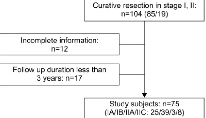
Figure 1. Patient population enrolled and excluded in this study.
낮지만 최근에 조기 폐암에서 수술 후 보조 항암 화학요법 을 통해서 생존율 및 무병생존기간(disease-free survival) 을 증가시켰다는 보고가 많아지고 있다4,5. 아직까지 조기 폐암에서 항암 화학요법의 효과는 확실히 입증되지 않았 고6, 항암 화학요법의 독성으로 인해 수술로 완전히 절제 된 I기, II기 폐암에서 항암 화학요법을 시행하는 것이 효 과적일지는 확실하지 않은 상태이다.
따라서 본 연구는 중앙대학교병원에서 폐암으로 진단 받고 근치적 절제술을 받은 폐암 I기, II기 환자에서 재발 을 일으킬 수 있는 요인을 찾아보기 위하여 재발한 군과 재발하지 않은 군을 비교 분석하였다.
대상 및 방법 1. 연구대상
1985년 7월부터 2005년 7월까지 중앙대학교병원 및 중 앙대학교 용산병원에서 비소세포 폐암 I기, II기로 확인되 어 근치적 절제술을 시행 받은 104명 중 3년 이상 추적 관찰한 75명을 대상으로 하였다. 추적 기간이 3년이 되지 않은 17명 및 추적 관찰에 실패하거나 충분한 정보가 제 공되지 않은 환자 12명은 배제하였다(Figure 1).
2. 연구방법
대상 환자의 의무기록 분석을 통하여 환자의 성별, 나 이, 병기 및 종양의 크기, 종양의 위치, 조직학적 분류, 분화도, 흡연력, 흡연량, 배아성 암항원(CEA), 증상 동반 여부, 수술 방법7 등을 재발과 연관성을 평가하였다.
병기는 American Joint Committee on Cancer 암 병기 를 기준으로 분석하였고8,9, 2007년에 제시된 새로운 병기 를 이용하여 새로운 분류법으로도 함께 분석하였다10.
새로운 병기는 이전에 비해서 원발 부위의 종양 크기를 세분화한 것이 특징으로 이전에는 지름이 3 cm 이하인 경우 T1으로 정의하였지만 개정에서는 T1a (2 cm 미만), T1b (2∼3 cm)으로 구분하였다. 직경이 3 cm 이상인 경 우로 정의했던 T2도 T2a (3∼5 cm), T2b (5∼7 cm)로 세분화하였고 T2에 해당하는 원발종양의 크기가 7 cm 이 상인 경우 T3로 다시 정의하였다11. 병기 분류를 위해 단 순 방사선촬영, 컴퓨터 단층촬영, 양전자방출단층촬영 등 영상적인 검사와 수술 후 조직 판독지를 검토하였다.
비소세포성 폐암의 각 조직학적 분류는 편평세포암, 선 암, 대세포암종으로 나누어 비교 분석하였으며 분화도는 고분화도, 중분화도, 저분화도군으로 나누어 살펴보았다.
수술방법은 구역절제술, 폐엽절제술, 폐절제술 등 세 가지 방법으로 비교하였다. 조직학적 분류에서 기관지 폐포암 은 선암으로 분류하였다. 또한 진단 당시 자각 증상이 있 어 병원에 내원한 경우와 우연히 발견된 경우 재발과 관련 이 있는지 살펴 보았다.
재발한 군은 수술한 이후 3년 이상 추적 관찰한 환자군 에서 의심되는 부위의 조직검사, 흉부방사선 혹은 흉부컴 퓨터 단층촬영을 통해 재발이 확인된 환자를 대상으로 하 였다12.
무질병 생존기간은 수술한 이후 재발이 발견될 때까지 기간으로 정의하였다.
3. 통계
어떤 인자가 중요하게 재발에 영향을 미치는지에 관해 분석하기 위해 카이제곱 검정을 통해 후향적으로 분석하 였다. 재발에 관한 다변량 분석은 로지스틱 회기 분석을 통해 살펴보았다. 무질병 생존기간은 Kaplan-Meier 방법 으로 관찰하였다. 모든 값은 SPSS 12.0 (SPSS Inc, Chica- go, IL, USA)로 분석하였고 p-value가 0.05 이하일 때 통 계적으로 유의한 것으로 보았다.
결 과 1. 대상군의 임상적 특성
폐 근치적 절제술을 받은 75명을 대상으로 연구하였다.
남자가 58명, 여자가 17명으로 남자가 많았고 평균 연령 은 61세(범위, 37∼76세)였다(Table 1). 이 중 재발된 환 자는 43명(57.3%)으로 수술한 부위에 국소 재발한 경우가 11명(25.6%), 원격 재발한 경우가 32명(74.4%)이었다. 연 구 대상 중 American Joint Committee on Cancer (AJCC)
Table 1. Demographics of study subjects
Variable Recurrence No recurrence p-value
No. 43 32
Sex (M/F) 24/11 24/5 0.192
Median age, 63 (37∼76) 61 (40∼74) 0.513 yr (range)
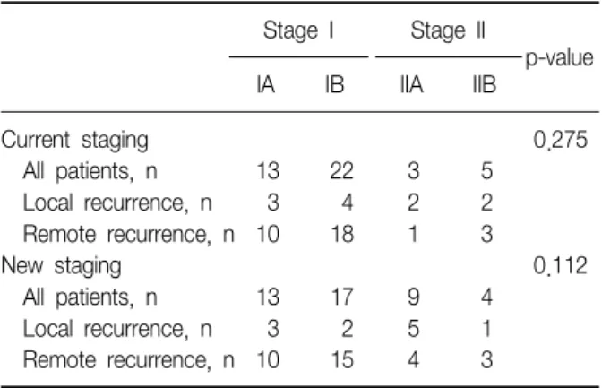
Table 2. Comparison of recurrence between stage I and stage II
Stage I Stage II
p-value
IA IB IIA IIB
Current staging 0.453
All patients, 25 39 3 8
n
Recurrence, 13 (52) 22 (56) 3 (100) 5 (80) n (%)
New staging 0.475
All patients, 25 31 13 5
n
Recurrence, 13 (52) 17 (55) 9 (69) 4 (80) n (%)
Table 3. Comparison of recurrence characteristics between local and remote recurrence
Stage I Stage II
p-value
IA IB IIA IIB
Current staging 0.275
All patients, n 13 22 3 5
Local recurrence, n 3 4 2 2 Remote recurrence, n 10 18 1 3
New staging 0.112
All patients, n 13 17 9 4
Local recurrence, n 3 2 5 1 Remote recurrence, n 10 15 4 3 암 병기 16판에 따라 병기를 나누면9, Ia 25명, Ib 39명,
IIa 3명, IIb 8명이었고 이 중 재발은 각각 13명(52%), 22 명(56%), 3명(100%), 5명(62.5%)에서 발생하였고 병기 I, II 간에 통계적으로 유의한 차이는 없었다(Table 2). 또한 재발한 환자 중 원격전이로 발견된 경우는 각각 10명 (77%), 18명(82%), 1명(33%), 3명(60%)이었고 유의한 차 이는 보이지 않았다(Table 3). AJCC암 병기 17판10으로는 Ia 25명, Ib 31명, IIa 13명, IIb 5명이었고 재발은 각각 13명(52%), 17명(54.8%), 9명(69.2%), 4명(80%)에서 발 생하였으며(Table 2), 그 중 원격전이는 10명(77%), 15명 (88%), 4명(44%), 3명(23%)으로 통계적으로 유의한 차이 는 없었다(Table 3).
종양의 크기의 평균은 3.9±1.6 cm이고 조직학적으로 편평세포암이 43명(57.3%), 선암이 31명(41.3%), 대세포 암이 1명(1.4%)으로 분포하고 있었다. 분화도는 중분화도 암이 36명(48%)으로 가장 많았고 저분화도암은 12명 (16%), 고분화도암은 12명(16%)이었으며 분화도가 언급 되지 않은 경우도 15명(20%)이었다. 조직 검사 결과 림프 혈관 내 종양 색전(lymphovascular tumor emboli)이 있 는지 여부도 관찰하였는데 언급이 되지 않은 환자가 55명
(73%)이었으며 언급된 20명(27%) 중에 6명(30%)에서 림 프혈관 내 종양 색전이 발견되었다.
수술 방법으로 폐엽절제술은 시행한 경우가 58명 (77.3%)으로 가장 많았고 폐전절제술로 한 경우는 15명 (20%)이었으며 구역절제술로 수술한 경우는 2명(2.7%)으 로 가장 적었다. 병변이 있던 위치로는 우상엽이 22명 (29.3%)으로 가장 많았고 우하엽은 18명(24%), 좌상엽은 14명(18.7%), 좌하엽은 14명(18.7%)이었고 우중엽이 7명 (9.3%)으로 가장 적었다.
진단 당시 증상이 있던 경우는 40명이었고 무증상으로 우연히 발견된 경우가 35명이었으며 기침이 19명으로 가 장 많았고 혈객담이 14명으로 그 다음으로 많았으며 흉통, 운동 시 호흡곤란을 주된 증상으로 한 경우가 각각 4명, 3명이었다.
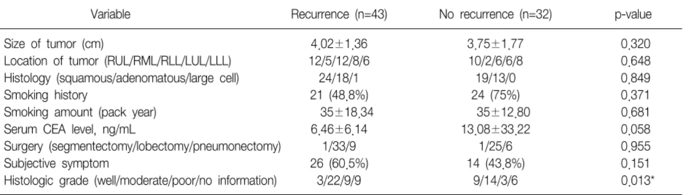
2. 각 인자와 재발과의 연관성
본 연구에 포함되었던 I기, II기의 환자군에서 나이, 종 양의 크기, 조직학적 분류, 림프혈관 내 종양 색전, 흡연 력, 흡연량, 배아성 암항원, 진단 시 증상이 있던 경우, 수술 방법은 재발하지 않은 군과 재발한 군의 차이는 통계 적으로 유의하지 않았다(Table 4). I기의 환자군만 따로 관찰하였을 때에도 역시 같은 결과를 얻었다(Table 5).
3. 분화도에 따른 재발률 비교
고분화도암은 12명 중 3명(25%)이 재발하였고 중분화 도암의 경우 36명 중 22명(61%)이 재발하였으며 저분화 도암은 12명 중 9명(75%)이 재발하여 저분화도암이 고분 화도암보다 재발률이 더 높았고 중분화도암 또한 고분화 도암보다 재발률이 통계적으로 유의하게 높았다(Figure
Table 5. Relationship between recurrence and clinicopathologic factors in stage I non-small cell lung cancer
Variable Recurrence (n=35) No recurrence (n=29) p-value
Size of tumor (cm) 4.06±1.42 3.48±1.33 0.884
Location of tumor (RUL/RML/RLL/LUL/LLL) 10/5/11/5/4 10/2/5/5/7 0.430
Histology (squamous/adenomatous/large cell) 19/15/1 17/12/0 0.816
Smoking history 14 (40%) 21 (72.4%) 0.184
Smoking amount (pack year) 40±20.04 37±12.30 0.635
Serum CEA level, ng/mL 7.60±6.39 13.48±35.88 0.083
Surgery (segmentectomy/lobectomy/pneumonectomy) 1/27/7 1/24/4 0.804
Subjective symptom 21 (60%) 11 (37.9%) 0.079
Histologic grade (well/moderate/poor/no information) 2/13/12/8 8/12/4/5 0.020*
CEA: carcioembryonic antigen; RUL: right upper lobe; RML: right middle lobe; RLL: right lower lobe; LUL: left middle lobe; LLL:
left lower lobe.
*p<0.05.
Table 4. Relationship between recurrence and clinicopathologic factors in stage I, II non-small cell lung cancer
Variable Recurrence (n=43) No recurrence (n=32) p-value
Size of tumor (cm) 4.02±1.36 3.75±1.77 0.320
Location of tumor (RUL/RML/RLL/LUL/LLL) 12/5/12/8/6 10/2/6/6/8 0.648
Histology (squamous/adenomatous/large cell) 24/18/1 19/13/0 0.849
Smoking history 21 (48.8%) 24 (75%) 0.371
Smoking amount (pack year) 35±18.34 35±12.80 0.681
Serum CEA level, ng/mL 6.46±6.14 13.08±33.22 0.058
Surgery (segmentectomy/lobectomy/pneumonectomy) 1/33/9 1/25/6 0.955
Subjective symptom 26 (60.5%) 14 (43.8%) 0.151
Histologic grade (well/moderate/poor/no information) 3/22/9/9 9/14/3/6 0.013*
CEA: carcioembryonic antigen; RUL: right upper lobe; RML: right middle lobe; RLL: right lower lobe; LUL: left middle lobe; LLL:
left lower lobe.
*p<0.05.
Figure 2. Recurrence rate of patients with stage I, II strati- fied by differentiation.
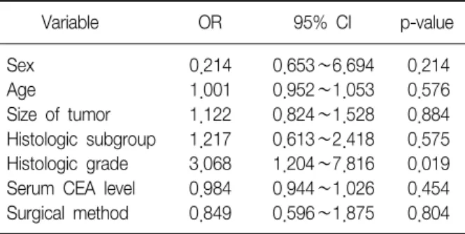
2). 다변량 분석으로 로지스틱 회기 분석한 결과 분화도가 재발의 위험도에 중요한 인자임을 알 수 있었다(p=0.019) (Table 6).
4. 림프혈관 내 종양 색전 유무에 따른 재발률 비교
총 75명 대상군 중에 림프혈관 내 종양 색전(lymph- ovascular tumor emboli)의 유무가 표시된 20명을 대상으 로 살펴보았다. 20명 중 재발한 환자는 12명, 재발하지 않은 환자는 8명이었다. 재발한 환자에서 림프혈관 내 종 양 색전이 발견된 경우가 4명(33%)으로 재발하지 않은 환 자 2명(25%)보다 많았으나 통계적인 유의성은 보이지 않 았다. I기 환자만을 대상으로 살펴보았을 때도 통계적으 로 유의성은 보이지 않았다(Table 7).
Table 6. Multivariate analysis for relationship between re- currence and histologic grade
Variable OR 95% CI p-value
Sex 0.214 0.653∼6.694 0.214
Age 1.001 0.952∼1.053 0.576
Size of tumor 1.122 0.824∼1.528 0.884 Histologic subgroup 1.217 0.613∼2.418 0.575 Histologic grade 3.068 1.204∼7.816 0.019 Serum CEA level 0.984 0.944∼1.026 0.454 Surgical method 0.849 0.596∼1.875 0.804 OR: odds ratio; CI: confidence interval.
Table 7. Relationship between recurrence and lymphovas- cular tumor emboli
Recurrence No recurrence Stage I, II (n=20)
Lymphovasular emboli, 4/12 (33%) 2/8 (25%) n (%)
Stage I (n=16)
Lymphovascular emboli, 3/9 (33%) 2/7 (29%) n (%)
Figure 3. The Kaplan-Meier survival curves in patients with stage I, II non-small cell lung cancer stratified by his- tologic grades.
5. 무질병 생존기간
Kaplan-Meier 방법으로 살펴 본 무질병 생존기간은 고 분화도군이 중분화도군, 저분화도군에 비해 의미 있게 높 았다(p=0.027) (Figure 3).
고 찰
본 연구는 폐암으로 진단받고 근치적 절제술을 받은 폐 암 I기, II기 환자 75명을 대상으로 재발을 일으킬 수 있는 요인을 분석하였다. 연구 결과 분화도가 폐암의 재발을 일으키는 중요한 위험인자임을 밝혀낼 수 있었다.
수술 후 폐암의 항암화학치료는 IA기 폐암에서는 권고 되지 않고 있고13,14 IB, II기 폐암에서는 권고되고 있고 생 존율을 높이고 재발률을 줄인다는 여러 연구가 있지만 Adjuvant Lung Cancer Project Italy (ALPI), International Adjuvant Lung Cancer (IALT) 연구에서는 II기암에서 항 암화학치료가 특별한 이득이 없음을 시사하였다6,14. 이런 상황에서 재발의 위험이 높은 인자를 알아낸다면 항암화 학치료를 위험인자를 갖고 있는 환자에게 시도할 때 더 많은 이득을 얻을 수 있다.
실제로 폐암은 항암치료를 해도 재발률이 매우 높고 항 암치료가 고려되지 않는 IA기 폐암의 경우에도 재발률이 높다. 따라서 IA기 폐암에서 재발 위험군을 찾아내는 것 은 중요하다. 본 연구에서 IA기 폐암의 수가 너무 적어 따로 살펴보지 못하였고 추후 이에 대한 연구가 필요하다.
본 연구에서 I기, II기 비소세포성암에서 조직의 분화도 가 근치적 수술 후 재발을 잘 일으키는 요인으로 밝혀져 이전에 다른 연구와 결과가 일치하였다12. 이전 연구에서 는 저분화도군에서 재발이 더 잘 발생하는 것으로 밝혔으 나 본 연구에서는 저분화도군 뿐 아니라 분화도에 따라 재발률이 증가하였다. 따라서 중분화도 및 저분화도암의 경우 수술 후 항암치료를 고려할 필요가 있다.
이전에 위험인자로 연구되었던 종양의 크기와 종양의 위치는 본 연구에서 통계적 유의성을 보이지 않았다7,15. 이전 연구에서 종양의 크기에 따라 5년 생존율의 차이가 있어 2 cm 미만의 경우 77%, 2∼3 cm은 71%, 3∼5 cm 58%, 5∼7 cm 49%, 7 cm 이상은 35%로 알려져 있다11. 실제로 재발과 연관이 있을 것으로 예상하였으나 재발한 군이 재발하지 않은 군보다 종양의 크기가 컸지만 통계적 으로 유의한 차이는 없었다. 이는 본 연구에 포함된 환자의 경우 종양의 크기가 대부분 3∼5 cm에 모여 있어 통계적 유의성이 없던 것으로 보이며 더 많은 환자를 대상으로 연 구가 필요하다. 또한 조직학적으로 림프 및 혈관 침윤이 있는 경우가 더 재발을 잘하는 것으로 알려져 있으나15-17, 림프 혈관 내 종양 색전은 본 연구에서는 재발과의 연관성 을 찾을 수 없었다. 림프 혈관 내 종양 색전이 관찰될수록 더 재발이 잘되는 경향을 보였으나 통계적으로 의미는 없
었으며 이는 본 연구에서 모든 환자를 대상으로 하지 않고 20명의 환자를 대상으로 관찰하여 통계상 문제점이 있을 것으로 생각된다.
본 연구의 가장 큰 제한점은 추적 관찰 기간이 연구 대 상마다 다르다는 것이다. 3년 이상 된 환자를 대상으로 하였지만 실제로 추적관찰을 시작한 1985년 환자는 23년 간 관찰하였지만 2005년부터 관찰한 환자는 3년 밖에 관 찰하지 못하였기 때문이다. 최소한 3년 이상 관찰했다는 점에 의의가 있다고 하겠으나 향후 장기간 관찰에 의한 결과 분석이 필요할 것으로 사료된다.
참 고 문 헌
1. Korea National Statistical Office. Annual report on the cause of death statistics, 2007. Daejeon: Korea National Statistical Office; 2008.
2. Hayat MJ, Howlader N, Reichman ME, Edwards BK.
Cancer statistics, trends, and multiple primary cancer analyses from the Surveillance, Epidemiology, and End Results (SEER) Program. Oncologist 2007;12:20-37.
3. Asamura H, Goya T, Koshiishi Y, Sohara Y, Tsuchiya R, Miyaoka E. How should the TNM staging system for lung cancer be revised? A simulation based on the Japanese Lung Cancer Registry populations. J Thorac Cardiovasc Surg 2006;132:316-9.
4. Visbal AL, Leighl NB, Feld R, Shepherd FA. Adjuvant chemotherapy for early-stage non-small cell lung can- cer. Chest 2005;128:2933-43.
5. Winton T, Livingston R, Johnson D, Rigas J, Johnston M, Butts C, et al. Vinorelbine plus cisplatin vs. ob- servation in resected non-small-cell lung cancer. N Engl J Med 2005;352:2589-97.
6. Scagliotti G. Multimodality approach to early-stage non-small cell lung cancer. Lung Cancer 2007;57 Suppl 2:S6-11.
7. Ou SH, Zell JA, Ziogas A, Anton-Culver H. Prognostic factors for survival of stage I nonsmall cell lung cancer patients: a population-based analysis of 19,702 stage I patients in the California Cancer Registry from 1989 to 2003. Cancer 2007;110:1532-41.
8. Mountain CF. Revisions in the international system for staging lung cancer. Chest 1997;111:1710-7.
9. Greene FL. AJCC cancer staging manual. 6th ed. New York: Springer-Verlag; 2002.
10. Goldstraw P, Crowley J, Chansky K, Giroux DJ, Gro- ome PA, Rami-Porta R, et al. The IASLC Lung Cancer Staging Project: proposals for the revision of the TNM stage groupings in the forthcoming (seventh) edition of the TNM Classification of malignant tumours. J Thorac Oncol 2007;2:706-14.
11. Rami-Porta R, Crowley JJ, Goldstraw P. The revised TNM staging system for lung cancer. Ann Thorac Cardiovasc Surg 2009;15:4-9.
12. Kobayashi N, Toyooka S, Soh J, Ichimura K, Yanai H, Suehisa H, et al. Risk factors for recurrence and un- favorable prognosis in patients with stage I non-small cell lung cancer and a tumor diameter of 20 mm or less. J Thorac Oncol 2007;2:808-12.
13. Tsuboi M, Ohira T, Saji H, Miyajima K, Kajiwara N, Uchida O, et al. The present status of postoperative ad- juvant chemotherapy for completely resected non-small cell lung cancer. Ann Thorac Cardiovasc Surg 2007;13:
73-7.
14. Pisters KM, Evans WK, Azzoli CG, Kris MG, Smith CA, Desch CE, et al. Cancer Care Ontario and American Society of Clinical Oncology adjuvant chemotherapy and adjuvant radiation therapy for stages I-IIIA resect- able non small-cell lung cancer guideline. J Clin Oncol 2007;25:5506-18.
15. Kita H, Koshiishi Y, Masui K, Fujita A, Ootsuka K, Furuyashiki G, et al. Risk factors of recurrence in re- sected stage I non-small cell lung cancer. Kyobu Geka 2007;60:883-7.
16. Fujisawa T, Yamaguchi Y, Saitoh Y, Hiroshima K, Ohwada H. Blood and lymphatic vessel invasion as prognostic factors for patients with primary resected nonsmall cell carcinoma of the lung with intrapul- monary metastases. Cancer 1995;76:2464-70.
17. Pechet TT, Carr SR, Collins JE, Cohn HE, Farber JL.
Arterial invasion predicts early mortality in stage I non-small cell lung cancer. Ann Thorac Surg 2004;78:
1748-53.