Ethanol Extract of Cynanchum wilfordii Produces Endothelium-Dependent Relaxation in Rat Aorta and Anti-inflammatory Activity in Human Aortic Smooth
Muscle Cells
Deok-Ho Choi1, Yun-Jung Lee1,2, Eun-Joo Kim1,2, Xiang Li1,2, Hye-Yoom Kim1,2, Sun-Mi Hwang1,2, Jung-Joo Yoon1,2, So-Min Lee1,2, Eun-Kyeong Min3,
Dae-Gill Kang1,2, Ho-Sub Lee1,2*
1College of Oriental Medicine and Professional Graduate School of Oriental Medicine, Wonkwang University, Iksan, Chonbuk, 570-749, Republic of Korea
2Hanbang Body-fluid Research Center, Wonkwang University, Shinyong-dong, Iksan, Chonbuk, 570-749, Republic of Korea
3Oriental Internal Medicine, Joonghwa Medical Center, Anyang, Gyeonggi, Republic of Korea Original Article
⋅Received:4 October 2010 ⋅Revised:30 October 2010 ⋅Accepted:25 November 2010
⋅Correspondence to:Ho-Sub Lee, OMD. PhD.
Tel:+82-63-850-6841, Fax:+82-63-850-7260, E-mail:[email protected]
Objective: The present study investigated the effect of ethanol extract of Cynanchum wilfordii (ECW) on vascular relaxation and vascular inflammation in rat artery isolated from rats and anti-inflammatory activity in human aortic smooth muscle cells (HASMC).
Methods: Vascular tone and guanosine 3’,5’-cyclic monophosphate (cGMP) production were examined in rat artery isolated from Sprague Dawley rats, in the presence of ECW. HASMC were incubated with tumor necrosis factor-alpha (TNF-α) or Angiotensin II for 24 h. Matrix metalloproteinase (MMP)-2 and anti-oxidant activity of ECW was investigated by pretreatment with ECW in HASMC.
Results: Cumulative treatment of ECW relaxed aortic smooth muscles of rats in a dose-dependent manner.
ECW-induced vasorelaxation was significantly decreased by pretreatment of L-arginine methyl ester (L-NAME) or oxadiazolo-quinoxalinone (ODQ). Furthermore, ECW treatment of thoracic aorta significantly increased cGMP production. Incubation of ECW with ODQ or L-NAME markedly decreased ECW-induced cGMP production. ECW treatment dose-dependently suppressed TNF-α- or Angiotensin II-induced increase in matrix metalloproteinase-2 expression in HASMC. Also, ECW exhibited 2,2-diphenyl-1-picrylhydrazyl radical scavenging activity in vitro and reduced TNF-α-induced increase in reactive oxygen species production in a dose-dependent manner.
Conclusions: Taken together, the results suggest that ECW exerts vascular relaxation via NO/cGMP signaling pathway and decreases MMP-2 expression via anti-oxidant activity.
Key Words : Cynanchum wilfordii, vascular relaxation, NO, cGMP, anti-oxidant activity, matrix metalloproteinase-2
Introduction
There is increasing evidence that vascular inflammation plays a key role in the pathogenesis of vascular disease and the atherosclerotic process1).
Endothelium-derived nitric oxide (NO) is a pleiotropic biological mediator that regulates diverse activities ranging from neuronal function to vascular homeostasis2). Especially, in vascular smooth muscle cells (SMCs), NO-mediated activation of guanylyl
cyclase (GC) catalyzes the formation of the second messenger guanosine 3’,5’-cyclic monophosphate (cGMP), leading to vasorelaxation in aorta. The endothelial production of NO depends on a delicate balance between NO production via endothelial nitric oxide synthase (eNOS) and inactivation by reactive oxygen species (ROS) such as superoxide (O2
-)3). Reduced endothelium derived NO is thought to contribute to endothelial dysfunction, atherosclerosis, and restenosis through the loss of the vasodilatory, anti-inflammatory, and antiproliferative properties of NO, respectively3-5).
ROS upregulate Matrix metalloproteinases (MMPs) expression in several tissues6,7). MMPs are a group of membrane-bound and extracellular proteinases that participate in the degradation of extracellular matrix (ECM) protein as well as several non-ECM proteins8). Over-expression of MMPs cannot only promote local inflammation by facilitating the infiltration of inflammatory cells, but can also lead to enhanced neointima formation by stimulating SMC migration and proliferation.
Radix of Cynanchum wilfordii (CW), which is known as ‘Hasuo’ in Korea and ‘Baishouwu’ in China, has long been used as a tonic and traditional herbal medicine for cardiovascular diseases. ‘Baishouwu’ is a common name of dried white root tubers from three Asclepiadacea plants, CW, C. bungei, and C.
auriculatumi. ‘Hasuo’ refers to the CW dried root tuber and Polygonum multiflora, a red root tuber dried from the Polygonaceae plant. CW exhibits gastroprotective effect on experimental gastric lesions in rats9), and protects cortical neurons from toxicity induced by H2O2
10). Gagaminine isolated from CW root inhibits aldehyde oxidase activity and lipid peroxidation in vitro11). However, there is currently little information to establish a pharmacological basis of action of CW dried root on vascular dysfunction associated with cardiovascular diseases. Thus, the present study was undertaken to clarify the effects of ethanol extract of CW (ECW) on vascular relaxation
in thoracic aorta of rats and anti-inflammatory activity in human aortic smooth muscle cells (HASMC).
Materials and methods
In Sprague Dawley rat, aortic rings for vascular tone were prepared for Measurement of vascular relaxation. Radioimmunoassay was conducted for cGMP level. For in vitro study, HASMC was cultured and their viability was performed by MTT assay. MMP-2 expression or activity was measured by western blotting analysis and gelatin zymography, respectively. For the study of anti-oxidant effect, we used 1,1-diphenyl-2-picrylhydrazyl (DPPH) and dichloro fluorescein diacetate (DCFDA).
1. Preparation of ECW
Cynanchum wilfordii (Max.) Hemsley was purchased from the Herbal Medicine Cooperative Association, Jeonbuk Province, Korea and authenticated by professor Tae-Oh Kwon, College of Life Sciences and Natural Resources, Wonkwang University.
Herbarium voucher specimen (HBN-021) was deposited in the herbarium of the Professional Graduate School of Oriental Medicine, Wonkwang University, Iksan, Jeonbuk, Korea. Tubers were cleaned and air-dried at room temperature. Dried tuber (1.2 kg) was extracted with 10 L of 95%
ethanol at 24℃ for 1 week. The extract was filtered through Whatman No. 3 filter paper and concentrated using a rotary evaporator (N-11, EYELA, Japan). The resulting extract (48.27 g) was lyophilized using a freeze-drier (FDU-1100, EYELA, Japan) and retained until required.
2. Preparation of aortic rings for vascular tone The animal procedures were in strict accordance with the National Institute of Health Guide for the Care and Use of Laboratory Animals (NIH
Publication No. 85-23, revised 1996) and were approved by the Institutional Animal Care and Utilization Committee for Medical Science of Wonkwang University (WKU09-039). Male Sprague Dawley rats were purchased from Samtako (Osan, Korea). The rats weighing 250-300 g were sacrificed by decapitation. The thoracic aorta was rapidly and carefully dissected and placed into ice-cold Kreb’s solution (118 mM NaCl, 4.7 mM KCl, 1.1 mM MgSO4, 1.2 mM KH2PO4, 1.5 mM CaCl2, 25 mM NaHCO3, and 10 mM glucose, pH 7.4). The aortae were removed free of connective tissue and fat and cut into rings with a width of approximately 3 mm.
All dissecting procedures were done with extreme care to protect the endothelium from inadvertent damage. In some aortic rings, the endothelial layer was mechanically removed by gently rubbing the luminal surface of the aortic ring back and forth several times with plastic tubing. Endothelial integrity or functional removal was verified by the presence or absence of the relaxant response to 1 μM acetylcholine (ACh) on 1 μM phenylephrine contracted vessels12).
3. Measurement of vascular relaxation
The aortic rings were suspended, by means of two L-shaped stainless-steel wires inserted into the lumen, in a tissue bath containing Kreb’s solution at 37 °C. Mixed gas containing 95% O2 and 5% CO2
was continuously bubbled through the bath. The baseline load placed on the aortic rings was 1.0 g.
Changes in isometric tension were recorded using a Grass FT 03 force displacement transducer (Grass Technologies, Quincy, MA, USA) connected to a Model 7E polygraph recording system (Grass Technologies). In the first set of experiments, the response to 1 μM ACh was confirmed as a positive control for endothelium-intact aortic rings contracted by 1 μM phenylephrine. The aortic rings were washed every 10 min with Kreb’s solution until the
tension returned to the basal level. The aortic relaxation by the cumulative addition of ECW was performed in the presence or absence of endothelium on various drugs such as Nω-Nitro-L-arginine methylester (L-NAME) and 1H-[1,2,4]oxadiazolo [4,3-a]quinoxalin-1-one (ODQ). The effect of vehicle, 0.01% dimethylsulfoxide (DMSO), was also tested. After each test, the aortic rings were washed three times with fresh Kreb’s solution and allowed to equilibrate for 30 min.
4. Radioimmunoassay of cGMP
The cGMP level was assayed in thoracic aorta treated with ECW or modulating agents with ECW.
The tissues were frozen rapidly and then homogenized using a Polytron homogenizer in 20 mM Tris-HCl buffer (pH 7.4). The homogenates were centrifuged at 3000 ×g for 10 min, and supernatants were extracted three times with two volumes of water-saturated diethylether and then concentrated with Speed-vac concentrator (Savant Instruments, Farmingdale, NY, USA). cGMP content was measured by an equilibrated radioimmunoassay as described previously13). In brief, standards or samples were introduced in a final volume of 100 μl of 50 mM sodium acetate buffer (pH 4.8). Then, 100 μl of a 1:1000 dilution of cGMP antiserum (Calbiochem-Novabiochem, San Diego, CA, USA) and iodinated cGMP (10,000 cpm/100 μl) were added in succession and incubated overnight at 4°C.
The bound form was separated from the free form by charcoal suspension. Results were expressed as picomoles of cGMP generated per mg protein.
5. HASMC cultures
HASMC purchased from Bio-Whittaker (San Diego, CA, USA) were cultured in SMC growth medium-2 containing 10% fetal bovine serum, 2 μg/ml human basic fibroblast growth factor, 0.5 μg/ml human epidermal growth factor, 50 mg/ml gentamicin, 50
mg/ml amphotericin-B, and 5 mg/ml bovine insulin.
For all experiments, early passage HASMC were grown to 70%-80% confluence and made quiescent by serum starvation for at least 24 h.
6. Cell viability assay
HASMC were counted and seeded onto gelatin-coated 96-well culture plates at a density of 5
× 104 cells/well. After incubation with various concentrations of ECW for 24 h, and then 100 μl of a 0.5 mg/ml solution of MTT was added to each well, and incubated at 37°C for an additional 4 h.
The absorbance of the solubilized formazan was read at 490 nm using a microplate reader (F-2500, Hitachi, Japan). HASMC incubated in control media were considered as 100% viable.
7. Western blot analysis for MMP-2 expression
The cell lysate protein (40 μg) was separated by 10% sodium dodecyl sulfate-polyacrylamide gel electrophoresis (SDS-PAGE) and transferred electro- phoretically to nitrocellulose membranes using a Mini-Protean II (Bio-Rad). A SDS-PAGE protein standard was used to check transfer efficiency and as a molecular weight marker. Membranes were blocked with 5% non-fat milk powder in 0.05%
Tween 20-PBS (PBST) for 1 h, and then were incubated with primary antibodies to MMP-2 or β-actin (Santa Cruz Biotechnology, Santa Cruz, CA, USA) at a final dilution of 1:1000 overnight at 4oC. The blot was washed several times with PBST and incubated with the appropriate horseradish peroxidase- conjugated secondary antibody for 1 h. After the membrane was washed several times with PBST and then detected by enhanced chemiluminescence (Amersham, Buckinghamshire, UK) procedure. The protein expression levels were determined by analyzing the signals captured on nitrocellulose membrane (Amersham) using a Chemi-Doc image
analyzer (Bio-Rad).
8. Gelatin zymography for MMP-2 activity Conditioned medium was electrophoresed in a polyacrylamide gel containing 1 mg/ml gelatin14). The gel was washed at room temperature for 2 h with 2.5% Triton X-100 and subsequently incubated in a buffer containing 10 mM CaCl2, 150 mM NaCl, and 50 mM Tris-HCl, pH 7.5 at 37°C overnight. The gel was stained with 0.2% Coomassie blue for 2 h and then washed in destaining buffer containing 30%
methanol and 10% acetic acid. The gel was photographed on a light box. Proteolysis was detected as a white zone on a dark blue field.
9. Free radical scavenging activity
The free radical scavenging activity of ECW was measured by 1,1-diphenyl-2-picrylhydrazyl (DPPH)15). Briefly, 0.2 mM solution of DPPH in ethanol was prepared and 1 ml of this solution was added to 1 ml of different concentrations (final concentration, 1-750 μg/ml) of the extract. The mixture was shaken vigorously and allowed to stand at room temperature for 30 min. Then the absorbance was measured at 517 nm using spectrophotometer (F-2500, Hitachi, Japan). Lower absorbance of the reaction mixture indicated higher free radical scavenging activity.
Vitamin C (0.1-100 μM) was used as the positive control.
10. Fluorescent measurement of intracellular ROS
A flow cytometric method was used to detect intracellular peroxide production in HASMC16). Cells were washed twice with PBS and incubated for 30 min with 5 mM dichloro fluorescein diacetate (DCFDA) (Molecular Probes, Eugene, OR, USA), a nonfluorescent compound that freely permeates cells.
Cells were then treated with trypsin to remove them from the surface of the culture flask. After
-40 -20 0 20 40 60 80 100
1 10 100 1000 ECW(μg/ml) Endothelium (+) Endothelium (-) A)
-40 -20 0 20 40 60 80 100
1 10 100 1000 ECW(μg/ml) Vehicle ODQ (10μM) L-NAME (10μM) B)
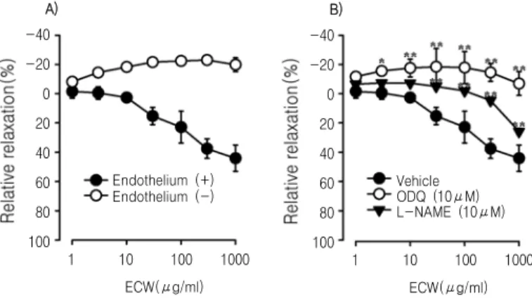
Fig. 1. Dose-response curves for the relaxation activity of ECW.
Dose-response curves (1-1000 μg/ml) are shown for endothelial-intact or denuded rat thoracic aorta precontracted with phenylephrine (A), and in the endothelium-intact rat thoracic aorta in absence or presence of 10 μM of L-NAME or ODQ (B).
Values are expressed as mean ± S.D. (n=6); **p<0.01, *p<0.05, vs. Vehicle.
centrifugation at 800 ×g for 2 min at room temperature, the supernatant was removed. Collected cells were suspended in 500 μl PBS. Fixed cells (1 x 10⁵) were analyzed on a FACScalibur (BD Biosciences, San Diego, CA).
11. Statistical analyses
Values are shown as mean ± SD. Statistical analyses were performed using analysis of variance followed by the Student’s t-test for unpaired data and one-way ANOVA followed by Bonferroni’s multiple-comparision test. Differences with a p value of <0.05 were considered statistically significant.
Results
1. Vascular relaxant effect of ECW
Cumulative treatment of ECW relaxed phenylephrine- contracted aortic SMCs in a dose-dependent manner in endothelial-intact rat thoracic aorta. The maximal relaxant effect of ECW was 44.1 ± 1.3% at the concentration of 1 mg/ml. Interestingly, ECW-induced vasorelaxation was abolished in rat aorta from which endothelium had been removed (Fig. 1A). Pretreatment with L-NAME (10 μM), an inhibitor of eNOS,
significantly attenuated ECW-induced relaxation in endothelial-intact thoracic aorta of rats. Similarly, pretreatment with 10 μM ODQ, an inhibitor of soluble guanylyl cyclase, attenuated the ECW-induced relaxation in endothelial-intact thoracic aorta of rats (Fig. 1B).
2. Effect of ECW on cGMP production in rat thoracic aorta
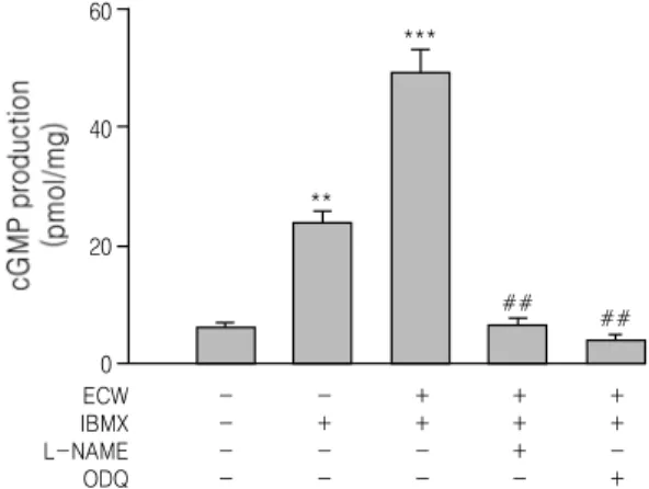
We investigated the effect of ECW on the production of cGMP in rat thoracic aorta. As shown in Fig. 2, IBMX (1 mM) significantly increased cGMP production in endothelial-intact thoracic aorta and treatment of 1 mg/ml ECW significantly increased cGMP production in endothelial-intact thoracic aorta pretreated with 1 mM IBMX.
Pretreatment of 10 μM ODQ or 10 μM L-NAME blocked the ECW-induced increase in cGMP accumulation in endothelial-intact thoracic aorta pretreated with 1 mM IBMX.
3. Effect of ECW on MMP-2 expression in HASMC
MTT assay determined that 1-500 μg/ml ECW had no adverse effect on cell viability (Fig. 3).
ECW IBMX L-NAME ODQ
- - - -
- + - -
+ + - -
+ + + -
+ + - + 60
40
20
0
**
***
## ##
Fig. 2. Effect of ECW on cGMP production in endothelium intact rat thoracic aorta.
ODQ or L-NAME (10 μM) were preincubated with 1 mg/ml in endothelial-intact thoracic aorta pretreated with 1 mM IBMX. Values are expressed as mean ± S.D. (n=4); **p<0.01, ***p<0.001 vs. Control; ##p<0.01, vs. HFCD.
120
100
80
60
40
20
0 Cont 1 10 100 500
BA (μM)
Fig. 3. Effect of ECW on HASMC viability.
Values are expressed as mean ± S.D. (n=6).
MMP-2 was also measured in HASMC using Western blot and zymography. In Western blot (Fig.
4A) expression of MMP-2 in HASMC was increased by treatment with 10 ng/ml TNF-α for 24 h.
Incubation of ECW attenuated the TNF-α-induced increase in MMP-2 expression in a dose-dependent manner in HASMC (Fig. 4A). In zymography, secretion of MMP-2 from HASMC into the culture medium was increased by treatment with TNF-α.
Incubation of ECW attenuated the TNF-α-induced increase in MMP-2 secretion in a dose-dependent
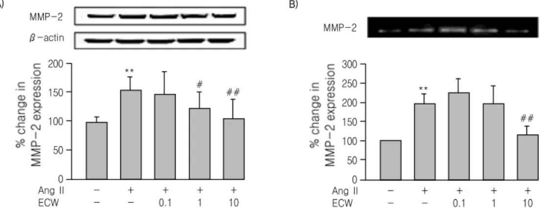
manner (Fig. 4B). Similarly, treatment of HASMC with 1 μM Angiotensin II (Ang II) for 24 h significantly increased expression and secretion of MMP-2 as determined using Western blot (Fig. 5A) and zymography (Fig. 5B). Interestingly, ECW treatment of HASMC attenuated the Ang II-induced increase in MMP-2 expression and secretion into culture medium.
4. Anti-oxidant effect of ECW in cultured HASMC
TNF-α ECW
- -
+ -
+ 0.1
+ 1
+ 10 300
250 200 150 100 50 0
**
#
##
A)
MMP-2 β-actin
TNF-α ECW
- -
+ -
+ 0.1
+ 1
+ 10 300
250 200 150 100 50 0
**
##
B)
MMP-2
## ##
Fig. 4. Effect of ECW on TNF-α-induced MMP-2 expression in cell lysates (A) and conditioned medium (B) of HASMC.
Representative zymography analysis and quantifications are shown. Values are expressed as a percentage of the density of blot (mean ± S.D.); **p<0.01, vs. Control; ##p<0.01, #p<0.05, vs. TNF-α.
Ang II ECW
- -
+ -
+ 0.1
+ 1
+ 10 200
150
100
50
0
**
# ##
A)
MMP-2 β-actin
Ang II ECW
- -
+ -
+ 0.1
+ 1
+ 10 300
250 200 150 100 50 0
**
##
B)
MMP-2
Fig. 5. Effect of ECW on Ang Ⅱ-induced MMP-2 expression in cell lysates (A) and conditioned medium (B) of HASMC.
Representative zymography analysis and quantifications are shown. Values are expressed as a percentage of the density of blot (mean ± S.D.); **p<0.01, vs. Control; ##p<0.01, #p<0.05, vs. Ang II.
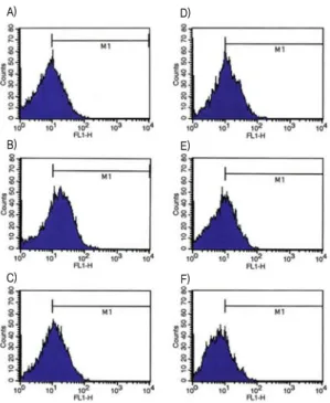
Stimulation of HASMC with 10 ng/ml TNF-α produced the highest levels of ROS production (data not shown). Incubation of HASMC with ECW reduced TNF-α-induced increase in ROS production in a dose-dependent manner (Figs. 6D-F). Also, n-acetylcystein (NAC) (5 mM) as positive control reduced the TNF-α-induced increase in ROS production (Fig. 6C).
5. In vitro DPPH scavenging activity of ECW
Treatment with ECW (Fig. 7A) had a dose- dependent scavenging activity by quenching DPPH radicals, as compared with Vitamin C as a positive control (Fig. 7B). The IC50 values (defined as the concentration of test compound required to produce 50% inhibition) for DPPH scavenging activity of ECW and ascorbic acid were 288.08 μg/mL and 9.83 μM, respectively.
A)
B)
C)
D)
E)
F)
Fig. 6. Effect of ECW on TNF-α-induced ROS production in HASMC.
Cells were incubated with various drugs for 24 h, (A) No TNF-α, (B) TNF-α (10 ng/ml), (C) TNF-α (10 ng/ml) + NAC (5 mM), (D) TNF-α (10 ng/ml) + ECW (0.1 μg/ml), (E) TNF-α (10 ng/ml) + ECW (1 μg/ml), (F) TNF-α (10 ng/ml) + ECW (10 μg/ml).
100
80
60
40
20
0
0.01 0.1 1 10 100 Vit. C (μM) 0.01 1 10 100 1000
ECW (μg/ml) 100
80
60
40
20
0
A) B)
Fig. 7. Effect of ECW (A) and Vitamin C (B) on DPPH radical scavenging in vitro.
Values are expressed as mean ± S.D. (n=6).
Discussion
In the present study, the cumulative treatment of ECW produced a dose-dependent and endothelium-
dependent vasorelaxation in rat aortic SMCs.
ECW-induced vasorelaxation was abolished in aortic rings that lacked endothelium. It is well-known that vascular endothelium produces NO, which is an
important regulating factor controlling vascular tone.
Endothelium-derived, NO-mediated activation of soluble GC (sGC) in vascular SMCs catalyzes the formation of the second messenger cGMP, leading to vasorelaxation3-5). In the present study, we found that NOS inhibition by pretreatment with L-NAME significantly inhibited ECW-induced vascular relaxation. Also, sGC inhibition by pretreatment with ODQ decreased ECW-induced vasorelaxation.
Furthermore, treatment of ECW increased the cGMP production in aortic smooth muscle, and pretreatment with ODQ and L-NAME significantly blocked the ECW-induced increase in cGMP production. These results suggest that ECW induces vascular relaxation through endothelium-derived NO/cGMP signaling pathway in thoracic aorta of rats.
Presently, TNF-α, which is a major pro- inflammatory cytokine, significantly evoked production of ROS in HASMC. ECW treatment of HASMC reduced the TNF-α-induced increase of ROS production in a dose-dependent manner. In addition, ECW exhibited a concentration-dependent DPPH scavenging activity in vitro. Free radicals and ROS are generated by all aerobic cells and have been shown to participate in many deleterious reactions, in particular, reduced formation of eNOS and inactivation of NO17,18) and increased oxidative stress19). Endogenous NO plays an important role in the regulation of blood pressure by maintaining vascular smooth muscle tone in a partially relaxed state and inhibiting platelet aggregation20). During hypertension, the endogenous vasodilatory effect of NO is inhibited due to interaction with ROS, specifically O2
-, resulting in increased vascular resistance and elevation of blood pressure21-23). It is well-known that antioxidant treatment has beneficial effects on NO metabolism and the pathogenesis observed in Ang II-induced hypertension24). Thus, ECW-induced anti-oxidant activity could improve vascular function by recovering a bioavailability of NO impaired by ROS.
MMPs are a family of membrane bound and extracellular proteinases that promote inflammation through a degradation of all ECM protein as well as several non-ECM proteins. Presently, we observed that the expression of MMP-2 was increased by TNF-α and Ang II in HASMC. Interestingly, incubation with ECW dose-dependently lowered this increased MMP-2 expression. Many studies have reported that ROS such as H2O2 and O2
- upregulate MMP-2 expression in several tissues. Presently, TNF-α in HASMC increased ROS production. Ang II activates the vascular NAD(P)H oxidase(s) resulting in the production of ROS including O2
- and H2O2
25). Our findings that ECW significantly inhibited an increase in TNF-α-induced ROS production and has DPPH scavenging activity in vitro suggests that ECW can downregulate an increase in MMP-2 expression induced by TNF-α and Ang II via anti-oxidant activity.
Considerable evidence suggests that over- expression of MMPs cannot only promote local inflammation by facilitating the infiltration of inflammatory cells, but can also lead to enhanced neointima formation by stimulating SMC migration and proliferation26,27). Inhibition of the expression and activity of MMPs can decrease lesion formation by inhibiting SMC migration28). Salvianolic acid B from Salvia miltiorrhiza inhibits TNF-α-induced MMP-2 upregulation in HASMC via suppression of NAD(P)H oxidase-derived ROS29). Also, it has been reported that an extract of Liuwei Dihuang has a protective effect on early diabetic nephropathy induced by streptozotocin via modulation of endothelin-ROS axis and MMP activity in rats30). Therefore, an anti-oxidant-mediated anti-inflammatory activity of ECW could be beneficial in the inhibition of vascular dysfunction associated with vascular inflmmation, atherosclerosis. In conclusion, our results have provided us with a better understanding of ECW in vascular relaxation and anti- inflammation.
Acknowledgement
This research was supported by a grant of Wonkwang University (2009).
References
1. Ross R. Atherosclerosis: an inflammatory disease. N Engl J Med. 1999;340:115-26.
2. Ignarro LJ, Buga GM, Wood KS, Byrns RE, Chaudhuri G. Endothelium-derived relaxing factor produced and released from artery and vein is nitric oxide. Proc Natl Acad Sci USA.
1987;84:9265-9.
3. Cai H, Harrison DG. Endothelial dysfunction in cardiovascular diseases: the role of oxidant stress. Circ Res. 2000;87:840-4.
4. Creager MA, Luscher TF, Cosentino F, Beckman JA. Diabetes and vascular disease: pathophy- siology, clinical consequences, and medical therapy: Part I. Circulation. 2003;108:1527-32.
5. Piatti P, Di Mario C, Monti LD, Fragasso G, Sgura F, Caumo A, et al. Association of insulin resistance, hyperleptinemia, and impaired nitric oxide release with in-stent restenosis in patients undergoing coronary stenting. Circulation.
2003;108:2074-81.
6. Werner E, Werb Z. Integrins engage mitochondrial function for signal transduction by a mechanism dependent on Rho GTPases. J Cell Biol. 2002;158:357-68.
7. Ra HJ, Parks WC. Control of matrix metalloproteinase catalytic activity. Matrix Biol.
2007;26:587-96.
8. Connor KM, Hempel N, Nelson KK, Dabiri G, Gamarra A, Belarmino J, et al. Manganese superoxide dismutase enhances the invasive and migratory activity of tumor cells. Cancer Res.
2007;67:10260-7.
9. Shan L, Liu RH, Shen YH, Zhang C, Wu DZ, Min L, et al. Gastroprotective effect of a traditional Chinese herbal drug “baishouwu” on experimental gastric lesions in rats. J Ethnopharmacol. 2006;107:389-94.
10. Lee MK, Yeo H, Kim J, Markelonis GH, Oh TH, Kim YC. Cynandione A from cynanchum wifordii protects cultured cortical neurons from toxicity induced by H2O2, L-glutamate, and kainite. J Neurosci Res. 2000;59:259-64.
11. Hwang BY, Kim SE, Kim YH, Kim HS, Hong YS, Ro JS, et al. Pregnane glycoside multidrug- resistance modulators from Cynanchum wilfordii.
J Nat Prod. 1999;62:640-3.
12. Furchgott RF, Zawadzki JV. The obligatory role of endothelial cells in the relaxation of arterial smooth muscle by acetylcholine. Nature.
1980;288:373-6.
13. Kim SZ, Kim SH, Park JK, Koh GY, Cho KW.
Presence and biological activity of C-type natriuretic peptide-dependent guanylate cyclase-coupled receptor in the penile corpus cavernosum. J Urol. 1998;159:1741-6.
14. Herron GS, Banda MJ, Clark EJ, Gavrilovic J, Werb Z. Secretion of metalloproteinases by stimulated capillary endothelial cells. Expression of collagenase and stromelysin activities is regulated by endogenous inhibitors. J Biol Chem.
1986;261:2814-8.
15. Hatano T, Kagawa H, Yasuhara T, Okuda T.
Two new flavonoids and other constituents in licorice root: their relative astringency and radical scavenging effects. Chem Pharm Bull.
1988;36:1090-2097.
16. Liu B, Bhat M, Nagaraj RH. αB-crystallin inhibits glucose-induced apoptosis in vascular endothelial cells. Biochem Biophys Res Commun. 2004;321:254-8.
17. Liu Y, Liu T, McCarron RM, Spatz M,
Feuerstein G, Hallenbeck JM, et al. Evidence for activation of endothelium and monocytes in hypertensive rats. Am J Physiol. 1996;270:
2125-31.
18. Ross R. Mechanism of disease: atherosclerosis:
an inflammatory disease. N Engl J Med.
1999;340:115-26.
19. Allen RG, Tresini M. Oxidative stress and gene regulation. Free Radic Biol Med. 1999;28 :463-99.
20. Moncada S, Palmer RMJ, Higgs EA. The discovery of nitric oxide as the endogenous nitrovasodilator. Hypertension. 1998;12:365-72.
21. Rajagopalan S, Kurx S, Munzel T, Freemaan BA, Griendling KK, Harrison DG. Angiotensin II-mediated hypertension in the rat increases vascular superoxide production via membrane NADH/NADPH oxidase activation: contribution to alteration of vasomotor tone. J Clin Invest.
1996;95:588-93.
22. Somers MJ, Mavromatis K, Galis ZS, Harrison DG. Vascular superoxide production and vasomotor function in hypertension induced by deoxycorticosterone acetate-salt. Circulation.
2000;101:1722-8.
23. Vaziri ND, Wang XQ, Oveisi F, Rad B.
Induction of oxidative stress by glutathione depletion causes severe hypertension in normal rats. Hypertension. 2000;36:142-6.
24. Muller DN, Dechend R, Mervaala E, Park J, Schmidt F, Fiebler A, et al. NF-κB inhibition ameliorates angiotensin II-induced inflammatory
damage in rats. Hypertension. 2000;35:193-201.
25. Hanna IR, Taniyama Y, Szocs K, Griendling KK.
NAD(P)H oxidase-derived reactive oxygen species as mediators of angiotensin II signaling.
Antioxid Redox Signal. 2002;4:899-914.
26. Lijnen HR, Soloway P, Collen D. Tissue inhibitor of matrix metalloproteinases-1 impairs arterial neointima formation after vascular injury in mice. Circulation Res. 1999;85:1186-91.
27. Mason DP, Kenagy RD, Hasenstab D, Bowen-Pope DF, Seifert RA, Coats S, et al.
Matrix metalloproteinase-9 overexpression enhances vascular smooth muscle cell migration and alters remodeling in the injured rat carotid artery. Circulation Res. 1999;85:1179-85.
28. Islam MM, Franco CD, Courtman DW, Bendeck MP. A nonantibiotic chemically modified tetracycline (CMT-3) inhibits intimal thickening.
Am J Pathol. 2003;163:1557-66.
29. Zhang HS, Wang SQ. Salvianolic acid B from saliva miltiorrhiza inhibits tumor necrosis factor-alpha (TNF-alpha)-induced MMP-2 upregulation in human aortic smooth muscle cells via suppression of NAD(P)H oxidase- derived reactive oxygen species. J Mol Cell Cardiol. 2006;41:138-48.
30. He H, Yang X, Zeng X, Shi M, Yang J, Wu L, et al. Protective effect of Liuwei Dihuang decoction on early diabetic nephropathy induced by streptozotocin via modulating ET-ROS axis and matrix metalloproteinase activity in rats. J Pharm Pharmacol. 2007;59:1297-305.