pISSN: 0378-6471 eISSN: 2092-9374 http://dx.doi.org/10.3341/jkos.2012.53.12.1807
= 증례보고 =
근시성 맥락막 신생혈관 환자에서 유리체강내 베바시주맙 주입방법에 따른 효과와 예후인자
임은혜⋅장윤수⋅유영주⋅유수진 건양대학교 김안과병원 안과학교실 명곡안연구소
목적: 근시성 맥락막신생혈관에서 유리체강내 베바시주맙 주입술 3회 연속 시행한 군(fixed regimen군)과 1회 주입 후 추가 주입이 이뤄진 군(PRN군)의 결과를 비교하고, 예후와 관련 있는 인자를 알아보고자 하였다.
대상과 방법: 2007년 1월부터 2009년 12월까지 유리체강내 베바시주맙 주입술을 시행 받은 fixed regimen군 14안, PRN군 9안을 대상 으로 시술 전과 1년 뒤의 최대교정시력, 중심황반두께, 신생혈관의 크기와 주입횟수 등을 조사하였다.
결과: 두 군 모두 시술 후 유의한 시력과 중심황반두께 호전을 나타냈다. 평균 주입횟수(p=0.16), 1년 후의 최대교정시력(p=0.83), 중심 황반두께(p=0.38) 모두 두 군 간 유의한 차이는 없었다. 시술 전 요인 중 나이가 시력 예후와 유의한 상관관계가 있는 것으로 나타났다 (p=0.01).
결론: 근시성 맥락막신생혈관의 유리체강내 베바시주맙 주입술에 있어 1회 주입 후 추가 치료를 고려하는 것도 3회 주입과 대등한 치료 효과를 얻을 수 있을 것으로 생각한다.
<대한안과학회지 2012;53(12):1807-1813>
■ 접 수 일: 2012년 5월 8일 ■ 심사통과일: 2012년 7월 22일
■ 게재허가일: 2012년 11월 13일
■ 책 임 저 자: 유 영 주
서울특별시 영등포구 영신로 136 김안과병원 안과
Tel: 1577-2639, Fax: 02-2671-6359 E-mail: [email protected]
* 이 논문의 요지는 2011년 대한안과학회 제105회 학술대회에서 구연으로 발표되었음.
병적 고도근시는 굴절력 -6.0디옵터, 안축장이 26 mm 이상으로 후극부의 점진적 맥락망막변성을 특징으로 한다.
근시성 맥락막신생혈관은 병적 고도근시 환자의 5-10%에 서 발병하며 갑작스러운 시력저하를 유발하는 가장 흔한 합병증이다.1,2 또한 병적 고도근시는 아시아 인종에서 더 흔하게 나타나며, 근시성 맥락막신생혈관은 질병 발생 10 년 후 96.3%에서 20/200 또는 그 이하의 시력을 보여 장기 간 시력예후가 불량한 것으로 알려졌다.2,3
이러한 근시성 맥락막신생혈관의 치료로 Verteporfin 광역학 치료는 2년 이상 장기간 치료 효과가 없는 것으로 나타났고, 레이저 광응고술 역시 광범위한 반흔과 암점을 남긴다는 단점이 있다.4,5 나이관련황반변성에 의한 맥락 막신생혈관에서 anti-VEGF의 치료효과가 보고되었기 에 근시성 맥락막신생혈관에서도 anti-VEGF 치료가 시 행되었고, 그 좋은 결과가 보고되고 있다.6-11현재근시 성 맥락막신생혈관의 치료로 anti-VEGF 중 Bevacizumab
(AVASTIN®, Genentech Inc., San Francisco, CA, USA) 이 널리 사용되고 있는데, 유리체강내 Bevacizumab 주입 술의 이상적인 용량과 초기 주입 횟수 등은 아직 논란의 여지가 있다.
이에 본 연구에서는 국내에서는 아직 유리체강내 Bevacizumab 주입술 용량과 용법에 대한 연구 결과가 발 표되지 않았기에, 1개월 간격으로 3회 주입술을 시행한 군 과, 1회 주입술 시행 후 상태에 따른 추가 주입을 고려한 군 의 치료 결과를 비교하고, 더불어 주입술의 치료효과를 예 측할 수 있는 인자를 알아보고자 하였다.
대상과 방법
2007년 1월부터 2009년 12월까지 본원에 내원하여 근시 성 맥락막신생혈관 진단을 받고 유리체강내 Bevacizumab 주입술을 시행 받은 후 1년 이상 추적관찰이 가능했던 21 명 23안의 의무기록을 후향적으로 분석하였다.
구면수치대응치로 -6디옵터 이상, 또는 후극부에 라커 크랙, 근시성맥락망막 위축, 후극포도종 등의 근시성 망막변 화가 있으며 안저검사에서 중심와하 또는 중심와곁 맥락막신 생혈관이 있고, 형광안저촬영상 형광 누출이 있는 환자를 근 시성 맥락막신생혈관이라 정의하고 유리체강내 Bevacizumab 주입술을 시행하였다. 매체 혼탁이 있거나 중심와외 맥락막 신생혈관, 연령관련황반변성, 당뇨망막병증, 외상, 맥락망
Table 1. Demographics
Fixed regimen (14 eyes) PRN (9 eyes) p-value*
Age (yrs) 49.4 ± 16.3 47.4 ± 14.1 0.38
Sex (M/F) 2/12 2/7
SE (D) -9.0 ± 6.0 -10.4 ± 3.0 0.97
BCVA (log MAR) 0.8 ± 0.5 0.6 ± 0.6 0.30
Baseline CMT (μm) 233.0 ± 48.1 244.5 ± 58.2 0.67
CNV size (mm) 1.31 ± 0.75 1.11 ± 0.28 0.92
Total injection time (Sec) 3.4 ± 0.9 1.5 ± 0.7 0.16
Subfoveal 8 6 0.85
Location of CNV (Number of eyes) Juxtafoveal 6 3 0.88
Values are presented as mean ± SD or number.
SE = spherical equivalent; BCVA = best-corrected visual acuity; CMT = central macular thickness.
*Mann-Whitney test.
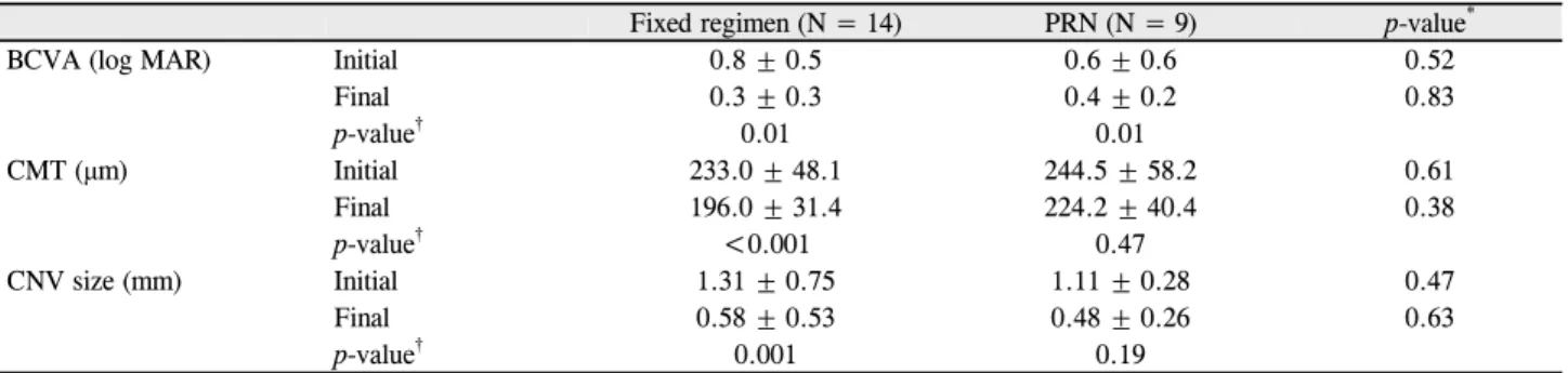
Table 2. Comparison of resulting parameters after IVB (over 1 year)
Fixed regimen (N = 14) PRN (N = 9) p-value*
BCVA (log MAR) Initial 0.8 ± 0.5 0.6 ± 0.6 0.52
Final 0.3 ± 0.3 0.4 ± 0.2 0.83
p-value† 0.01 0.01
CMT (μm) Initial 233.0 ± 48.1 244.5 ± 58.2 0.61
Final 196.0 ± 31.4 224.2 ± 40.4 0.38
p-value† <0.001 0.47
CNV size (mm) Initial 1.31 ± 0.75 1.11 ± 0.28 0.47
Final 0.58 ± 0.53 0.48 ± 0.26 0.63
p-value† 0.001 0.19
Values are presented as mean ± SD.
IVB = Intravitreal Bevacizumab injection; BCVA = best-corrected visual acuity; CMT = central macular thickness.
*Mann-Whitney test; †Paired t-test.
막염 등 기타 망막 질환이 있는 경우, 맥락막신생혈관에 대 하여 다른 치료를 시행받은 과거력이 있는 경우는 연구에 서 제외하였다.
모든 환자에게서 시술 전, 최종 내원 시 최대교정시력과 중심황반두께, 맥락막신생혈관의 크기를 측정하였고, 시술 전 신생혈관의 위치, 장액망막박리 유무, 망막하출혈 유무 와 시행받은 주입술의 총 횟수를 조사하였다. 시력은 스넬 렌 차트를 이용하여 측정한 후 logMAR (logarithm of the minimal angle of resolution)로 변환하였고 빛간섭단층촬 영과 형광안저촬영을 바탕으로 황반두께와 맥락막신생혈관 의 크기를 측정하였다. 연구 진행 중 OCT 기종이 바뀌게 되어 Stratus OCT (Carl Zeiss Meditec, USA)로 측정한 중심망막두께는 40 μm를 더하여 SD-SLO/OCT (OTI, Toronto, Canada)로 측정한 중심망막두께와 비교하였다.12
1개월 간격 3회 연속 시행한 군(fixed regimen group) 14안, 1회 주입 후 추가 주입술을 시행한 군(PRN group) 9안이 포함되었는데, 첫 번째 군은 1개월 간격으로 1.25 mg bevacizumab을 3회 주사하였고, 두 번째 군은 1.25 mg bevacizumab 1회 주사한 후 추적관찰 중 맥락막 신생혈관 의 활동성이 의심되는 경우 형광안저촬영과 빛간섭단층촬
영을 시행하여 추가 주입술을 고려하였다.
일차 결과로 두 군의 최대교정시력, 중심황반두께와 맥 락막신생혈관 크기를 SPSS version 18.0 프로그램을 이용 하여 통계적 분석을 시행하여 두 군 간 결과를 비교하였고, 이차 결과로 예후에 영향을 주는 인자를 알아보았다.
결 과
총 21명 23안 중 fixed regimen군 14안, PRN군 9안이 대상이 되었고 fixed regimen군의 평균나이는 49.4 ±16.3 세, PRN군은 47.4 ± 14.1세였다. 구면수치대응치, 술 전 최대교정시력, 중심황반두께, 맥락막신생혈관의 크기와 신 생혈관의 위치 등은 두 군간 통계학적으로 유의한 차이를 보이지 않았다. 총 주입 횟수는 fixed regimen군(3.4 ±0.9 회)에서 PRN군(1.5 ±0.7) 보다 많은 횟수를 주입한 것으 로 나타났으나, 통계학적으로 유의하지는 않았다(p=0.16) (Table 1).
시술 전 최대 교정시력은 fixed regimen군에서 0.8 ± 0.5, PRN군에서 0.6 ± 0.6였고, 최종 내원 시 두 군 각각 0.3 ±0.3, 0.4 ±0.2로 향상되어, 두 군 모두 통계학적으
A D
B E
C F
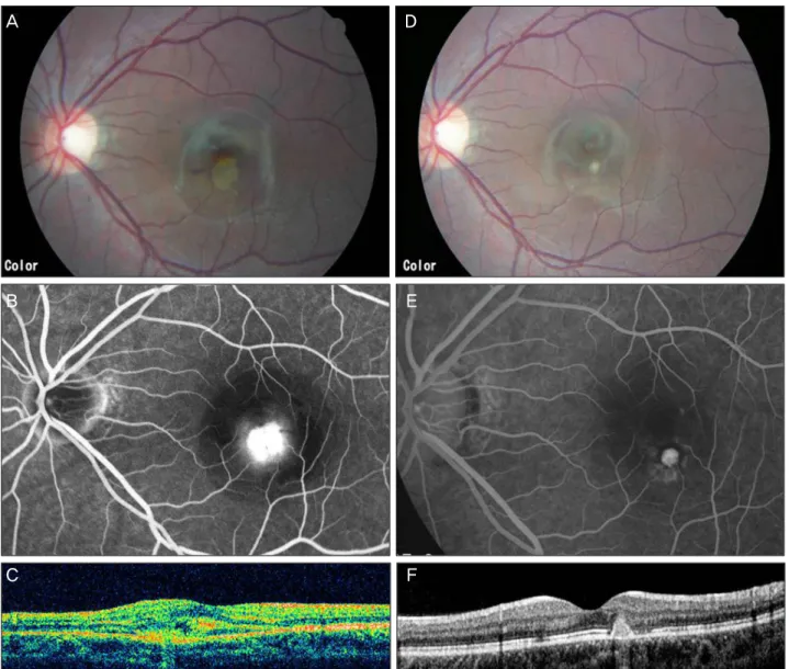
Figure 1. Left eye of 19-year-old woman in fixed regimen group treated by IVB. The refractive error was -8.5 D and initial best-cor-
rected visual acuity was 20/50. (A) Initial fundus photograph of the patients. (B) Early phase of fluorescein angiogram showing a classic juxtafoveal CNV with leakage. (C) Optical coherence tomography findings of the macula demonstrating a juxtafoveal CNV with retinal thickening and intraretinal fluid. (D) At 1 year after treatment, the patient’s visual acuity improved to 20/25. (E) Late phases of fluorescein angiogram shows staining of the CNV with no evidence of leakage. (F) Post-treatment OCT shows formation of a scar with resolution of the intraretinal fluid.로 유의한 최대교정시력의 향상을 가져왔으나(p=0.01) (Fig. 1, 2), 최종 내원 시 최대교정시력을 비교했을 때 두 군 간의 유의한 차이를 나타내지 못했다(p=0.83) (Table 2).
중심황반두께는 시술 전 fixed regimen군에서 233.0 ± 48.1 μm, PRN군에서 244.5 ±58.2 μm였고, 최종 내원 시 각각 196.0 ±31.4 μm, 224.2 ±40.4 μm로 호전되었는데, fixed regimen군의 경우 시술 전에 비해 유의하게 호전되 었으나(p<0.001) (Fig. 1), PRN 군에서는 그 호전정도가 통계학적으로 유의하지 않았다(p=0.47) (Fig. 2). 또한 최 종 내원 시 두 군의 차이는 통계학적으로 유의하지 않았다 (p=0.44) (Table 2).
근시성 맥락막신생혈관의 크기를 살펴보면, 시술 전 fixed regimen군은 1.31 ± 0.75 mm, PRN군은 1.11 ± 0.28 mm로 측정되었고, 최종 내원 시 두 군 각각 0.58 ± 0.53 mm, 0.48 ±0.26 mm로 그 크기가 감소하였다. 그러 나 fixed regimen군의 경우 통계학적으로 유의하게 감소하 였으나(p=0.001), PRN군에서는 유의하지 않았으며, 최종 내원 시 두 군 간 비교에서도 통계학적인 유의성은 없었다 (p=0.19) (Table 2) (Fig. 1, 2).
이차 결과로 bevacizumab 주입술의 치료 효과에 영향을 주는 요인을 알아보았는데, 여러가지 인자들 가운데 나이가 많을수록 최종 최대교정시력이 좋지 않았던 것으로 나타났
A D
B E
C F
Figure 2. Left eye of 29-year-old man in PRN group treated by IVB. The refractive error was -7 D and initial best-corrected visual
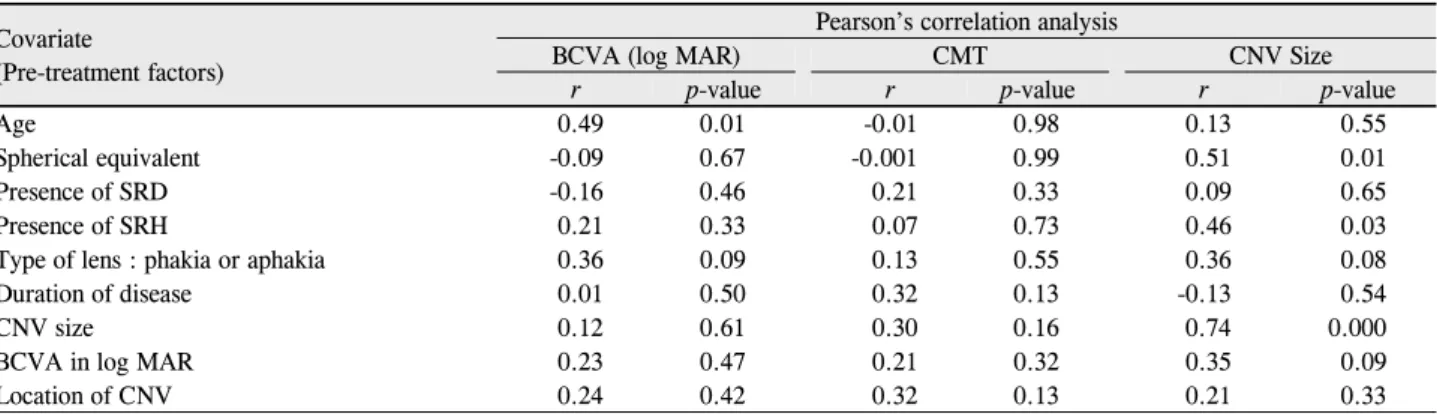
acuity was 20/30. He received only one intravitreal bevacizumab injection. (A) Initial fundus photograph showing subfoveal hemorrhage. (B) Early phase of fluorescein angiogram showing a juxtafoveal CNV with leakage. (C) Optical coherence tomography of the macula demonstrating a juxtafoveal CNV with retinal thickening and intraretinal fluid. (D) At 1 year after treatment, the pa- tient’s visual acuity improved to 20/25. (E) Early phases of fluorescein angiogram shows staining of the CNV slightly decreased in size. (F) Post-treatment OCT shows resolution of the intraretinal fluid.Table 3. Correlation analysis to access the influence of each pre-treatment factor at 1 year after IVB for mCNV
Covariate(Pre-treatment factors)
Pearson’s correlation analysis
BCVA (log MAR) CMT CNV Size
r p-value r p-value r p-value
Age 0.49 0.01 -0.01 0.98 0.13 0.55
Spherical equivalent -0.09 0.67 -0.001 0.99 0.51 0.01
Presence of SRD -0.16 0.46 0.21 0.33 0.09 0.65
Presence of SRH 0.21 0.33 0.07 0.73 0.46 0.03
Type of lens : phakia or aphakia 0.36 0.09 0.13 0.55 0.36 0.08
Duration of disease 0.01 0.50 0.32 0.13 -0.13 0.54
CNV size 0.12 0.61 0.30 0.16 0.74 0.000
BCVA in log MAR 0.23 0.47 0.21 0.32 0.35 0.09
Location of CNV 0.24 0.42 0.32 0.13 0.21 0.33
고(p=0.01), 구면수치 대응치가 작을수록 최종 맥락막 신 생혈관의 크기가 작았던 것으로 조사되었다(p=0.01) (Table 3).
고 찰
근시성 맥락막신생혈관의 치료로 레이저 광응고술, Verteporfin
광역학치료, 황반하 수술, 유리체강내 anti-VEGF 주입술 등이 고려될 수 있다. 레이저 광응고술의 경우 치료가 성공 적이라 하더라도, 중심와곁 맥락막신생혈관의 경우 레이저 반흔이 시간이 경과하면서 커지는 경향을 보이기 때문에 시력호전에는 도움이 안 된다는 단점이 있으며 중심와밑 맥락막신생혈관의 치료로는 사용될 수 없다는 한계점을 갖 는다.5,13 광역학 치료의 경우를 보면 Verteporfin in pho- todynamic therapy (VIP) study group이 12개월을 추적 관찰하여 대조군과 비교한 연구에서는 통계적으로 유의한 시력의 호전이 있었으나, 24개월까지 그 효과가 유지되지 않는다는 또 다른 결과가 보고되어, 광역학 치료의 장기간 효과는 의문점으로 남아있다.4,14한편 국내 연구에는 치료 후 6개월 간의 추적관찰 결과 Verteporfin 광역학치료는 근 시성 맥락막신생혈관의 환자에게 병변 안정화 및 안정화에 효과가 있었다고 발표하였다.15 이렇듯 광역학 치료 이후 장기간의 시력의 유지가 어려운 것은 치료 후 광범위한 황 반 위축이 진행되기 때문인 것으로 여겨진다. 황반하 수술 의 경우 다양한 결과들이 보고되고 있는데, Ruiz-Moreno and de la Vega16은 수술 후에도 유의한 시력 변화는 없었 다고 보고하였고, Uemura and Thomas17은 수술 후 맥락막 신생혈관이 재발되고, 망막색소상피의 위축이 올 수 있다고 하였으며, Bottoni et al18은 45%에서 시력호전이 있었던 성공적인 결과를 보고한 바 있다.
근시성 맥락막신생혈관의 치료에서 bevacizumab의 용 량, 주입횟수와 추적관찰기간 등에서 매우 다양한 연구들이 시행되었다.19-23Gharbiya et al9과 Chan et al22은 1.25 mg 용량으로 매달 3회 주입하여 1년간의 추적관찰한 후 시력 호전을 나타냈다는 보고하였다. 또한 Wu and Chen23은 9 안의 근시성 맥락막신생혈관을 대상으로 한 연구에서 2.5 mg 단 1회 주입으로 평균 14.9개월의 경과 관찰 기간 동안 3줄 이상의 시력호전이 있었으며 단 1안에서 재발을 보고하 였다. 반면 Ruiz-Moreno and Montero19은 총 19안을 대상 으로 한 연구에서 1개월 간격으로 1.25 mg의 Bevacizumab 을3회 주입한 후 최대교정시력이 0.54 ±0.25에서 1년 후 0.40 ±0.35 (p=0.04)로 유의하게 호전되었으나 2년 후에 는 0.47 ±0.31 (p=0.20)로 유의한 호전은 없었다고 보고 하였다. 주입횟수에 관한 연구를 살펴보면, Ikuno et al10은 1 mg 1회 주사 후 악화 시 추가 주사를 고려하는 방식으로 치료하여 1년간 평균 2.4 ±1.4회 주사하여 총 63안에서 logMAR 시력 0.57 ±0.43에서 1년 후 0.33 ±0.34로 유 의한(p<0.01) 호전을 나타냈다고 하였다. Ruiz-Moreno et al20은 1.25 mg의 Bevacizumab을 1개월 간격 3회 주사 한 19안과, 1회 주사 후 추가 주사를 고려한 20안을 1년간 추적관찰하여 비교한 전향연구를 발표하였는데, 시력과 중
심황반두께의 호전에는 두 군 간 유의한 차이는 없었으나, 두번째 군에서 1.7 ± 1.2회 주입술을 받은 것으로 나타나 첫 번째 군(3.2 ± 0.4회)과 유의한 차이를 보였으며(p= 0.00), 1년의 경과관찰 기간 동안 첫 번째 군에서는 재발한 경우가 4안이었던 것에 비해, 두 번째 군에서는 15안으로 유의한 차이가 있었다고 발표하였다(p=0.001).
국내에서는 Kim et al11은 20안을 대상으로 하여 2.5 mg 1회 주사 후 1년간 경과 관찰을 하는 후향연구를 하였으며 경과관찰 기간 동안 최대교정시력과 중심황반두께 모두 유 의한 호전을 보였으며 첫 주사 후 뚜렷한 효과가 없을 경우 재주사를 시행하여 1년간 평균 1.5회의 추가 치료를 시행 하였다. 반면 Seo and Chang24은 6안을 대상으로 2.5 mg의 bevacizumab 1회 주사를 시행하여 2년 동안 시술전과 비교 하여 통계학적으로 유의한 시력 호전을 유지하였다.
본 연구에서는 1.25 mg의 용량으로 초기 주입 횟수를 다 르게 한 두 군의 결과를 비교하였으며 이전의 연구들과 큰 차이는 없는 결과를 얻었다. Fixed regimen군에서 1년간의 경과관찰 기간 동안 평균 3.4 ±0.9회로 1.5 ±0.7회를 주 입한 PRN군보다 많은 수의 주입술이 필요했으나, 통계적 인 유의성은 없었고(p=0.16), 치료 후 두 군 간 시력 호전 이나, 황반두께 호전, 맥락막 신생혈관의 크기 감소 등 유의 한 차이는 없는 것으로 나타나 1회 주입 후 환자의 상태에 따라 추가 주입을 고려하는 것도 3회 주입술과 대등한 치료 효과를 가져올 수 있을 것으로 생각한다. 다만 3회 주입술 을 시행한 경우 최대교정시력, 황반두께, 맥락막 신생혈관 크기 등이 모두 시술 전에 비해 유의한 호전을 나타내었다.
근시성 맥락막신생혈관 환자에서 치료를 받지 않고 5년 이상 경과 관찰을 한 경우 최종 시력이 20/40 이상인 경우 와 관련된 요인으로 나이가 젊을 경우, 신생혈관의 크기가 작을 경우 그리고 초기시력이 좋을수록 예후가 좋은 것으 로 보고되어 있다.25Nakanishi et al26는 bevacizumab 주사 후 2년간 경과 관찰하며 필요에 따른 추가 주사 치료 계획 을 한 결과 최종 시력에는 처음 신생혈관의 크기만이 통계 적으로 유의한 연관성(p=0.008)을 보여 주는 것을 발표하 였다. 반면 본 연구 결과 나이가 주사치료 후 최종 시력 예 후에 영향을 주는 것으로 나타났다.
따라서 초기 치료 계획의 결정은 환자의 경제 상태와 내 원 가능 기간 등의 환자 개인 요인을 고려하여 연속 주사를 하거나 1회 주사 후 경과 관찰을 할 수 있을 것으로 생각하 며 또한 본 연구 결과에서 보여주는 예후에 영향을 주는 요 인인 나이와 다른 연구에서 관련성 있는 인자로 밝혀진 맥 락막 신생혈관 크기, 최대교정시력 등의 인자를 고려하여 치료계획을 수립할 수 있을 것으로 판단된다.
본 연구 결과에서 보여 주듯이 1회 주입술로도 효과는 충
분할 수 있으나, 만약 추적관찰이 어려워 신생혈관의 활동 성 여부를 판단하기 어려운 경우 단 1회 주입만으로는 치료 가 부족하여 시력저하가 나타날 위험성이 있으며 실제로 본 연구에서도 PRN군 9안 중 4안에서 추가 주입술이 시행 되었다. 따라서 3회 주입술 또는 1회 주입술의 정확한 적응 증을 알기 위해서는 추후 좀더 많은 환자를 대상으로 한 장 기간의 전향적인 연구가 필요할 것으로 생각한다. 또한 본 연구에서는 주입 횟수에 따른 차이만을 보았는데 신생혈관 의 크기와 중심망막두께가 시술 전에 비해 유의한 호전을 보인 경우는 3회 연속 주입군이었으므로 1회 주사를 하는 경우 bevacizumab 용량에 따라서 주사 횟수를 줄이며 여러 번 주사와 대등한 치료효과가 있는 지를 확인하는 연구도 필요할 것이라고 생각한다.
참고문헌
1) Cohen SY, Laroche A, Leguen Y, et al. Etiology of choroidal neo- vascularization in young patients. Ophthalmology 1996;103:
1241-4.
2) Yoshida T, Ohno-Matsui K, Yasuzumi K, et al. Myopic choroidal neovascularization: a 10-year follow-up. Ophthalmology 2003;
110:1297-305.
3) McCarty CA, Livingston PM, Taylor HR. Prevalence of myopia in adults: implications for refractive surgeons. J Refract Surg 1997;13:229-34.
4) Blinder KJ, Blumenkranz MS, Bressler NM, et al. Verteporfin ther- apy of subfoveal choroidal neovascularization in pathologic my- opia: 2-year results of a randomized clinical trial--VIP report no. 3.
Ophthalmology 2003;110:667-73.
5) Brancato R, Pece A, Avanza P, Radrizzani E. Photocoagulation scar expansion after laser therapy for choroidal neovascularization in degenerative myopia. Retina 1990;10:239-43.
6) Spaide RF, Laud K, Fine HF, et al. Intravitreal bevacizumab treat- ment of choroidal neovascularization secondary to age-related macular degeneration. Retina 2006;26:383-90.
7) Rich RM, Rosenfeld PJ, Puliafito CA, et al. Short-term safety and efficacy of intravitreal bevacizumab (Avastin) for neovascular age-related macular degeneration. Retina 2006;26:495-511.
8) Bashshur ZF, Bazarbachi A, Schakal A, et al. Intravitreal bev- acizumab for the management of choroidal neovascularization in age-related macular degeneration. Am J Ophthalmol 2006;142:
1-9.
9) Gharbiya M, Allievi F, Mazzeo L, Gabrieli CB. Intravitreal bev- acizumab treatment for choroidal neovascularization in pathologic myopia: 12-month results. Am J Ophthalmol 2009;147:84-93. e1.
10) Ikuno Y, Sayanagi K, Soga K, et al. Intravitreal bevacizumab for choroidal neovascularization attributable to pathological myopia:
one-year results. Am J Ophthalmol 2009;147:94-100. e1.
11) Kim KH, Jung JH, Lee JE, Oum BS. Clinical effect of intravitreal bevacizumab injection in myopic choroidal neovascularization. J Korean Ophthalmol Soc 2010;51:359-65.
12) Forte R, Cennamo GL, Finelli ML, de Crecchio G. Comparison of time domain Stratus OCT and spectral domain SLO/OCT for as- sessment of macular thickness and volume. Eye (Lond) 2009;
23:2071-8.
13) Ruiz-Moreno JM, Montero JA. Long-term visual acuity after argon green laser photocoagulation of juxtafoveal choroidal neo- vascularization in highly myopic eyes. Eur J Ophthalmol 2002;12:
117-22.
14) Vip Study Group. Photodynamic therapy of subfoveal choroidal neovascularization in pathologic myopia with verteporfin: 1-year results of a randomized clinical trial-VIP report no. 1.
Ophthalmology 2001;108:841-52.
15) Chung EJ, Oh HS, Koh HJ, et al. Photodynamic therapy in prac- tice: A review of experiences with myopic CNV in Korean patients. J Korean Ophthalmol Soc 2005;46:664-70.
16) Ruiz-Moreno JM, de la Vega C. Surgical removal of subfoveal cho- roidal neovascularisation in highly myopic patients. Br J Ophthalmol 2001;85:1041-3.
17) Uemura A, Thomas MA. Subretinal surgery for choroidal neo- vascularization in patients with high myopia. Arch Ophthalmol 2000;118:344-50.
18) Bottoni F, Perego E, Airaghi P, et al. Surgical removal of subfoveal choroidal neovascular membranes in high myopia. Graefes Arch Clin Exp Ophthalmol 1999;237:573-82.
19) Ruiz-Moreno JM, Montero JA. Intravitreal bevacizumab to treat myopic choroidal neovascularization: 2-year outcome. Graefes Arch Clin Exp Ophthalmol 2010;248:937-41.
20) Ruiz-Moreno JM, Montero JA, Amat-Peral P. Myopic choroidal neovascularization treated by intravitreal bevacizumab: compar- ison of two different initial doses. Graefes Arch Clin Exp Ophthalmol 2011;249:595-9.
21) Sakaguchi H, Ikuno Y, Gomi F, et al. Intravitreal injection of bev- acizumab for choroidal neovascularisation associated with patho- logical myopia. Br J Ophthalmol 2007;91:161-5.
22) Chan WM, Lai TY, Liu DT, Lam DS. Intravitreal bevacizumab (Avastin) for myopic choroidal neovascularisation: 1-year results of a prospective pilot study. Br J Ophthalmol 2009;93:150-4.
23) Wu PC, Chen YJ. Intravitreal injection of bevacizumab for myopic choroidal neovascularization: 1-year follow-up. Eye (Lond) 2009;23:2042-5.
24) Seo YS, Chang MH. Long-term Therapeutic effect of Intravitreal Bevacizumab (Avastin) on Myopic Choroidal Neovascularization.
J Korean Ophthalmol Soc 2011;52:34-40.
25) Hayashi K, Ohno-Matsui K, Yoshida T. Characteristics of patients with a favorable natural course of myopic choroidal neovascularization.
Graefes Arch Clin Exp Ophthalmol 2005;243:13-9.
26) Nakanishi H, Tsujikawa A, Yodoi Y, et al. Prognostic factors for visual outcomes 2-years after intravitreal bevacizumab for myopic choroidal neovascularization. Eye (Lond) 2011;25:375-81.
=ABSTRACT=
Comparison of Two Doses of IVB and Prognostic Factor on Myopic CNV : 1-Year Outcome
Eun Hae Lim, MD, Yoon Soo Jang, MD, Young Ju Lew, MD, PhD, Su Jin Yoo, MD
Myung-Gok Eye Research Institute, Department of Ophthalmology, Kim’s Eye Hospital, Konyang University, Seoul, Korea
Purpose: To compare the efficacy of 3 consecutive monthly intravitreal bevacizumab (IVB) injections (fixed regimen group) with a single IVB injection (PRN group) on patients with myopic choroidal neovascularization (CNV) and to de- termine the prognostic factors associated with IVB injection outcomes.
Methods: 23 Twenty-three eyes in 21 patients with myopic CNV (14 eyes in the fixed regimen group and 9 eyes in the PRN group) treated with IVB were studied retrospectively. Best corrected visual acuity (BCVA), central macular thickness (CMT), the size of CNV prior to the initial IVB injection, CMT at the completion of treatment, and the number of IVB in- jections during the study period was measured.
Results: IVB resulted in improved BCVA and decreased CMT in both groups, and the differences before and after IVB in- jections were significantly correlated. Average injection time in the fixed regimen group and PRN group was 3.4 ± 0.9 and 1.5 ± 0.7 respectively, and was not statistically significant (p = 0.16). Differences between the groups in BCVA (p = 0.83) and CMT (p = 0.38) were not significantly correlated. Among the variables measured prior to IVB injection that affected fi- nal BCVA was age (p = 0.01).
Conclusions: In the present study, a single injection of IVB compared to 3 consecutive IVB injections in patients with my- opic CNV resulted in similar outcomes. In the future, these results can be considered as a reference when designing treat- ments for myopic CNV patients.
J Korean Ophthalmol Soc 2012;53(12):1807-1813
Key Words: Bevacizumab, Myopic choroidal neovascularization
Address reprint requests to Young Ju Lew, MD, PhD Department of Ophthalmology, Kim’s Eye Hospital
#136 Yeongsin-ro, Yeongdeungpo-gu, Seoul 150-902, Korea Tel: 82-1577-2639, Fax: 82-2-2671-6359, E-mail: [email protected]