Collagenase-induced Arthritis Rat Model에서 Thermal Hyperalgesia에 대한 電鍼의 鎭痛效果 및 기전연구:
Adrenergic Mechanism에 對한 硏究
서병관․박동석․백용현 경희대학교 한의과대학 침구학교실
목적 : Collagenase-induced osteoarthritis(OA) 동물 모델에서 전침의 adrenergic mechanism을 연구하고 자 한다.
방법 : Collagenase-induced arthritis(CIA)를 유발하기 위하여 5주령의 male Sprague-Dawley rat의 뒷다 리 좌측 무릎 관절에 0.05ml의 4mg/ml collagenase solution을 intra-articular 주입하고, 다시 4일 후에 같은 부위에 같은 농도의 collagenase solution을 intra-articular boosting injection 시행한 뒤, gross, histopathological features 및 biomarker activity 변화를 관찰하였다. 예비실험을 통하여 CIA rat model에서 진통효과를 발휘하는 것으로 확인한, 足三里 (ST36)에 대한 저빈도 train pulse EA stimulation (2Hz, 0.07 mA, 0.3ms)을 침치료 방법으로 적용하였다. 전침의 진통기전을 확인하기 위하여, α1-adrenergic antagonist (prazosin, 1 mg/kg, i.p.), α2-adrenergic receptor antagonist (yohimbine, 2mg/kg, i.p.), α1-adrenergic
1)
The Analgesic Effect and the Mechanism of Electroacupuncture on Thermal Hyperalgesia in the Rat Model of Collagenase-induced Arthritis:
Mediation by Adrenergic Receptors
Seo Byung-kwan, Park Dong-suk and Baek Yong-hyeon
Department of Acupuncture & Moxibustion, College of Oriental Medicine, Kyung Hee University
* This research was supported by Basic Science Research Program through the National Research Foundation of
Korea(NRF) funded by the Ministry of Education, Science and Technology(2009-0069462).
․Acceptance : 2011. 2. 23. ․Adjustment : 2011. 4. 3. ․Adoption : 2011. 4. 6.
․Corresponding author : Baek Yong-hyeon, Department of Acupuncture and Moxibustion, Kyung Hee University at Gangdong, #149, Sangil-Dong, Gangdong-Gu, Seoul, Republic of Korea 134-727.
Tel. 82-2-440-6224 E-mail : [email protected]
국문초록
Original Article
receptor agonist(phenylephrine, 2mg/kg, i.p.), α2-adrenergic receptor agonist(clonidine, 40μg/kg, i.p.)을 전 침시행 20분 전에 복강 내로 전처치하였다. Tail flick unit(Ugo Basile Model 7360)을 이용하여 열자극에 대 한 통증역치를 측정하였다.
결과 : 퇴행성관절염 징후(gross, histopathological features)와 통증역치의 변화가 최대값을 나타내는 CIA 유발 4주차에 저빈도 전침자극(train pulse, 2Hz, 0.07mA, 0.3ms)을 족삼리(ST36)에 적용하였으며, 족삼 리 전침의 진통효과는 α2-adrenergic receptor antagonist(yohimbine, 2mg/kg, i.p.)전처치에 의해 억제되었으 나, α1-adrenergic antagonist(prazosin, 1 mg/kg, i.p.)전처치에는 억제되지 않았다. 또 α2-adrenergic receptor agonist(clonidine, 40μg/kg, i.p.)의 전처치를 통하여 유의한 synergistic analgesic effect가 관찰되었 으나, α1-adrenergic receptor agonist(phenylephrine, 2mg/kg, i.p.)의 전처치는 전침의 진통효과에 synergistic effect를 미치지 않는 것으로 나타났다.
결론 : 저빈도 족삼리 전침은 CIA로 유발된 염증성 통증에 대하여 진통효과를 발휘하며, 이는 α2- adrenergic receptor에 의하여 매개되는 것으로 보이며 α1-adrenergic receptor는 영향을 미치지 않는 것으로 사료된다.
핵심 단어 : electroacupuncture, collagenase-induced arthritis, adrenergic receptor, antinociceptive mechanism
Ⅰ. Introduction
Osteoarthritis(OA) is a progressive disease that affects joint cartilage and causes pain and disability. With an alteration of tissue homeostasis, an imbalance of anabolic and catabolic processes consequently leads to the loss of articular cartil- age1,2). In the pathogenesis of OA, prostaglandin E2 (PGE2) plays an important role in joint inflam- mation, tissue destruction, and inflammatory pain3). PGE2 is associated with the breakdown of cartilage extracellular matrix via upregulation of metallo- proteinases(MMPs), bone resorption, osteophyte formation, and regulation of other proinflammatory cytokines, nitric oxide(NO), and connective tissue- degrading enzymes4,5). Prostagladin E2(PGE2) is produced via cyclooxygenase(COX) enzymes from arachidonic acid, following the stimulation of pro- inflammatory cytokines, such as interleukin (IL)-1β and tumor necrosis factor(TNF)-α, which are secreted from activated synoviocytes, mononuclear cells, or by articular cartilage itself and are related to the inflammatory process in OA6-8).
Previous researches have been elucidated thera-
peutic properties of various modalities of acupunc- ture which alleviate pain, reduce inflammation and pathogenesis of OA9-11). Electroacupuncture (EA) improves OA of knee either on its own or as an adjunctive therapy12), and acupuncture is significantly superior to non-penetrating sham acupuncture13). EA can treat more effectively OA, relieve joint pain and improve joint function than conventional treatment such as oral administration of Diclofenac Sodium and injection of hyaluronic acid11,14,15). And the therapeutic effect of EA on OA partially achieved by modulation of parameters such as IL-1beta, IL-6, TNF-α, PGE(2α) and MMP-315,16).
Repeated application of EA suppresses the peripheral inflammation response with acupoint specificity17-19), and the analgesic effect of EA was partially mediated by the descending modulation of nociceptive processing correlated with various neural substrates, such as adrenergic systems in the neuropathic pain model and ankle sprain model20-22).
The current study attempts to establish rat model of OA with pathophysiological similarity to human OA properties and investigate the antinoci- ceptive effect of EA and its adrenergic mechanisms.
Ⅱ. Materials and Methods
A. Subjects
Five-week-old male Sprague Dawley rats(Samtaco, Osan, Korea, 200g) were housed under specific pathogen-free conditions under controlled tempera- ture(22 ± 1℃), humidity(55 ± 5%), and a 12:12 light-dark cycle(light on 6:00 AM to 6:00 PM) with food and water available ad libitum. Rats were randomly assigned to each experimental group with the initial injection of collagenase. All of the experimental procedures were conducted according to National Institute of Health guide for the Care and Use of Laboratory Animals and the ethical guidelines of the International Association for the Study of Pain(IASP). For the adaptation to the restraint, the experimental rats were properly fitted into a plastic holder(5.3 × 15, 6.3 × 18 cm in diameter
× length) with their tails protruding outside for 60 min/day for 7 days. Adaptation was conducted before TFL assessment and EA treatment.
B. The induction of collagenase-induced arthritis rat model
After one week of adaptation to the experimental environment, Male Sprague-Dawley rats were intra-articularly(i.a.) injected with 0.05 ml of 4 mg/ml type II collagenase solution(Clostridium histolyticum, type II; enzyme activity 425U/mg) in the left knee, followed by a boost injection 4days after first injection.
C. Electroacupuncture
Followed by acclimation, EA treatment was performed at the end of the fourth week after initiation of CIA. The Zusanli(ST36) acupoint was selected based on a human equivalent method of bone-length measurement at the anterior tibialis muscle of left hind limb. Two disposable sterile stainless-steel acupuncture needles(0.25mm in
diameter and 40mm in length) were inserted 5mm deep into the Zusanli acupoint and into another point about 5 mm distally and caudally from the Zusanli acupoint. For EA, negative electrode was connected to the Zusanli acupoint, and train pulses (2Hz, 0.07mA, 0.3ms) generated by an electrical stimulator(Nihon Kohden) were applied to the inserted needle for 30 minutes.
D. Pretreatment with agonists and antagonists
The α1-adrenergic antagonist(prazosin, 1mg/kg, i.p.), α2-adrenergic receptor antagonist(yohimbine, 2mg/kg, i.p.), α1-adrenergic receptor agonist(phen- ylephrine, 2mg/kg, i.p.), α2-adrenergic receptor agonist(clonidine, 40μg/kg, i.p.) were dissolved in sterile 0.9% NaCl(normal saline, NS) and 10%
DMSO (dimethyl sulfoxide) and were injected intra- peritoneally 20 minutes before EA treatment.
E. Histopathological Analysis &
Determination of PGE2 and COX-1 and -2 activity
At the end of the fourth week, the rats were sacrificed for histopathological analysis. The left knee joints were dissected out and fixed in 10%
phosphate-buffered formalin for 2days, decalcified in Calci-Clear Rapid solution(National Diagnostics, Atlanta, GA, USA) for 10days, and then embedded in paraffin wax. A standard frontal section of 5μm was prepared and stained with hematoxylin and eosin(H & E staining). The following six param- eters were evaluated in the histological analysis:
loss of the superficial layer, erosion of cartilage, fibrillation and/or fissures, disorganization of chon- drocytes, loss of chondrocytes, and cluster formation.
All sections were graded using a Biocom microscope with an Axio Cam camera(Carl Zeiss, Germany) by two independent observers who were blinded to the treatment groups.
After sacrifice, the serum from each subject was pre-diluted to 1 : 10, 1 : 50, and 1 : 200, and then
COX-2 inhibitor(DuP-697) was added in the test samples for the measurement of COX-1 activity.
For the measurement of COX-2 activity, the serum was serially diluted to 1 : 10, 1 : 50, 1 : 200, and then COX-1 inhibitor(SC-560) was added. COX-1 and COX-2 concentrations in the diluted serum samples were analyzed by colorimetric assay(Cayman kit, Ann Arbor, MI, USA).
The activity of PGE2 was determined by enzyme- immunoassay kit(Cayman, Ann Arbor, MI, USA).
Briefly, cells were grown in 6-well tissue culture plates until they were 80~90% confluent. The growth medium was replaced with serum-free medium M199. After 24 hours, culture supernatant was individually collected, filtered through 0.22 mm fiber, and frozen until the time of assay. Concen- trations of PGE2 were measured at 405 nm by an ELISA reader.
F. Assessment of pain threshold
The pain threshold to thermal stimuli was assessed at the tail of a rat restrained in a plastic holder by the tail flick unit(Ugo Basile Model 7360, Comrio, Italy), according to D’Amour and Smith, 1941. The mean reaction time was assessed as the time lapse between the initiation of irradiation of infrared bulb of tail flick unit and the evasion of tail from three consecutive measurements at baseline, 10, 20, 30, 45 and 60 minutes. Preliminary exploratory experiments set the intensity as basal latency of 10 seconds in normal rats and the maximum cut-off latency as 20 seconds to avoid thermal injury. All the assessment was performed after acclimation to the laboratory room environ- ment and restraint. Tail flick latency was expressed in seconds. The change of TFL was calculated as a percentage of change of tail flick latency :
Acquired TFL change(%) = post ATx TFL - baseline TFL
× 100 baseline TFL
G. Statistical analysis
The results are presented as the mean ± S.D.
The significance of the statistical differences was determined using the non-parametric Friedman’s rank test followed by Dunnett’s post hoc test in a group, non-parametric Mann-Whitney U test between two groups, and non-parametric Kruskal-Wallis ANOVA followed by Dunnett’s post hoc test among groups. p<0.05 was considered statistically significant.
Ⅲ. Results
A. Gross and histological arthritic features
The severity of joint edema and movement im- pairment and the change of pain threshold increased continuously and reached the maximum arthritic features from the fourth week. From the cartilage surfaces of the femoral condyle and tibial plateau of collagenase injected joints obtained sacrificed animals at the fourth week from first injection, the irregu- larity with superficial and deep fibrillation, loss of superficial-layer cells, erosion, and disorganization of chondrocytes were observed (data not shown).
B. Serological examinations
The activities of PGE2, COX-1, and COX-2 were evaluated from serum samples of the CIA group and normal control group at the end of the fourth week, and the results are shown in Fig. 1 &
2. In the CIA group, PGE2 production was signifi- cantly increased, as compared to the control group.
COX-1 and COX-2 activities were also both sig- nificantly increased, as compared to the control group.
C. Inhibition of thermal hyperalgesia of electroacupuncture
The low frequency Zusanli EA treatment
Fig. 1. Activity of PGE2 assayed by ELISA in serum from rats that received an intra-articular injection of normal saline(control) or 0.05ml of 4mg/ml collagenase solution(CIA)
PGE2 production was significantly elevated at 4weeks after CIA induction.
*** : p<0.001. Mann-Whitney U test.
□ control (n=10)
■ CIA (n=10)
Fig. 2. Activity of COX-1 and COX-2, assayed by colorimetric assay in serum from the rats with intra- articular injection with normal saline(control) or 0.05ml of 4mg/ml collagenase solution(CIA)
COX-1 and COX-2 activities were significantly elevated at 4weeks after CIA induction.
*** : p<0.001. Mann-Whitney U test.
significantly prolonged the decrement in TFL caused by collagenase injection and the increment in TFL was observed from the beginning of EA stimulation, reached the maximum value at 30 minutes and diminished at the end of the EA stimulation, but the latency difference between EA treated rats and non-treated control rats lasted for 60minutes after initiation(data not shown).
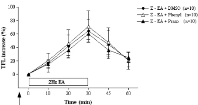
Fig. 3. Intraperitoneal pretreatment(arrow) of α1-adren- ergic antagonist prazosin (Prazo, 1 mg/kg, i.p., n=10) and α1-adrenergic receptor agonist phenylephrine (Phenyl, 2mg/kg, i.p., n=10) did not showed any significant changes of increment of tail flick latency(TFL) caused by low frequency(2-Hz) Zusanli(ST36) electroacupuncture.
DMSO, prazosin, phenylephrine were administered 20minutes before the initiation of EA stimulation.
Fig. 4. Intraperitoneal pretreatment(arrow) of α2-ad- renergic receptor antagonist yohimbine(Yohim, 2mg/kg, i.p., n=10) significantly inhibited the analgesic effects of 2-Hz Zusanli(ST36) electroacupuncture(Z-EA+DMSO, n=10)
There were significant differences in TFL at 10, 20, 30, 45, 60min after the initiation of EA stimulation between the Z-EA+
Yohim and the Z-EA+DMSO group. And the synergistic increment in TFL was observed by pretreatment of α2-adrenergic receptor agonist clonidine(Clonid, 40μg/kg, i.p., n=10), and it is significantly differentat 30minutes comparing the Z-EA+DMSO.DMSO, yohim- bine, clonidine were administered 20minutes before the initiation of EAstimulation.
** : p<0.01; *** : p<0.001 indicate the significant differences between Z-EA+Yohim and vehicle control.
# : p<0.05 indicates the significant difference between Z-EA+Clonid and vehicle control(Kruskal-Wallis ANOVA followed by Dunnett's post hoc test).
D. Adrenergic mechanism study
The effects of adrenergic receptor agonists and antagonists on EA-induced analgesia in CIA are shown in Fig. 3, 4. The antinociceptive effect of Zusanli EA was significantly blocked by intraper- inoneal(i.p.) pretreatment of α2-adrenergic receptor antagonist yohimbine(Yohim, 2mg/kg, i.p., n=10), but not by α1-adrenergic antagonist prazosin (Prazo, 1 mg/kg, i.p., n=10). There were significant differences in TFL at 10, 20, 30, 45, 60min after the initiation of EA stimulation between the Z-EA + Yohim and the Z-EA + DMSO group. The synergistic increament in TFL was observed by pretreatment of α2-adrenergic receptor agonist clonidine(Clonid, 40μg/kg, i.p., n=10) with statistical significance, but not observed by α1-adrenergic receptor agonist phenylephrine(Phenyl, 2mg/kg, i.p., n=10) respect- ively. Comparing between the Z-EA + DMSO and Z-EA + Clonid, there was significant synergistic increase in TFL at 30minutes.
Ⅳ. Discussion
Performing wide variety of studies on the therapeutic application of EA in human and animals have elucidated the analgesic effect and its mech- anism of action for treating musculoskeletal disorders.
Diverse modalities of acupuncture have been used extensively to treat OA of knee because of its safety and efficacy13,23). It is reported that EA is more effective modalities than manual acupunc- ture24) because EA is easily standardized by various stimulation parameters25). EA is significantly more effective in treating OA of knee than placebo and diclofenac in a single-blind, placebo controlled trial11) and can be a therapeutic options superior to oral administration of Diclofenac Sodium for OA patients needed to improve joint functions14) and intra-articular injection of hyaluronic acid15). In addition to alleviate pain in OA of knee(visual analogue scale(VAS) and Western Ontario and
McMaster osteoarthritis index(WOMAC)), EA also reduces stiffness(WOMAC) in patients with OA of knee, either on its own or as an adjunct therapy, with beneficial maintenance after one month from treatment12). EA not only improves the intensity of pain and the ability of movement but also inhibits activity of IL-1beta, IL-6, PGE(2α), MMP-3 and TNF-α15,16) and raises synovia SOD activity and lower MDA and NO content, which may contribute to their effect in relieving knee OA26). However, in spite of the fluent investigations proving the effectiveness of EA for treating OA, there are some controversies, limitations and shortcomings about the antinociceptive properties of EA in rigid RCTs27,28), and the need to investigate the analgesic effect of EA and its therapeutic mechanisms using more sophisticated animal models reflecting histology and pathophysiology of OA have been raised.
Numerous studies have observed the analgesic effect of EA and its mechanisms in various animal models such as neuropathic pain, inflammatory pain, post-herpetic neuralgia, and referred pain29), these animal models have some limitations which do not reflect the pathogenesis of OA. Degeneration of articular cartilage plays a most important role in the pathogenesis of OA; therefore, the evaluation of histopathological changes of cartilage with pain-re- lieving efficacy is crucial for assessing the therapeutic effect and exploring its mechanisms. Transgenic modifications30) and surgical procedures with load- bearing31) are complicated and take a significant amount of time to induce OA-like changes. Intra- articular injection of chemical substances, like papain32), needs a large injection volume, and its mechanism related to cartilage degeneration is unknown. CIA models have been reported to result in rapid, con- venient and reproducible human OA-like changes within a short period with articular cartilage de- generation by direct digestion of cartilage and inflammatory reaction of joint tissues at an early stage33). Gross, histopathological features and biomarker analysis changes consistent with human OA characteristics, including cartilage destruction
and related clinical features of pain, swelling, and movement impairment34,35) have been demonstrated in various species such as rabbits and mice36). In the present study, we chose the rats for the precise assessment of pain-related behavior and the convenient application of EA.
In current study, the intra-articular injection of collagenase demonstrated the disease characteristics of OA not only such gross features as joint stiffness, swelling, movement impairment, pain but also histological features as the irregularity with super- ficial and deep fibrillation, loss of superficial-layer cells, erosion, and disorganization of chondrocytes.
Also the activities of PGE2, COX-1, and COX-2 were significantly increased. From these results we could postulate the success of manifestation of osteoarthritic features at 4 week after intra-articular collagenase injections in rats, in accordance with the previous studies37).
Our preliminary experiments showed inhibition of thermal hyperalgesia of EA on ST36 in a short term follow up. It is generally accepted that repeated application of EA suppresses the peripheral inflammation response with acupoint specificity17-19), and the analgesic effect of EA was partially mediated by the descending modulation of nociceptive pro- cessing correlated with various neural substrates20-22), such as adrenergicsy stems in the neuropathic pain model and ankle sprain model38,39). Local adminis- tration of EA to adjunctive points may be sufficient to treat knee OA. Taechaarpornkul and colleagues reported that the two point group received low-frequency(3Hz) of EA treatment at ST35 and EX-LE4(Neixiyan) and the six point group received the same EA treatment at ST35, EX-LE4, ST36, SP9, SP10 and ST34 have been showed no significant difference statistically in mean WOMAC score, number of celecoxib capsule taken as rescue medicine, global assessment of global change and body weight change40). Huang and colleagues reported that EA on Zusanli(ST36), Liangqiu(ST34) can raise synovia SOD activity and lower MDA and NO content, which may contribute to their effect in relieving knee OA in the rabbit26). Also previous
researches have shown that EA into Zusanli(ST36) elicits anti-hyperalgesia more effectively through the peripheral nerve receptors and the analgesic signals relayed to the cerebro-cerebellar and limbic systems41,42).
The results of the present study demonstrate that the antinociceptive effect of Zusanli EA was blocked by intraperitoneal(i.p.) pretreatment of α2- adrenergic receptor antagonist (yohimbine, 2mg/kg, i.p.), but not by α1-adrenergic antagonist (prazosin, 1 mg/kg, i.p.). Synergistic effect was observed by pretreatment of α2-adrenergic receptor agonist (clonidine, 40μg/kg, i.p.) with statistical significance, but not observed by α1-adrenergic receptor agonist (phenylephrine, 2mg/kg, i.p.).
Our results confirmed those of Baek43), Kim38), Kim44) and Koo45). Kim and colleagues reported that intrathecal pretreatment of yohimbine(α2-adren- oceptor antagonist, 30μg) significantly blocked the relieving effect of 2 Hz EA on cold allodynia but not by prazosin(α1-adrenoceptor antagonist, 30μg)38). Koo and colleagues reported that intrathecal administration of yohimbine, an α(2)-adrenergic antagonist, reduced the EA-induced analgesia in a dose-dependent manner, whereas terazosin, an α (1)-adrenergic antagonist, did not produce any effect39). Baek and colleagues reported that intra- peritoneal pretreatment of yohimbine blocked the analgesic effect of bee venom pharmacopuncture into Zusanli(ST36) in the adjuvant-induced arthritis model46). Kim and colleagues reported that intrathecal pretreatment of prazocin did not modified the antinociceptive effect of apipuncture but intrethecal pretreatment of idazoxan(an α2 adrecepter antagonist) reversed apipuncture-induced antinociception44). On the contrary, Su and colleagues47) reported that an activation of α1-or α2-adrenoceptors would decrease the analgesic effect of EA, which injection into the lateral cerebral ventricles with α1-(prazocin) and α2- (Yohimbine) adrenergic antagonist enhanced the analgesic effect of EA and with the α1-(Clonidine) and α2-(methoxamine) adrenergic agonist decreased the analgesic effect of EA.
The role of adrenergic system in EA analgesia is
postulated due to the complication of adrenergic receptors, the acupoint being applied and the condition being treated. And the inhibition of peripheral adrenergic mechanisms is proportional to the effectiveness of the acupuncture analgesia induction48). And the frequency selected and the roles of post-ganglionic sympathetic nervous system are postulated as another major factors in acupunc- ture analgesia, the suppressive effects of low frequency EA on carrageenan-induced paw inflam- mation are mediated by sympathetic post-ganglionic neurons, while the suppressive effects of high frequency EA are mediated by the sympatho- adrenal medullary axis49). Another study suggests that EA produces anti-inflammatory effect in the release of catecholamines from post-ganglionic nerveterminals, which act on beta-adrenoceptors on immune cells to inhibit their migration50). Collec- tively speaking, further research on adrenergic mechanisms in acupuncture analgesia will be needed in the variety of stimulation parameters, acupoints, diseases, administration of antagonists and agonists, and observation of long-term effect and repeated treatments.
In result, we observed that maximal osteoarth- ritic features(gross, histopathological features) and maximal altered pain threshold were achieved in the fourth week after induction of arthritis by intra- articular injection of collagenase and low-frequency EA stimulation delivered into Zusanli(ST36) acupoint reduced the raised pain threshold. And the antinociceptive effect of Zusanli EA was blocked by intraperinoneal(i.p.) pretreatment of α2-adrenergic receptor antagonist yohimbine, but not by α 1-adrenergic antagonist prazosin. Synergistic effect was observed by pretreatment of α2-adrenergic receptor agonist clonidine, but not by α1-adrenergic receptor agonist phenylephrine.
Ⅴ. Conclusion
The results of the present study demonstrate
that low frequency EA(2-Hz) can relieve inflam- matory pain in rats with collagenase-indueced arthritis and the analgesic effect of EA can be mediated by α2-adrenergic receptor, but not α 1-adrenergic receptor.
Ⅵ. References
1. Das SK, Farooqi A. Osteoarthritis. Best Pract Res Clin Rheumatol. 2008 ; 22(4) : 657-75.
2. Poole AR. An introduction to the pathophysiology of osteoarthritis. Front Biosci. 1999 ; 4 : 662-70.
3. Shimpo H, Sakai T, Kondo S, Mishima S, Yoda M, Hiraiwa H, Ishiguro N. Regulation of prostaglandin E(2) synthesis in cells derived from chondrocytes of patients with osteoarthritis.
J Orthop Sci. 2009 ; 14(5) : 611-7.
4. Li M, Thompson DD, Paralkar VM. Prostaglan- din E(2) receptors in bone formation. Int Orthop.
2007 ; 31(6) : 767-72.
5. Miller SB. Prostaglandins in health and disease:
an overview. Semin Arthritis Rheum. 2006 ; 36(1) : 37-49.
6. Fernandes JC, Martel-Pelletier J, Pelletier JP.
The role of cytokines in osteoarthritis patho- physiology. Biorheology. 2002 ; 39(1-2) : 237-46.
7. Malemud CJ, Islam N, Haqqi TM. Pathophys- iological mechanisms in osteoarthritis lead to novel therapeutic strategies. Cells Tissues Organs.
2003 ; 174(1-2) : 34-48.
8. Park JY, Pillinger MH, Abramson SB. Pros- taglandin E2 synthesis and secretion: The role of PGE2 synthases. Clin Immunol. 2006 ; 119(3) : 229-40.
9. Kumar AM, Wen XL. Acupuncture treatment for osteoarthritic pain and inflammation of the knee.
Altern Ther Health Med. 2002 ; 8(6) : 126, 128.
10. Ng MM, Leung MC, Poon DM. The effects of electro-acupuncture and transcutaneous electrical nerve stimulation on patients with painful osteoarthritic knees: a randomized controlled trial with follow-up evaluation. J Altern Complement
Med. 2003 ; 9(5) : 641-9.
11. Sangdee C, Teekachunhatean S, Sananpanich K, Sugandhavesa N, Chiewchantanakit S, Pojcham- arnwiputh S, Jayasvasti S. Electroacupuncture versus diclofenac in symptomatic treatment of osteoarthritis of the knee: a randomized con- trolled trial. BMC Complement Altern Med. 2002 ; 2 : 3.
12. Tukmachi E, Jubb R, Dempsey E, Jones P. The effect of acupuncture on the symptoms of knee osteoarthritis-an open randomised controlled study. Acupunct Med. 2004 ; 22(1) : 14-22.
13. Jubb RW, Tukmachi ES, Jones PW, Dempsey E, Waterhouse L, Brailsford S. A blinded randomised trial of acupuncture(manual and electroacupuncture) compared with a non-pene- trating sham for the symptoms of osteoarthritis of the knee. Acupunct Med. 2008 ; 26(2) : 69-78.
14. Sheng XP, Fan TY. Comparative observation on hip osteoarthritis treated with electroacupuncture and medication. Zhongguo Zhen Jiu. 2010 ; 30(12) : 982-4.
15. Wu MX, Li XH, Lin MN, Jia XR, Mu R, Wan WR, Chen RH, Chen LH, Lin WQ, Huang CY, Zhang XR, Hong KD, Li L, Liu XX. Clinical study on the treatment of knee osteoarthritis of Shen-Sui insufficiency syndrome type by electro- acupuncture. Chin J Integr Med. 2010 ; 16(4) : 291-7.
16. Xu FY, Gan JH, Li WP, Yang M, Liu X. Effect of electroacupuncture on the level of IL-1beta and TNF-alpha in patients with osteoarthritis.
Zhongguo Zhen Jiu. 2009 ; 29(7) : 529-31.
17. Kim HW, Roh DH, Yoon SY, Kang SY, Kwon YB, Han HJ, Lee HJ, Choi SM, Ryu YH, Beitz AJ, Lee JH. The anti-inflammatory effects of low- and high-frequency electroacupuncture are mediated by peripheral opioids in a mouse air pouch inflammation model. J Altern Complement Med. 2006 ; 12(1) : 39-44.
18. Yang EJ, Koo ST, Kim YS, Lee JE, Hwang HS, Lee MS, Choi SM. Contralateral electroacupunc- ture pretreatment suppresses carrageenan-induced inflammatory pain via the opioid-mu receptor.
Rheumatol int. 2010 Epub ahead of print.
19. Zhang RX, Lao L, Wang X, Fan A, Wang L, Ren K, Berman BM. Electroacupuncture attenuates inflammation in a rat model. J Altern Comple- ment Med. 2005 ; 11(1) : 135-42.
20. Gebhart GF. Descending modulation of pain.
Neurosci Biobehav Rev. 2004 ; 27(8) : 729-37.
21. Takeshige C, Sato T, Mera T, Hisamitsu T, Fang J. Descending pain inhibitory system involved in acupuncture analgesia. Brain Res Bull. 1992 ; 29(5) : 617-34.
22. Zhao ZQ. Neural mechanism underlying acu- puncture analgesia. Prog Neurobiol. 2008 ; 85(4) : 355-75.
23. Selfe TK, Taylor AG. Acupuncture and osteo- arthritis of the knee: a review of randomized, controlled trials. Fam Community Health. 2008 ; 31(3) : 247-54.
24. Ulett GA, Han S, Han JS. Electroacupuncture:
mechanisms and clinical application. Biol Psych- iatry. 1998 ; 44(2) : 129-38.
25. Ma SX. Neurobiology of Acupuncture: Toward CAM. eCAM. 2004 ; 1(1) : 41-7.
26. Huang J, Zhuo LS, Wang YY, Feng WQ. Effects of electroacupuncture on synovia free radicals in rabbits with knee osteoarthritis. Zhen Ci Yan Jiu. 2008 ; 33(2) : 116-9.
27. Manheimer E, Cheng K, Linde K, Lao L, Yoo J, Wieland S, van der Windt DA, Berman BM, Bouter LM. Acupuncture for peripheral joint osteoarthritis. Cochrane Database Syst Rev.
2010 ; 20(1) : CD001977.
28. Manheimer E, Linde K, Lao L, Bouter LM, Berman BM. Meta-analysis: acupuncture for osteoarthritis of the knee. Ann Intern Med. 2007 ; 146(12) : 868-77.
29. Guilbaud G. Use of animal models of clinical pain.
Neurophysiol Clin. 1990 ; 20(5) : 301-21.
30. Saamanen AK, Salminen HJ, Dean PB, De Crom- brugghe B, Vuorio EI, Metsaranta MP. Osteo- arthritis-like lesions in transgenic mice harboring a small deletion mutation in type II collagen gene.
Osteoarthritis Cartilage. 2000 ; 8(4) : 248-57.
31. Colombo C, Butler M, O’Byrne E, Hickman L,
Swartzendruber D, Selwyn M, Steinetz B. A new model of osteoarthritis in rabbits. I.
Development of knee joint pathology following lateral meniscectomy and section of the fibular collateral and sesamoid ligaments. Arthritis Rheum. 1983 ; 26(7) : 875-86.
32. Farkas T, Bihari-Varga M, Biro T. Thermo- analytical and histological study of intra-articular papain-induced degradation and repair of rabbit cartilage. II. Mature animals. Ann Rheum Dis.
1976 ; 35(1) : 23-6.
33. Hollander AP, Pidoux I, Reiner A, Rorabeck C, Bourne R, Poole AR. Damage to type II collagen in aging and osteoarthritis starts at the articular surface, originates around chondrocytes, and extends into the cartilage with progressive degeneration. J Clin Invest. 1995 ; 96(6) : 2859-69.
34. Aigner T, Sachse A, Gebhard PM, Roach HI.
Osteoarthritis: Pathobiology—targets and ways for therapeutic intervention. Adv Drug Deliv Rev. 2006 ; 58(2) : 128-49.
35. Van den Berg WB. Lessons from animal models of osteoarthritis. Curr Opin Rheumatol. 2001 ; 13(5) : 452-6.
36. Kikuchi T, Sakuta T, Yamaguchi T. Intra-articular injection of collagenase induces experimental osteoarthritis in mature rabbits. Osteoarthritis Cartilage. 1998 ; 6(3) : 177-86.
37. Huh JE, Baek YH, Kim YJ, Lee JD, Choi DY, Park DS. Protective effects of butanol fraction from Betula platyphyla var. japonica on cartilage alterations in a rabbit collagenase-induced osteoarthritis. J Ethnopharmacol. 2009 ; 123(3) : 515-21.
38. Kim SK, Park JH, Bae SJ, Kim JH, Hwang BG, Min BI, Park DS, Na HS. Effects of electro- acupuncture on cold allodynia in a rat model of neuropathic pain: mediation by spinal adrenergic and serotonergic receptors. Exp Neurol. 2005 ; 195(2) : 430-6.
39. Koo ST, Lim KS, Chung K, Ju H, Chung JM.
Electroacupuncture-induced analgesia in a rat model of ankle sprain pain is mediated by spinal alpha-adrenoceptors. Pain. 2008 ; 135(1-2) : 11-9.
40. Taechaarpornkul W, Suvapan D, Theppanom C, Chanthipwaree C, Chirawatkul A. Comparison of the effectiveness of six and two acupuncture point regimens in osteoarthritis of the knee: a randomised trial. Acupunct Med. 2009 ; 27(1) : 3-8.
41. Kim JH, Min B-I, Na HS, Park DS. Relieving effects of electroacupuncture on mechanical allodynia in neuropathic pain model of inferior caudal trunk injury in rat: mediation by spinal opioid receptors. Brain Res. 2004 ; 998(2) : 230-6.
42. Yu XJ, Zhan R, Huang H, Ding GH. Analysis on the difference of afferent mechanism of analgesic signals from manual acupuncture and electroacupuncture of “Zusanli”(ST36). Zhen ci yan jiu. 2008 ; 33(5) : 310-5.
43. Baek YH, Choi DY, Park DS. The study on the analgesic effect and its mechanism of electro- acupuncture in the rat model of adjuvant- induced arthritis. JKAMS. 2003 ; 20(3) : 117-30.
44. Kim HW, Kwon YB, Han HJ, Yang IS, Beitz AJ, Lee JH. Antinociceptive mechanisms asso- ciated with diluted bee venom acupuncture (apipuncture) in the rat formalin test: involve- ment of descending adrenergic and serotonergic pathways. Pharmacol Res. 2005 ; 51(2) : 183-8.
45. Koo ST, Lim KS, Chung K, Ju H, Chung JM.
Electroacupuncture-induced analgesia in a rat model of ankle sprain pain is mediated by spinal alpha-adrenoceptors. Pain. 2008 ; 135(1-2) : 11-9.
46. Baek YH, Huh JE, Lee JD, Choi Do Y, Park DS. Antinociceptive effect and the mechanism of bee venom acupuncture(Apipuncture) on inflam- matory pain in the rat model of collagen-induced arthritis: Mediation by alpha2-Adrenoceptors. Brain Res. 2006 ; 1073-1074 : 305-10.
47. Su S, Zheng S, Su C. Effects of four adrenergic drugs on electroacupuncture analgesia. Zhen Ci Yan Jiu. 1992 ; 17(3) : 175-8.
48. Cocchi R. Acupuncture and adrenergic mechanisms of the autonomous nervous system. Minerva Med. 1983 ; 74(42) : 2533-6.
49. Kim HW, Uh DK, Yoon SY, Roh DH, Kwon YB, Han HJ, Lee HJ, Beitz AJ, Lee JH.
Low-frequency electroacupuncture suppresses carrageenan-induced paw inflammation in mice via sympathetic post-ganglionic neurons, while high-frequency EA suppression is mediated by the sympathoadrenal medullary axis. Brain Res Bull. 2008 ; 75(5) : 698-705.
50. Kim HW, Kang SY, Yoon SY, Roh DH, Kwon YB, Han HJ, Lee HJ, Beitz AJ, Lee JH. Low- frequency electroacupuncture suppresses zymosan- induced peripheral inflammation via activation of sympathetic post-ganglionic neurons. Brain Res.
2007 ; 1148 : 69-75.