pISSN 2383-899X eISSN 2234-7631
서 론
비만은 대사성 질환(당뇨병, 심혈관계 질환 등)의 유병률과 밀접한 관련이 있기 때문에 산업화된 국가에서는 널리 퍼져있는 건강문제로 인식되고 있다.1 흡수된 에너지와 소비된 에너지의 불균형으로 에너 지의 소비보다 공급이 많아질 때 과량의 에너지는 지방 형태로 지방 조직에 저장된다. 지방이 지속적으로 체내 지방조직에 축적되면 지방 조직이 증가되어 결국 비만으로 이어지게 된다.
비만은 식생활 습관, 활동량 부족, 스트레스 및 내분비계의 변화 등
여러 요인에 의해 발생한다. 특히 남성들은 나이 및 성선기능저하증 (hypogonadism) 등에 의해 생식선 성 스테로이드 호르몬인 테스토 스테론이 감소되면서 지방조직의 무게가 증가되고, 고인슐린증, 심혈 관질환, 고지혈증 및 제2형 당뇨병 등을 포함한 대사증후군의 발병률 을 증가시킨다.2-5 그러나 이러한 남성들이 테스토스테론을 보충 받으 면 제지방무게(fat-free mass)는 증가되는 반면, 지방무게는 감소되고 비만과 관련된 대사증후군의 발병이 감소되는 경향을 나타낸다.6,7 따 라서 테스토스테론은 남성에서 지방조직 대사를 조절하는 중요한 인 자로 인식되고 있다. 또한 성 스테로이드 호르몬과 성 스테로이드 호
PPARγ에 의해 유도된 지방세포생성에 대한 테스토스테론의 억제효과
정선효*
목원대학교 의생명·보건학부
The Inhibitory Effect of Testosterone on PPARγ-induced Adipogenesis
Sunhyo Jeong*
Division of biomedical Engineering & Health Science Management, Mokwon University, Daejeon, Korea
Background: Peroxisome proliferator-activated receptor γ (PPARγ) plays a major role in adipocyte differentiation. Testosterone is well known
for inhibiting adipocyte metabolism in men. To investigate the inhibitory mechanism of testosterone on adipogenesis, this study evaluated the effects of testosterone on PPARγ expression and activity in adipocytes using in vitro approaches.
Methods: After differentiated 3T3-L1 adipocytes were treated with PPARγ agonist troglitazone and sex hormone testosterone, the effects of tes-
tosterone on troglitazone-induced triglyceride accumulation and expression of genes involved in adipogenesis were investigated. We also inves- tigated whether testosterone regulates troglitazone-induced PPARγ reporter activity in 3T3-L1 preadipocytes.
Results: Testosterone decreased triglyceride accumulation in differentiated 3T3-L1 cells compared with the vehicle treated control group. Tes-
tosterone also decreased the expression of PPARγ mRNA as well as PPARγ dependent adipocyte-specific genes, such as adipocyte fatty acid binding protein and tumor necrosis factor α. Moreover, testosterone treatment inhibited triglyceride accumulation, and the expression of PPARγ and adipocyte-specific genes caused by troglitazone in differentiated 3T3-L1 cells. Testosterone decreased troglitazone-induced PPARγ reporter activity. Also, treatment with testosterone led to an inhibition of troglitazone-induced PPARγ reporter activity in PPARγ and androgen receptor (AR) expressed 3T3-L1 preadipocytes.
Conclusion: These results suggest that testosterone interferes with the actions of PPARγ on adipogensis by an AR-dependent component. In
addition, this study may have provided valuable molecular and biological insights regarding testosterone therapy in obese hypogonadal men.
Key words: Testosterone, Troglitazone, PPARγ, Adipogenesis, 3T3-L1 adipocytes
Corresponding author Sunhyo Jeong http://orcid.org/0000-0001-9575-0354 Division of biomedical Engineering & Health Science Management, College of Techno- Sciences, Mokwon University, 88 Doanbuk-ro, Seo-gu, Daejeon 35349, Korea Tel +82-42-829-7595 Fax +82-42-829-7590 E-mail [email protected] Received Nov. 19, 2015 Reviewed Dec. 17, 2015 Accepted Feb. 22, 2016
Copyright © 2016 Korean Society for the Study of Obesity
This is an Open Access article distributed under the terms of the Creative Commons Attribution Non-Commercial License (http://creativecommons.org/licenses/by-nc/4.0/) which permits unrestricted non-commercial use, distribution, and reproduction in any medium, provided the original work is properly cited.
르몬의 수용체가 지방조직에서 높게 발현되기 때문에, 지방조직은 성 스테로이드 호르몬의 주요 표적기관이 된다.8,9
지방조직 증가는 지방세포로의 지방축적으로 지방세포의 크기가 비대해지는 과정과 미분화된 지방전구세포의 증식과 분화로 성숙한 지방세포의 수가 증가되는 과정을 통해 일어난다.10 이러한 과정 중 지 방전구세포가 성숙한 지방세포로 분화되는 지방세포생성(adipogen- esis)는 지방세포의 형태적 변화, 지질의 축적 및 지방대사 조절과 관 련된 유전자 발현의 변화와 관련이 있으며11, 지방세포생성에 관한 연 구는 3T3-L1 지방전구세포를 이용한 in vitro model이 널리 사용되고 있다.12 3T3-L1 지방전구세포에 fetal calf serum과 3-isobutyl-1-meth- ylxanthine (IBMX), dexamethasone 및 insulin이 포함된 지방세포 분화배지인 MDI medium을 처리하면 지방세포로 분화되는 지방세 포생성 과정이 일어난다. 이러한 MDI medium이 처리된 3T3-L1 지방 전구세포는 세포 속에 중성지방이 축적되고 지방세포 특이적 유전자 들이 발현되면서 성숙한 지방세포로 분화된다.13
지방세포생성은 CCAAT/enhancer binding proteins (C/EBPs), peroxisome proliferator-activated receptor γ (PPARγ) 등의 tran- scription factors에 의해 조절된다. 특히 PPARγ는 지방세포생성 과정 을 조절하는 주요한 transcription factor로 알려져 있으며 지방세포생 성 과정에 대한 PPARγ의 중요성은 지방전구세포를 이용한 in vitro에 서 널리 연구되고 있다.14-16 PPARγ는 지방세포생성의 후기단계에서 발현이 증가되며 지방세포 특이적 유전자의 발현을 촉진시킴으로써 지방세포 특이적 기능을 조절한다.17
PPARγ는 ligand에 의해 활성화되는 transcription factor로써 지방 세포 특이적 유전자의 발현과 지방세포 분화에 중요한 역할을 담당 하며, PPARγ는 백색지방과 갈색지방에서 높은 수준으로 발현된다.18 리간드에 의해 활성화된 PPARγ는 retinoic X receptor와 heterodi- mer를 이룬 후, 지질대사와 관련된 지방세포 특이적 유전자들의 pro- moters/enhancers에 존재하는 peroxisome proliferator response ele- ments (PPREs)에 결합하여 PPARγ-표적유전자의 발현을 조절한다.19 Thiazolidinediones (TZDs; troglitazone과 rosiglitazone)은 PPARγ agonist로서 지방세포생성과 lipid accumulation을 촉진시키며 지방 세포생성 동안 PPARγ-responsive genes의 발현을 유도한다.20,21 지방 조직에서 대부분의 PPARγ-표적유전자는 adipocyte fatty acid bind- ing protein (aP2)과 fatty acid synthase 등의 lipogenic pathway와 직 접적인 관련이 있는 유전자들이다.11,22 또한 지방세포 분화과정 동안 PPARγ는 지방세포에서 생성되고 분비되는 tumor necrosis factor α (TNFα)와 leptin 등의 지방세포 유전자들(adipogenic genes)을 유도 한다.23,24
지방세포생성은 PPARγ뿐만 아니라 성 스테로이드 호르몬에 의해 서도 영향을 받는다. Dehydroepiandrosterone을 비롯한 남성 성 스
테로이드 호르몬에 의해 지방전구세포가 지방세포로 분화되는 과정 이 억제된다고 보고되고 있다.25-27 그러나 남성 성 스테로이드 호르몬 이 남성의 지방조직 대사에 주요한 요인이 됨에도 불구하고 남성의 지 방조직 대사에 대한 남성 성 스테로이드 호르몬의 작용기전이 분자세 포생물학적 측면에서 명확히 규명되지 않고 있다.
따라서 본 연구는 3T3-L1 지방전구세포를 이용한 in vitro 연구를 통해 남성 호르몬인 테스토스테론이 troglitazone에 의해 유도된 PPARγ 활성에 미치는 영향과 그 작용기전을 규명함으로써, 테스토스 테론에 의한 지방세포생성 조절에 대한 정확한 분자세포생물학적 조 절기전을 규명하여 남성 호르몬 생성의 이상으로 발생되는 비만과 관 련된 대사질환의 예방 및 치료에 유익한 기초자료를 제공하고자 한다.
방 법
1. 3T3–L1 지방전구세포 배양 및 지방세포로의 분화유도
3T3-L1 지방전구세포(ATCC, Manassas, VA, USA)는 6-well plates 에서 bovine calf serum (Invitrogen, Carlsbad, CA, USA)이 포함된 Dulbecco’s modified Eagle’s medium (DMEM) 배지(Gibco-Brl, Grand Island, NY, USA)로 배양되었다. 2일 후 10% fetal bovine se- rum (FBS)이 포함된 DMEM배지에 0.5 mmol/L IBMX, 1 μmol/L dexamethasone 및 1 μg/mL insulin이 첨가된 MDI medium으로 교 체하여(day 0) 2일 동안 지방세포분화를 유도한 후(day 2), 2일마다 10% FBS가 포함된 DMEM 배지로 교체하여 6일 동안 배양하였다 (day 6). 배양 6일째에 세포의 total RNA를 추출하였고 Oil red O 염색 액으로 염색하였다. Vehicle, 테스토스테론 및 troglitazone은 MDI medium을 첨가한 2일 동안 처리하였다.2. 분화된 3T3–L1 세포에서의 중성지방 축적의 분석
세포를 phosphate buffered saline (PBS)으로 세척하고 10% form- aldehyde로 1시간 동안 고정시킨 후 PBS로 세척하고 4% Oil red O 염 색액으로 2시간 동안 염색하였다. 세포를 PBS로 세척한 후 중성지방 축적 정도를 광학현미경으로 분석하였다.
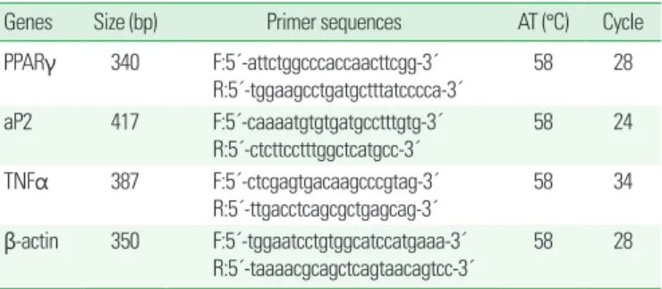
3. Target genes 발현 분석
Total RNA는 Trizol (Invitrogen, Avenue Carlsbad, USA)을 사용 하여 추출하였으며, 역전사-중합효소연쇄반응(reverse transcrip- tion-polymerase chain reaction, RT-PCR)을 이용하여 mRNA의 발 현 양을 측정하였다. Complementary DNA는 total RNA 2 μg과 re- verse primer 0.5 μg을 혼합하여 최종 약 14 μL를 준비하여 75°C에서 15분 동안 열처리(heating)한 후, 5분 동안 얼음 속에 보관하였다. 여 기에 5X M-MLV reaction buffer, 10 mM dNTP mixture, 200 units
M-MLV RT (Promega, Madison, WI, USA)를 첨가하여 최종 양이 25 μL가 되게 한 후, 42°C에서 1시간 동안 반응시켰다. RT reaction 5 μL에 10X reaction buffer (Mg2+ 포함), 10 mM dNTP, 5 unites Taq polymerase (Solgent, Daejeon, South Korea), 그리고 10 μM primer 를 첨가하여 최종 50 μL가 되게 한 후, RTC- 100TM Programmable Thermal Controller (MJ Research, INC., Waltham, MA, USA)를 이 용하여 PCR을 실시하였다. Table 1은 primer sequences와 PCR con- ditions이다.
4. Transient transfection 및 luciferase assay
pSG5-mPPARγ gene과 PPRE3-tk-luc reporter gene의 발현벡터 (expression vector)는 Dr. Gonzalez (National Cancer Institute, NIH, Bethesda, MD)로부터 받은 것이며, pSG5-AR gene constructs의 발 현벡터(expression vector)는 Dr. Cynthia Heinlein (University of Rochester Medical Center, Rochester, NY)로부터 받은 것이다. Mu- rine preadipocyte cell line 3T3-L1 cells는 6-well tissue culture plates (2×10⁴ cells/well)에서 10% fetal bovine serum (Gibco-Brl, Grand Is- land, NY, USA), penicillin G (100 U/mL), streptomycin sulfate (100 μg/mL), amphotericin B (0.25 μg/mL) 및 2-mercaptoethanol (50 μ M)이 포함된 DMEM 배지로 24시간 동안 배양한 후 transfecton하였 다. 모든 transfection의 경우 well당 각각 200 ng의 plasmid가 사용되 었고 lipofectamine (Life technologies, Rockville, MD, USA)을 사용
하여 transfection하였다. 반응 6시간 후 신선한 배지 1 mL를 첨가하 고 vehicle, 테스토스테론 및 troglitazone을 처리하였다. 처리 24시간 후 배지를 제거하고 배양한 세포를 수확하였다. Luciferase와 β-ga- lactosidase 활성은 Promega사(Promega, Madison, WI, USA)의 kit 인 Reporter Lysis Buffer가 포함된 Luciferase Assay System과 β-ga- lactosidase Enzyme Assay System을 사용하여 측정하였다.
5. 통계분석
모든 값은 mean±standard deviation (SD)으로 표시하였다. 통계 분석은 Tukey’s multiple-comparison test에 의한 one-way ANOVA 를 실시하였다. 유의수준은 P<0.05로 설정하였다.
결 과
1. 3T3-L1 세포의 분화에 대한 troglitazone과 테스토스테론의 영향
3T3-L1 지방전구세포가 지방세포로 분화되었는지를 조사하기 위 해서, 중성지방을 염색하는 Oil red O 염색을 이용하여 3T3-L1 세포 에서 중성지방의 축적을 측정하였다(Fig. 1). 3T3-L1 지방전구세포는 중성지방의 축적이 나타나지 않았으며, 지방세포로 분화된 3T3-L1 세포에서 vehicle이 처리된 대조군 세포와 troglitazone이 처리된 세포 는 중성지방의 축적이 확연히 관찰되었다. 그러나 테스토스테론이 처 리된 세포는 vehicle이 처리된 세포에 비해 중성지방의 축적이 현저히 감소되었으며, 특히 troglitazone과 테스토스테론이 동시 처리된 세포 는 troglitazone이 처리된 세포에 비해 중성지방의 축적이 감소되었다.
2. 지방세포 특이적인 유전자의 발현에 대한 troglitazone과 테스토스테론의 영향
3T3-L1 세포에서 troglitazone 처리에 의해 증가된 triglyceride droplets 축적이 테스토스테론에 의해 감소된 것이 PPARγ와 PPARγ- 의존적인 지방세포 특이적 유전자의 mRNA 발현의 변화에 의한 것 인지를 조사하였다. Troglitazone이 처리된 세포는 vehicle이 처리된 Table 1. Sequences of oligonucleotide primers and polymerase chain reaction
conditions
Genes Size (bp) Primer sequences AT (°C) Cycle PPARγ 340 F:5´-attctggcccaccaacttcgg-3´
R:5´-tggaagcctgatgctttatcccca-3´ 58 28 aP2 417 F:5´-caaaatgtgtgatgcctttgtg-3´
R:5´-ctcttcctttggctcatgcc-3´
58 24
TNFα 387 F:5´-ctcgagtgacaagcccgtag-3´
R:5´-ttgacctcagcgctgagcag-3´
58 34
β-actin 350 F:5´-tggaatcctgtggcatccatgaaa-3´
R:5´-taaaacgcagctcagtaacagtcc-3´ 58 28 AT, annealing temperature; bp, base pair.
Fig. 1. Differentiation of 3T3-L1 preadipocytes into adipocytes. Microphotographs were obtained on days 0 and 6. Oil red O staining was performed to identify lipid droplet formation (magnification, × 10).
Tro; 10 μmol/L troglitazone; T; 10 μmol/L testosterone.
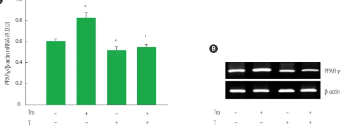
대조군 세포에 비해 PPARγ mRNA 발현이 16.8% 증가하였다 (P<0.05) (Fig. 2). 그러나 테스토스테론이 처리된 세포는 vehicle이 처 리된 대조군 세포에 비해 PPARγ mRNA 발현이 15.3% 감소되었으며, troglitazone이 처리된 세포에 비해 troglitazone과 테스토스테론이 동시 처리된 세포는 troglitazone에 의해 유도된 PPARγ mRNA 발현 이 38.2% 감소되었다(P<0.05) (Fig. 2). PPARγ-의존적인 지방세포 특
이적 유전자인 aP2와 TNFα의 mRNA 발현은 대조군 세포에 비해 troglitazone이 처리된 세포에서 각각 28.7%와 25.1%씩 증가되었지 만, 테스토스테론이 처리된 세포는 대조군 세포에 비해 각각 15.1%와 18.7%씩 감소되었다(P<0.05) (Fig. 3). Troglitazone에 의해 증가된 aP2와 TNFα의 mRNA 발현은 troglitazone과 테스토스테론의 동시 처리에 의해 각각 36.8%와 34.1%씩 감소되었다(P<0.05) (Fig. 3).
B
PPARγ
β-actin
Tro T
–
– –
+ –
+ + + A
Tro T
–
– –
– +
+
+ +
PPARγ/β-actin mRNA (R.D.U)
1.0
0.8
0.6
0.4
0.2
0
*
* †
Fig. 2. Effect of Co-treatment with troglitazone (Tro) and testosterone (T) on the mRNA expression of PPARγ in differentiated 3T3-L1 adipoctyes. (A) Cells were treated with vehicle, 10 μmol/L troglitazone (Tro), 10 μmol/L testosterone (T) or 10 μmol/L Tro plus 10 μmol/L T. Total cellular RNA was extracted from differentiated cells on day 6. All values are expressed as mean± SD of R.D.U. (relative density units) using β-actin. (B) Representative RT-PCR photographs from an independent experiment.
*P< 0.05, as compared with control group, †P< 0.05, as compared with Tro group.
B
aP2
TNF-α β-actin
Tro T
–
– –
+ – +
+ + Tro
T –
– –
+ –
+
+ + aP2/β-actin mRNA (R.D.U)
0.8
0.6
0.4
0.2
0
*
* †
A
Tro T
–
– –
+ –
+
+ +
TNFα/β-actin mRNA (R.D.U)
0.8
0.6
0.4
0.2
0
*
*
†
Fig. 3. Effect of Co-treatment with troglitazone (Tro) and testosterone (T) on the mRNA expression of PPARγ target genes involved in adipogenesis in differentiat- ed 3T3-L1 adipoctyes. (A) Cells were treated with vehicle, 10 μmol/L troglitazone (Tro), 10 μmol/L testosterone (T) or 10 μmol/L Tro plus 10 μmol/L T. Total cellular RNA was extracted from differentiated cells on day 6. All values are expressed as mean± SD of R.D.U. (relative density units) using β-actin. (B) Representative RT- PCR photographs from an independent experiment.
*P< 0.05, as compared with control group; †P< 0.05, as compared with Tro group.
3. PPARγ report gene의 발현에 대한 troglitazone과 테스토스테론의 영향
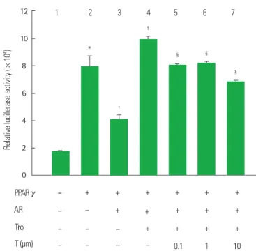
Troglitazone에 의해 유도된 PPARγ mRNA 발현과 PPARγ-의존적 인 지방세포 특이적 유전자 mRNA 발현에 대한 테스토스테론의 억 제적 영향을 이해하기 위한 분자세포생물학적 조절기전을 조사하기 위해, 3T3-L1 preadipocytes를 PPARγ expression constructs, andro- gen receptor (AR) expression constructs 및 luciferase reporter gene construct (PPRE3-tk-luc)로 transient transfection시켰다(Figs. 4 and 5). PPARγ로 transfection시킨 세포는 PPARγ로 transfection시키지 않은 세포에 비해 luciferase reporter activity가 증가되었다(P<0.05) (Fig. 4, lane 2 vs lane 1). PPARγ로 transfection시킨 세포에서 trogli- tazone 처리는 PPARγ transfection에 의해 유도된 luciferase reporter activity를 86.6% 증가시켰다(P<0.05) (Fig. 4, lane 3 vs lane 2). 그러나 troglitazone과 테스토스테론의 동시처리는 troglitazone에 의해 유도 된 luciferase reporter activity를 테스토스테론 처리농도에 의존적으 로 감소시겼으며(P<0.05) (Fig. 4, lane 4, 5, and 6 vs lane 3), 특히 tro- glitazone과 10 μM 테스토스테론 동시처리에 의해 44.8% 감소시켰다 (P<0.05) (Fig. 4, lane 6 vs lane 3). 따라서 이러한 결과는 테스토스테 론이 troglitazone에 의해 활성화된 PPARγ의 전사작용을 억제시켰
음을 보여주었다.
또한 PPARγ와 AR로 동시에 transfection시킨 세포는 PPARγ로 transfection시킨 세포에 비해 luciferase reporter activity가 48.2% 감 소되었다(P<0.05) (Fig. 5, lane 3 vs lane 2). PPARγ와 AR로 동시에 transfection시킨 세포에서 troglitazone 처리는 vehicle 처리에 비해 luciferase reporter activity를 139.9% 증가시켰지만(P<0.05) (Fig. 5, lane 4 vs lane 3), troglitazone 처리에 의해 증가된 luciferase reporter activity는 troglitazone과 테스토스테론의 동시처리에 의해 감소되었 으며(P<0.05) (Fig. 5, lane 5, 6, and 7 vs lane 4), 특히 troglitazone과 10 μM 테스토스테론 동시처리에 의해 30.5% 감소되었다(P<0.05) (Fig. 5, lane 7 vs lane 4).
고 찰
본 연구는 지방세포로 분화된 3T3-L1 세포를 이용한 in vitro 연구 를 통해 PPARγ 활성에 의해 유도된 지방세포생성에 대한 남성 호르몬 인 테스토스테론의 억제적 작용을 조사하였다. 테스토스테론은 분화 된 지방세포에서의 중성지방 축적을 감소시켰으며 PPARγ와 PPARγ- Fig. 4. Effect of Co-treatment with troglitazone (Tro) and testosterone (T) on the
PPARγ reporter gene expression in 3T3-L1 preadipoctyes with PPARγ. 3T3-L1 pre- adipocytes were transiently transfection with pSG5-mPPARγ gene and PPRE3-tk- luc reporter gene. Cells were treated with a 10 μmol/L troglitazone (Tro), or 10 μmol/L Tro plus testosterone (T). After incubation for 24 hours, cells were harvest- ed, lysed and subsequently assays for luciferases and β-galactosidase activities.
All values are expressed as mean± SD of relative luciferase units/β-galactosidase activity.
*P< 0.05, as compared with lane 1; †P< 0.05, as compared with lane 2; ‡P< 0.05, as compared with lane 3.
Relative luciferase activity (×104)
Tro T (μm)
– –
–
– – – 0.1 1 10
+ + + +
+ + + +
+ 18
16 14 12 10 8 6 4 2 0
*
‡ ‡
‡
†
1 2 3 4 5 6
PPARγ
Fig. 5. Effect of Co-treatment with troglitazone (Tro) and testosterone (T) on the PPARγ reporter gene expression in 3T3-L1 preadipoctyes with PPARγ and AR.
3T3-L1 preadipocytes were transiently transfection with pSG5-mPPARγ gene, PPRE3-tk-luc reporter gene and pSG5-AR gene. Cells were treated with a 10 μmol/
L troglitazone (Tro), or 10 μmol/L Tro plus testosterone (T). After incubation for 24 hours, cells were harvested, lysed and subsequently assays for luciferases and β-galactosidase activities. All values are expressed as mean±SD of relative lucif- erase units/β-galactosidase activity.
*P< 0.05, as compared with lane 1; †P< 0.05, as compared with lane 2; ‡P< 0.05, as compared with lane 3; §P< 0.05, as compared with lane 4.
Tro T (μm)
–
– –
– –
–
– – – – 0.1 1 10
+ + + +
+
+ + + + +
+ + +
+ +
PPARγ AR Relative luciferase activity (×104)
12
10
8
6
4
2
0
*
‡
§
§ §
†
1 2 3 4 5 6 7
의존적인 지방세포 특이적 유전자의 mRNA 발현도 감소시켰다. 또한 PPARγ agonist인 troglitazone 처리에 의해 지방세포에 축적된 중성 지방이 테스토스테론 처리에 의해 감소되었고, troglitazone 처리에 의해 증가된 PPARγ와 PPARγ-의존적인 지방세포 특이적 유전자의 mRNA 발현도 테스토스테론 처리에 의해 감소되었다. 추가적으로 PPARγ를 발현시킨 3T3-L1 지방전구세포뿐만 아니라 PPARγ와 AR 을 발현시킨 3T3-L1 지방전구세포에서도 troglitazone에 의해 유도된 PPARγ reporter activity는 테스토스테론 처리에 의해 억제되었다.
비만은 생활습관의 영향뿐만 아니라 성 스테로이드 호르몬의 변화 와도 밀접한 관련이 있다. 여성의 경우 여성 호르몬인 estrogen의 생성 이 감소되는 폐경시기가 되면 급속도로 복부비만이 진행된다.28,29 여 성뿐만 아니라 남성도 나이가 들어감에 따라 남성 호르몬의 분비가 점점 감소되고, 이 시기부터 남성은 근육과 뼈의 양이 감소되는 데 비 해 체지방의 양이 증가된다.2 남성 성 스테로이드 호르몬인 androgens 는 근육, 지방, 연골 및 뼈 세포로 분화가 가능한 C3H 10T1/2 pluripo- tent cells가 근육세포로 분화되는 것을 촉진하였지만 지방세포로의 분화는 억제하였다.30 또한 epididymal preadipocytes의 primary cul- ture에서의 androgens 처리는 지방분화의 후기단계 표지자(marker) 인 glycerol 3-phosphate dehydrogenase activity를 감소시켰다.8 이러 한 연구결과는 지방세포생성에 대한 테스토스테론의 억제작용을 보 여준 본 연구결과를 뒷받침해 주고 있으며 남성 호르몬이 직·간접적 인 조절기전을 통해 지방세포분화를 억제한다는 것을 시사하고 있다.
본 연구에서 PPARγ agonist인 troglitazone에 의해 증가된 중성지 방 축적 및 PPARγ mRNA 발현과 PPARγ-의존적인 지방세포 특이적 인 유전자들의 mRNA 발현이 troglitazone과 테스토스테론의 동시 처리에 의해 감소되었다. 이러한 연구결과는 테스토스테론이 trogli- tazone에 의해 활성화된 PPARγ의 작용을 억제시킴으로써 지방세포 생성 과정을 억제한다는 것을 시사하고 있다.
Androgens (; 테스토스테론, dihydrotestosterone)의 작용은 an- drogen receptor (AR)와의 결합을 통해 조절된다.31 AR의 리간드인 androgen과의 결합으로 활성화된 AR은 표적유전자에 존재하는 an- drogen response elements와 결합하여 표적유전자의 전사를 조절한 다.32 그러나 테스토스테론은 AR에 의존적인 메커니즘뿐만 아니라 AR에 비의존적인 메커니즘을 통해서도 작용한다.33
Bourghardt 등34의 연구에 의하면, 테스토스테론 처리는 고환이 절 제된 mice의 wild mice와 AR knockout (ARKO) mice 모두에서 동맥 경화증을 개선하였다. 또한 콜레스테롤이 포함된 먹이를 섭취한 AR mutant testicular feminized mice (Tfm)에서 테스토스테론 처리는 지방선조(fatty streak) 형성을 감소시켰다.35 반면 콜레스테롤을 섭취 한 토끼에서 AR blocker인 flutamide 처리는 동맥경화성 플라크 영역 (atherosclerotic plaque area)을 감소시키는 테스토스테론의 영향을
억제하였다.36 이러한 연구결과들은 동맥경화증에 대한 테스토스테 론의 생리학적 작용이 AR-의존적 또는 AR-비의존적 조절기전에 의 해 조절된다는 것을 시사하고 있다.
그러나 지방세포생성에 대한 테스토스테론의 생리학적 작용은 AR-의존적 조절기전에 의해 조절된다. 10T1/2 cells에서 androgens (;
테스토스테론, dihydrotesotosterone) 처리에 의해 감소되었던 지방 세포의 수와 지방세포생성의 주요 조절인자인 C/EBPα 단백질 발현 이 AR antagonist인 bicalutamide 처리에 의해 지방세포생성에 대한 androgens의 이러한 효과가 차단됨으로써, 지방세포생성에 대한 테 스토스테론의 작용이 AR-의존적 조절기전에 의해 조절된다는 것이 보고되고 있다.30 또한 지방세포생성에 관한 연구에 널리 사용되고 있 는 3T3-L1 지방전구세포는 AR이 낮은 수준으로 발현되고 있으며, 3T3-L1 지방전구세포를 지방세포로 분화시키는 동안 AR의 발현량 은 증가된다.37 따라서 본 연구에 사용된 3T3-L1 지방전구세포에는 낮은 수준의 AR이 발현되어 있으므로, PPARγ를 transfection시킨 3T3-L1 지방전구세포에서 AR을 transfection시키지 않은 경우뿐만 아니라 AR을 transfection시킨 경우에도 troglitazone에 의해 유도된 PPARγ reporter activity가 테스토스테론 처리에 의해 억제된 본 연구 결과들은, 테스토스테론이 AR-의존적 조절기전에 의해 지방세포생 성에 대한 PPARγ의 작용을 억제조절한다는 것을 시사하고 있다.
AR과 결합한 테스토스테론이 troglitazone에 의해 유도된 PPARγ 의 전사활성을 억제시킨 본 연구결과에 대한 분자세포생물학적 조절 기전의 가능성은 발표된 몇몇 논문들에 의해 설명될 수 있다. PPARγ 와 성 호르몬 수용체(; estrogen receptor 또는 androgen receptor)는 리간드에 의해 활성화되는 transcription factor이다. 이러한 수용체들 의 전사작용의 활성은 transcriptional coactivators에 의해 증가되며 이러한 작용은 리간드의 존재에 의존적이다.38,39 최근 연구결과에 의 하면 AR의 대표적인 coactivators인 ARA70은 AR뿐만 아니라 PPARγ 의 coactivators로서 작용한다고 보고되고 있고, dihydro테스토스테 론에 의해 활성화된 AR이 PPARγ-ARA70 transactivation을 방해함 으로써 지방세포에서 PPARγ-mediated response와 AR-mediated re- sponse 간에 negative signaling cross-talk가 일어난다는 것을 제시하 고 있다.40 또한 androgen은 Wnt signaling pathway에 관여하여 지방 세포분화를 억제한다. Androgen이 AR에 결합함으로써 발생하는 세 포 내의 현상으로 AR과 β-catenin이 결합한 후 이 결합체가 세포의 핵 속으로 이동하여 Wnt signaling pathway에 관여해서 TCF4/LEF 의 활성을 촉진시킴으로써 지방분화의 주요 인자인 PPARγ, C/EBP-α 및 C/EBP-δ의 억제와 관련된 지방세포생성를 억제하였다.41
따라서 이러한 연구결과들을 바탕으로, 테스토스테론에 의해 활 성화된 AR에 의해 troglitazone에 의해 유도된 PPARγ reporter activ- ity가 억제된다는 본 연구결과는, 테스토스테론이 AR의 직·간접적인
조절기전에 의해 troglitazone에 의해 유도된 PPARγ activity의 작용 을 억제조절한다는 것을 시사하고 있다.
결론적으로 본 연구는 테스토스테론이 AR-의존적 조절기전에 의 해 troglitazone에 의해 유도된 PPARγ의 작용을 억제시킴으로써 지 방세포생성를 억제한다는 것을 제시하고 있다. 또한 본 연구는 테스 토스테론이 남성의 지방조직 대사와 비만조절에 중요한 요인임을 시 사하고 있고, 더 나아가 테스토스테론 감소로 인해 발생하는 지방조 직 대사 및 비만과 관련된 대사증후군의 치료 및 예방에 대한 분자세 포생물학적 조절기전을 이해하는 데 기초자료를 제공할 것으로 기대 된다.
요 약
배경: Peroxisome proliferator-activated receptor γ (PPARγ)는 지질 대사와 지방세포분화에 대해 중요한 역할을 한다. 또한 테스토스테 론은 남성의 지방세포 대사작용을 억제하는 것으로 알려져 있다. 따 라서 본 연구는 테스토스테론에 의한 지방세포생성의 억제조절기전 을 규명하기 위해, in vitro 연구를 이용하여 지방세포에서의 PPARγ expression과 activity에 대한 테스토스테론의 영향을 조사하였다.
방법: 분화된 3T3-L1 지방세포에 troglitazone과 테스토스테론을 처 리한 후, troglitazone에 의해 유도된 중성지방 축적과 지방세포생성 과 관련된 유전자들의 발현에 대한 테스토스테론의 영향을 조사하 였다. 또한 테스토스테론이 3T3-L1 지방전구세포에서 troglitazone에 의해 유도된 PPARγ reporter activity를 조절하는지에 대해서도 조사 하였다.
결과: 테스토스테론은 분화된 3T3-L1 지방세포에서 중성지방 축적 을 감소시켰다. 그리고 테스토스테론은 PPARγ mRNA의 발현뿐만 아니라 PPARγ에 의존적인 지방세포 특이적 유전자인 adipocyte fat- ty acid binding protein과 tumor necrosis factor α의 발현을 억제하였 다. 테스토스테론의 처리는 분화된 3T3-L1 지방세포에서 trogli- tazone에 의해 유도된 중성지방 축적뿐만 아니라 PPARγ와 지방세포 특이적 유전자들의 발현을 억제하였다. 또한 PPARγ와 androgen re- ceptor (AR)가 발현되는 3T3-L1 지방전구세포에서 테스토스테론은 troglitazone에 의해 유도된 PPARγ reporter activity도 감소시켰다.
결론: 본 연구결과는 테스토스테론이 AR-의존적 조절기전에 의해 지 방세포생성에 대한 PPARγ의 작용을 방해한다는 것을 시사하고 있으 며, 남성의 비만조절에 대한 테스토스테론 치료요법의 유익한 분자생 물학적 정보를 제공할 것으로 사료된다.
중심단어: 테스토스테론, troglitazone, PPARγ, 지방세포생성, 3T3-L1 지방세포
Conflicts of Interest
The researcher claims no conflicts of interest.
References
1. Hursting SD, Dunlap SM. Obesity, metabolic dysregulation, and cancer: a growing concern and an inflammatory (and microenvi- ronmental) issue. Ann N Y Acad Sci 2012;1271:82-7.
2. Blouin K, Després JP, Couillard C, Tremblay A, Prud’homme D, Bouchard C, et al. Contribution of age and declining androgen levels to features of the metabolic syndrome in men. Metabolism 2005;54:1034-40.
3. Kapoor D, Malkin CJ, Channer KS, Jones TH. Androgens, insulin resistance and vascular disease in men. Clin Endocrinol (Oxf) 2005;63:239-50.
4. Martin AC. Osteoporosis in men: a review of endogenous sex hormones and testosterone replacement therapy. J Pharm Pract 2011;24:307-15.
5. Mori Y, Hoshino K, Yokota K, Yokose T, Tajima N. Increased vis- ceral fat and impaired glucose tolerance predict the increased risk of metabolic syndrome in Japanese middle-aged men. Exp Clin Endocrinol Diabetes 2005;113:334-9.
6. Tenover JL. Experience with testosterone replacement in the el- derly. Mayo Clin Proc 2000;75:S77-81.
7. Wang C, Swerdloff RS, Iranmanesh A, Dobs A, Snyder PJ, Cun- ningham G, et al. Transdermal testosterone gel improves sexual function, mood, muscle strength, and body composition parame- ters in hypogonadal men. J Clin Endocrinol Metab 2000;85:2839- 53.
8. Dieudonne MN, Pecquery R, Leneveu MC, Giudicelli Y. Opposite effects of androgens and estrogens on aipogenesis in rat preadipo- cytes: evidence for sex and site-related specificities and possible involvement of insulin-like growth factor 1 receptor and peroxi- some proliferator-activated receptor gamma2. Endocrinology 2000;141:649-56.
9. Pedersen SB, Børglum JD, Eriksen EF, Richelsen B. Nuclear estra- diol binding in rat adipocytes. Regional variations and regulatory influences of hormones. Biochim Biophys Acta 1991;1093:80-6.
10. Liu L, Li Y, Tollefsbol TO. Gene-environment interactions and epigenetic basis of human diseases. Curr Issues Mol Biol 2008;10:
25-36.
11. Rosen ED, Spiegelman BM. Molecular regulation of aipogenesis.
Annu Rev Cell Dev Biol 2000;16:145-71.
12. Green H, Meuth M. An established pre-adipose cell line and its differentiation in culture. Cell 1974;3:127-33.
13. Cornelius P, MacDougald OA, Lane MD. Regulation of adipocyte development. Annu Rev Nutr 1994;14:99-129.
14. Butterwith SC. Molecular events in adipocyte development. Phar- macol Ther 1994;61:399-411.
15. Hu E, Tontonoz P, Spiegelman BM. Transdifferentiation of myo- blasts by the adipogenic transcriprion factors PPARγ and C/EBPα. Proc Natl Acad Sci U S A 1995;92:9856-60.
16. MacDougald OA, Lane MD. Transcriptional regulation of gene expression during adipocyte differentiation. Annu Rev Biochem 1995;64:345-73.
17. Ntambi JM, Young-Cheul K. Adipocyte differentiation and gene expression. J Nutr 2000;130:3122S-6S.
18. Kliewer SA, Forman BM, Blumberg B, Ong ES, Borgmeyer U, Mangelsdorf DJ, et al. Differential expression and activation of a family of murine peroxisome proliferator-activated receptors. Proc Natl Acad Sci U S A 1994;91:7355-9.
19. Rosen ED, Walkey CJ, Puigserver P, Spiegelman BM. Transcrip- tional regulation of adipogenesis. Genes Dev 2000;14:1293-307.
20. Gimble JM, Robinson CE, Wu X, Kelly KA, Rodriguez BR, Kliew- er SA, et al. Peroxisome proliferator-activated receptor-gamma ac- tivation by thiazolidinediones induces adipogenesis in bone mar- row stromal cells. Mol Pharmacol 1996;50:1087-94.
21. Sottile V, Seuwen K. Bone morphogenetic protein-2 stimulates adipogenic differentiation of mesenchymal precursor cells in syn- ergy with BRL 49653 (rosiglitazone). FEBS Lett 2000;475:201-4.
22. Spiegelman BM, Choy L, Hotamisligil GS, Graves RA, Tontonoz P.
Regulation of adipocyte gene expression in differentiation and syndromes of obesity/diabetes. J Biol Chem 1993;268:6823-6.
23. Kern PA, Saghizadeh M, Ong JM, Bosch RJ, Deem R, Simsolo RB. The expression of tumor necrosis factor in human adipose tissue. Regulation by obesity, weight loss, and relationship to lipo- protein lipase. J Clin Invest 1995;95:2111-9.
24. Zhang Y, Proenca R, Maffei M, Barone M, Leopold L, Friedman JM. Positional cloning of the mouse obese gene and its human homologue. Nature 1994;372:425-32.
25. Gordon GB, Newitt JA, Shantz LM, Weng DE, Talalay P. Inhibi-
tion of the conversion of 3T3 fibroblast clones to adipocytes by dehydroepiandrosterone and related anticarcinogenic steroids.
Cancer Res 1986;46:3389-95.
26. Lea-Currie YR, Monroe D, Mcintosh MK. Dehydroepiandros- terone and related steroids alter 3T3-L1 preadipocyte proliferation and differentiation. Comp Biochem Physiol C Pharmacol Toxicol Endocrinol 1999;123:17-25.
27. McIntosh M, Hausman D, Martin R, Hausman G. Dehydroepian- drosterone attenuates preadipocyte growth in primary cultures of stromal-vascular cells. Am J Physiol 1998;275(2 Pt 1):E285-93.
28. Fukunaga T, Abe T, Ishida Y, Kondoh M. Subcutaneous fat and muscle distribution patterns in middle and old aged Japanese. J Therm Biol 1993;18:303-6.
29. Jeong S, Han M, Lee H, Kim M, Kim J, Nicol CJ, et al. Effects of fenofibrate on high-fat diet-induced body weight gain and adipos- ity in female C57BL/6J mice. Metabolism 2004;53:1284-9.
30. Singh R, Artaza JN, Taylor WE, Gonzalez-Cadavid NF, Bhasin S.
Androgens stimulate myogenic differentiation and inhibit aipo- genesis in C3H 10T1/2 pluripotent cells through an androgen re- ceptor-mediated pathway. Endocrinology 2003;144:5081-8.
31. Dieudonne MN, Pecquery R, Boumediene A, Leneveu MC, Giu- dicelli Y. Androgen receptors in human preadipocytes and adipo- cytes: regional specificities and regulation by sex steroids. Am J Physiol 1998;274(6 Pt 1):C1645-52.
32. Claessens F, Denayer S, Van Tilborgh N, Kerkhofs S, Helsen C, Haelens A. Diverse roles of androgen receptor (AR) domains in AR-mediated signaling. Nucl Recept Signal 2008;6:e008.
33. Liu PY, Death AK, Handelsman DJ. Androgens and cardiovascu- lar disease. Endocr Rev 2003;24:313-40.
34. Bourghardt J, Wilhelmson AS, Alexanderson C, De Gendt K, Ver- hoeven G, Krettek A, et al. Androgen receptor-dependent and in- dependent atheroprotection by testosterone in male mice. Endo- crinology 2010;151:5428-37.
35. Nettleship JE, Jones TH, Channer KS, Jones RD. Physiological tes- tosterone replacement therapy attenuates fatty streak formation and improves high-density lipoprotein cholesterol in the Tfm mouse: an effect that is independent of the classic androgen re- ceptor. Circulation 2007;116:2427-34.
36. Li S, Li X, Li Y. Regulation of atherosclerotic plaque growth and stability by testosterone and its receptor via influence of inflam- matory reaction. Vascul Pharmacol 2008;49:14-8.
37. Hartig SM, Feng Q, Ochsner SA, Xiao R, McKenna NJ, McGuire SE, et al. Androgen receptor agonism promotes an osteogenic gene program in preadipocytes. Biochem Biophys Res Commun 2013;434:357-62.
38. Arany Z, Sellers WR, Livingston DM, Eckner R. E1A-associated p300 and CREB-associated CBP belong to a conserved family of coactivators. Cell 1994;77:799-800.
39. Yeh S, Chang C. Cloning and characterization of a specific coacti- vator, ARA70, for the androgen receptor in human prostate cells.
Proc Natl Acad Sci U S A 1996;93:5517-21.
40. Heinlein CA, Ting HJ, Yeh S, Chang C. Identification of ARA70 as a ligand-enhanced coactivator for the peroxisome proliferator- activated receptor gamma. J Biol Chem 1999;274:16147-52.
41. Singh R, Artaza JN, Taylor WE, Braga M, Yuan X, Gonzalez-Ca- david NF, et al. Testosterone inhibits adipogenic differentiation in 3T3-L1 cells: nuclear translocation of androgen receptor complex with beta-catenin and T-cell factor 4 may bypass canonical Wnt signaling to down-regulate adipogenic transcription factors. Endo- crinology 2006;147:141-54.