221 http://www.jchestsurg.org
JCS
Journal of Chest SurgeryCase Report
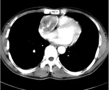
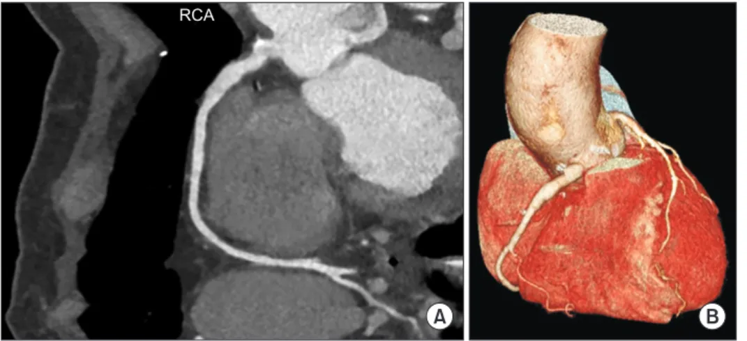
Eighteen Years of Follow-up after Resection of a Giant Coronary Artery Aneurysm and Reconstruction with a Vein Graft
Yelee Kwon, M.D. 1 , Chong Bin Park, M.D., Ph.D. 2 , Pil Je Kang, M.D. 1 , Won Chul Cho, M.D., Ph.D. 2
1