Clinicopathological features and post-resection outcomes of inflammatory pseudotumor of the liver
Kibong Oh1, Shin Hwang2, Chul-Soo Ahn2, Ki-Hun Kim2, Deok-Bog Moon2, Tae-Yong Ha2, Gi-Won Song2, Dong-Hwan Jung2, and Seung-Mo Hong3
1Department of Surgery, Anyang SAM Hospital, Anyang, Departments of 2Surgery and 3Pathology, Asan Medical Center, University of Ulsan College of Medicine, Seoul, Korea
Backgrounds/Aims: Hepatic inflammatory pseudotumor (HIPT) is a rare disease characterized by chronic infiltration of inflammatory cells and area of fibrosis. The objective of this retrospective observational study was to investigate clin- icopathological features and outcomes of patients who underwent hepatic resection (HR) for HIPT. Methods: From 2009 to 2018, seven patients with HIPT underwent HR, accounting for 0.06% of 11,979 adults who underwent HR at our center. Results: These seven patients included five men and two women. Their mean age was 62.3±11.6 years.
In four patients with hepatitis B virus (HBV)-associated liver cirrhosis or chronic hepatitis, liver masses were suspected of hepatocellular carcinoma (HCC) or combined HCC-cholangiocarcinoma based on imaging studies. In three patients without HBV infection, two patients were suspected of HCC, for whom liver biopsy was not performed. One patient was suspected of liver abscess or HIPT, for whom percutaneous liver biopsy was performed and the mass was diag- nosed with HIPT. However, this patient underwent HR owing to abdominal pain. No patient presented with abnormally elevated levels of alpha-fetoprotein, protein induced by vitamin K absence or antagonist-II, or CA19-9. During a mean follow-up period of 76.4±34.8 months, no patient experienced recurrence of HIPT. Conclusions: HIPT, a rare form of liver disease, is often misdiagnosed as malignant liver tumor. Active histological diagnosis is warranted for patients with suspected HIPT to avoid unnecessary operation. HR can be indicated in case of diagnostic ambiguity of HIPT or under a clinical diagnosis of malignant liver tumor. (Ann Hepatobiliary Pancreat Surg 2021;25:34-38)
Key Words: Abscess; Inflammation; Liver mass; Benign disease; Hepatocellular carcinoma
Received: September 13, 2020; Revised: September 18, 2020; Accepted: September 20, 2020 Corresponding author: Shin Hwang
Department of Surgery, Asan Medical Center, University of Ulsan College of Medicine, 88 Olympic-ro 43-gil, Songpa-gu, Seoul 05505, Korea Tel: +82-2-3010-3930, Fax: +82-2-3010-6701, E-mail: [email protected]
Copyright Ⓒ 2021 by The Korean Association of Hepato-Biliary-Pancreatic Surgery
This is an Open Access article distributed under the terms of the Creative Commons Attribution Non-Commercial License (http://creativecommons.org/
licenses/by-nc/4.0) which permits unrestricted non-commercial use, distribution, and reproduction in any medium, provided the original work is properly cited.
Annals of Hepato-Biliary-Pancreatic Surgery ∙ pISSN: 2508-5778ㆍeISSN: 2508-5859
INTRODUCTION
Inflammatory pseudotumor (IPT) is a rare disease char- acterized by chronic infiltration of inflammatory cells and area of fibrosis.1 It is also known as inflammatory myofi- broblastic tumor or plasma cell granuloma.2 Its etiology is uncertain, although infectious condition, autoimmune phe- nomenon, and systemic inflammatory response syndrome have been suggested to play roles in its pathogenesis.2-5 Recently, IPTs are more frequently recognized than before probably due to frequent abdominal imaging studies.
Despite recent development of radiologic imaging stud- ies, it is still difficult to differentiate hepatic inflammatory pseudotumor (HIPT) from malignant tumors (such as hep- atocellular carcinoma [HCC], intrahepatic cholangiocarci-
noma [ICC], and metastatic cancer6,7) and liver abscess showing incomplete liquefaction and granulation.8,9 Treat- ment for HIPT has not been established yet.9-12 Due to its diagnostic ambiguity, some patients undergo surgical resec- tion and other patients are managed with antibiotics, non- steroidal anti-inflammatory drugs, or no medication.2,7,9,13-15
The objective of this study was to investigate clinicopatho- logical features and outcomes of patients who underwent hepatic resection (HR) for HIPT.
MATERIALS AND METHODS
Patient selection
Liver resection database at our institution was extensi- vely searched to identify patients diagnosed with HIPT.
Table 1. Profiles of seven patients diagnosed with inflammatory pseudotumor of the liver
Case
No. Sex Age (yrs)
HBV infec- tion
Clinical diagnosis
Liver biopsy
Preoper- ative AFP (ng/ml)
Preoper- ative PIVKA-
II (mAU/
ml)
Preoper- ative CA19-9 (ng/ml)
Extent of hepatic
resec- tion
Tumor size (cm)
Tumor num-
ber Fol- low-up
period (mos)
Survival status
1 F 83 No Abscess,
HIPT, ICC
HIPT 2.5 27 15.7 LL 3.5 1 123 Alive
2 M 70 No HCC ND 3.2 17 ND RL 5.0 1 94 Alive
3 F 56 Yes HCC,
cHCC-CC
ND 3.2 27 2 LLS 2.3 1 91 Alive
4 M 46 Yes HCC No mass 4.7 22 ND RAS 0.7 1 89 Alive
5 M 60 No HCC ND 2.1 32 2.8 RL 5.5 1 79 Alive
6 M 61 Yes HCC ND 2 19 13.6 LMS 1.5 1 33 Alive
7 M 60 Yes Metastasis,
HCC
ND 4.8 36 10.4 LLS 2.5, 1.4 2 26 Alive
HBV, hepatitis B virus; HIPT, hepatic inflammatory pseudotumor; ICC, intrahepatic cholangiocarcinoma; HCC, hepatocellular carcinoma; cHCC-CC, combined hepatocellular carcinoma-cholangiocarcinoma; ND, not done; AFP, alpha-fetoprotein; PIVKA-II, protein induced by Vitamin K absence or antagonist-II; CA19-9, carbohydrate antigen 19-9; LL, left lobectomy; RL, right lobec- tomy; LLS, left lateral sectionectomy; RAS, right anterior sectionectomy; LMS, left medial sectionectomy
Of 11,976 patients who underwent HR for various in- dications from January 2009 to December 2018,14 seven (0.06%) were pathologically diagnosed with HIPT using resected liver specimens. Medical records of these patients were retrospectively reviewed. This study protocol was approved by the Institutional Review Board (IRB) of our institution. The requirement for informed consent was waived due to the retrospective nature of this study. This study was performed in accordance with ethical guidelines of the World Medical Association Declaration of Helsinki 2013. Patients were followed up until August 2020 based on medical record review with the assistance of the National Health Insurance Service.
Preoperative evaluation, surgical procedures, and postoperative follow-up
Routine preoperative evaluation was performed for pri- mary liver tumors as described previously.16 In general, patients who underwent HR for benign diseases were fol- lowed up every 6 months during 3 years after surgery and every 1-2 years thereafter as part of routine health screen- ing.
Statistical analysis
Numerical data are presented as means and standard deviations. All statistical analyses were performed using
SPSS version 22 (IBM, New York, NY, USA).
RESULTS
Patient demographics and preoperative diagnosis
Clinicopathological features of seven patients with HIPT are described in Table 1. These seven patients included five men and two women. Their mean age was 62.3±11.6 years. Four (57.1%) patients were positive for hepatitis B virus (HBV) infection. However, none of them was pos- itive for hepatitis C virus infection or had alcoholic liver disease. Liver lesions were detected during routine health screening or HBV-associated HCC screening in five pa- tients and during general examination for abdominal pain in two patients.
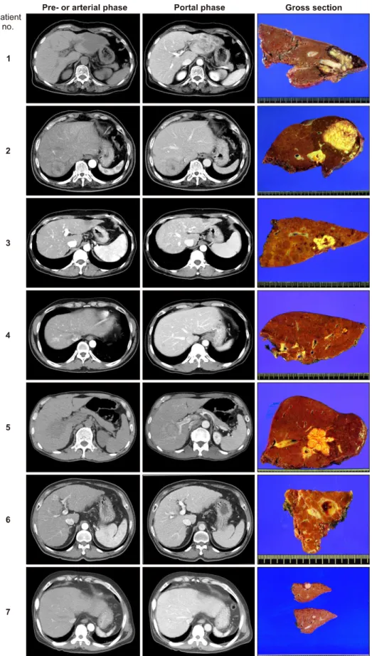
In four patients with HBV-associated liver cirrhosis or chronic hepatitis, liver masses were suspected of HCC or combined HCC-cholangiocarcinoma on imaging studies (Fig. 1). One of them (Case No. 4) underwent percuta- neous liver biopsy; however, no mass was identified. In three patients without HBV infection, two patients were suspected of HCC, thus liver biopsy was not performed.
In contrast, one patient (Case No. 1) was suspected of liv- er abscess, ICC or HIPT. Percutaneous liver biopsy was performed for this patient and the mass was diagnosed
Fig. 1. Preoperative computed tomography findings and gross photographs of surgical speci- mens obtained after initial hepa- tectomy. Number denotes Case number.
with HIPT. However, the patient underwent HR instead of observation owing to persistence of abdominal pain.
Mean preoperative serum concentrations of alpha-feto-
protein (AFP; 3.2±1.2 ng/ml; reference: 7.5 ng/ml), protein induced by vitamin K absence or antagonist-II (PIVKA-II;
25.7±6.9 mAU/ml; reference: 40 mAU/ml), and carbohy-
drate antigen 19-9 (CA19-9; 8.9±6.2 ng/ml; reference: 37 ng/ml) were within normal ranges. None of these patients had abnormal levels of these tumor markers.
Outcomes after hepatic resection
The extent of HR was decided based on tumor location and size. Two patients underwent right hepatectomy. One each underwent right anterior sectionectomy, left hepatec- tomy, and left medial sectionectomy. Two underwent left lateral sectionectomy. All patients recovered uneventfully from HR without any major complications. During a mean follow-up period of 76.4±34.8 months, no patient experi- enced recurrence of HIPT. Currently, all seven patients are alive and doing well.
DISCUSSION
HIPT is a rare disease. It does not show specific symp- toms, laboratory findings, or radiologic features. A Korean multicenter study9 has suggested that dynamic computed tomography (CT) and gadolinium (Gd)-enhanced magnetic resonance imaging (MRI) can provide clues to the diag- nosis of HIPT in patients with liver mass and normal tu- mor marker levels. Because there is not pathognomonic finding, clinical suspicion and subsequent histological di- agnosis are necessary to make accurate diagnosis for HIPT.
In this study reflecting the reality, the majority of liver masses were clinically diagnosed with HCC. Thus, percu- taneous liver biopsy was performed in only 2 of 7 patients.
The etiology of HIPT is unknown yet, although inflam- matory pattern of pathology and its associated laboratory findings have been suggested to have possible association with underlying infection via hepatobiliary tract in several studies.2,5 Some studies have shown that 68%-80% of pa- tients have hepatobiliary tract disease.14,17-20 Abdominal pain, fever, and elevated inflammatory markers (including er- ythrocyte sedimentation rate, C-reactive protein, and leu- kocyte count) are common in HIPT cases.2,9,14,17,18 These find- ings suggest the association of an underlying infectious con- dition with HIPT.
HIPT presents as a large solitary mass. It occurs pre- dominantly in the right lobe, although multicentricity has been also described.2,17,21-23 A Korean multicenter has also shown that the mean size of mass is 4.4 cm and that the majority of patients have a single tumor with right liver
predominance.9 Results of the present study showed that a solitary mass of variable size without locational predom- inance was the common finding.
In the literature, HIPT has shown inconsistent appear- ance on radiologic studies without specific image find- ings.2,8,9,17 The majority of HIPT patients show poorly de- fined hypoattenuating lesion in pre-contrast CT, poorly de- fined peripheral enhancement at arterial phase, and poorly defined hyperattenuating lesions with internal hypoattenua- ting area at equilibrium phase during dynamic CT imaging.
Pre-contrast MRI shows low signal intensity lesion at T1- weighted image and relatively homogenous high signal in- tensity lesion at T2-weighted image. Gd-enhanced MRI shows poorly defined peripheral rim-like enhancement at the arterial phase. However, similar enhancement patterns during CT or MRI can be observed in other lesions such as atypical HCC, ICC, metastatic tumors and abscess.6,7,10 Because of such ambiguity in imaging diagnosis, histo- logical diagnosis is essential to diagnose HIPT accurately.
It is known that the clinical course and prognosis of HIPT are favorable with conservative treatment.2,5,8,9,14,17,23
In the present study, no recurrence occurred during a mean follow-up period of 76.4±34.8 months. In seven patients of this study, six received HR under the clinical diagnosis of liver malignancy. In contrast, one patient received HR instead of observation despite preoperative pathological diagnosis of HIPT. The primary reason to choose HR in this patient was the presence of abdominal pain. If HIPT is pathologically diagnosed in the percutaneous liver biop- sy, it is reasonable to manage it with supportive care or observation for a while. If the mass grows or other abnor- mal findings are found, more aggressive treatment includ- ing surgical resection should be taken into account.
This study has several limitations, including its retro- spective design and inclusion of a small number of pa- tients treated at a single center. Multi-center studies are needed in the future to evaluate characteristics of this very rare disease.
In conclusion, HIPT is a rare form of liver disease that is often misdiagnosed as malignant liver tumor. Active histological diagnosis is warranted for patients with sus- pected HIPT to avoid unnecessary operation. HR can be indicated in case of diagnostic ambiguity of HIPT or un- der a clinical diagnosis of malignant liver tumor.
CONFLICT OF INTEREST
The authors have no conflicts of interest to disclose.
ORCID
Kibong Oh: https://orcid.org/0000-0002-4661-4046 Shin Hwang: https://orcid.org/0000-0002-9045-2531 Chul-Soo Ahn: https://orcid.org/0000-0002-3844-3646 Ki-Hun Kim: https://orcid.org/0000-0002-4016-0995 Deok-Bog Moon: https://orcid.org/0000-0002-8209-3540 Tae-Yong Ha: https://orcid.org/0000-0001-9932-0212 Gi-Won Song: https://orcid.org/0000-0002-4235-0434 Dong-Hwan Jung: https://orcid.org/0000-0001-5984-023X Seung-Mo Hong: https://orcid.org/0000-0002-8888-6007
AUTHOR CONTRIBUTIONS
Conceptualization: SH. Data curation: CSA, KHK, DBM, TYH, GWS, DHJ. Methodology: KO, SMH. Visualization:
SH. Writing - original draft: KO, SH. Writing - review &
editing: SH.
REFERENCES
1. Torzilli G, Inoue K, Midorikawa Y, Hui AM, Takayama T, Ma- kuuchi M. Inflammatory pseudotumors of the liver: prevalence and clinical impact in surgical patients. Hepatogastroenterology 2001;48:1118-1123.
2. Horiuchi R, Uchida T, Kojima T, Shikata T. Inflammatory pseu- dotumor of the liver. Clinicopathologic study and review of the literature. Cancer 1990;65:1583-1590.
3. Yamamoto H, Yamaguchi H, Aishima S, Oda Y, Kohashi K, Oshiro Y, et al. Inflammatory myofibroblastic tumor versus IgG4- related sclerosing disease and inflammatory pseudotumor: a com- parative clinicopathologic study. Am J Surg Pathol 2009;33:1330- 1340.
4. Zen Y, Fujii T, Sato Y, Masuda S, Nakanuma Y. Pathological classification of hepatic inflammatory pseudotumor with respect to IgG4-related disease. Mod Pathol 2007;20:884-894.
5. White JE, Chase CW, Kelley JE, Brock WB, Clark MO. Inflam- matory pseudotumor of the liver associated with extrahepatic in- fection. South Med J 1997;90:23-29.
6. Kitajima K, Shiba H, Nojiri T, Uwagawa T, Ishida Y, Ichiba N, et al. Intrahepatic cholangiocarcinoma mimicking hepatic in- flammatory pseudotumor. J Gastrointest Surg 2007;11:398-402.
7. Ishida H, Tatsuta M, Furukawa H, Ohta H, Hashimoto K,
Hayashi N, et al. Multiple inflammatory pseudotumors mimick- ing liver metastasis from colon cancer: report of a case. Surg Today 2000;30:530-533.
8. Fukuya T, Honda H, Matsumata T, Kawanami T, Shimoda Y, Muranaka T, et al. Diagnosis of inflammatory pseudotumor of the liver: value of CT. AJR Am J Roentgenol 1994;163:1087-1091.
9. Park JY, Choi MS, Lim YS, Park JW, Kim SU, Min YW, et al. Clinical features, image findings, and prognosis of inflam- matory pseudotumor of the liver: a multicenter experience of 45 cases. Gut Liver 2014;8:58-63.
10. Nam KJ, Kang HK, Lim JH. Inflammatory pseudotumor of the liver: CT and sonographic findings. AJR Am J Roentgenol 1996;
167:485-487.
11. Yoon KH, Ha HK, Lee JS, Suh JH, Kim MH, Kim PN, et al.
Inflammatory pseudotumor of the liver in patients with recurrent pyogenic cholangitis: CT-histopathologic correlation. Radiology 1999;211:373-379.
12. Di Vita G, Soresi M, Patti R, Carroccio A, Leo P, Franco V, et al. Concomitant inflammatory pseudotumor of the liver and spleen. Liver 2001;21:217-222.
13. Vassiliadis T, Vougiouklis N, Patsiaoura K, Mpoumponaris A, Nikolaidis N, Giouleme O, et al. Inflammatory pseudotumor of the liver successfully treated with nonsteroidal anti-inflammatory drugs: a challenge diagnosis for one not so rare entity. Eur J Gastroenterol Hepatol 2007;19:1016-1020.
14. Koea JB, Broadhurst GW, Rodgers MS, McCall JL. Inflammato- ry pseudotumor of the liver: demographics, diagnosis, and the case for nonoperative management. J Am Coll Surg 2003;196:
226-235.
15. Yamaguchi J, Sakamoto Y, Sano T, Shimada K, Kosuge T.
Spontaneous regression of inflammatory pseudotumor of the liv- er: report of three cases. Surg Today 2007;37:525-529.
16. Hwang S, Ha TY, Song GW, Jung DH, Ahn CS, Moon DB, et al. Quantified risk assessment for major hepatectomy via the indocyanine green clearance rate and liver volumetry combined with standard liver volume. J Gastrointest Surg 2015;19:1305- 1314.
17. Park KS, Jang BK, Chung WJ, Cho KB, Hwang JS, Kang YN, et al. Inflammatory pseudotumor of liver--a clinical review of 15 cases. Korean J Hepatol 2006;12:429-438.
18. Ahn KS, Kang KJ, Kim YH, Lim TJ, Jung HR, Kang YN, et al. Inflammatory pseudotumors mimicking intrahepatic chol- angiocarcinoma of the liver; IgG4-positivity and its clinical significance. J Hepatobiliary Pancreat Sci 2012;19:405-412.
19. Sakai T, Shiraki K, Yamamoto N, Kawakita T, Ohmori S, Itoh I, et al. Diagnosis of inflammatory pseudotumor of the liver. Int J Mol Med 2002;10:281-285.
20. Toda K, Yasuda I, Nishigaki Y, Enya M, Yamada T, Nagura K, et al. Inflammatory pseudotumor of the liver with primary sclerosing cholangitis. J Gastroenterol 2000;35:304-309.
21. Anthony PP. Inflammatory pseudotumour (plasma cell granuloma) of lung, liver and other organs. Histopathology 1993;23:501-503.
22. Anthony PP, Telesinghe PU. Inflammatory pseudotumour of the liver. J Clin Pathol 1986;39:761-768.
23. Shek TW, Ng IO, Chan KW. Inflammatory pseudotumor of the liver. Report of four cases and review of the literature. Am J Surg Pathol 1993;17:231-238.