Received September 30, 2013, Revised March 27, 2014, Accepted for publication April 4, 2014
Corresponding author: Beom Joon Kim, Department of Dermatology, Chung-Ang University College of Medicine, 102 Heukseok-ro, Dongjak-gu, Seoul 156-755, Korea. Tel: 82-2-6299-1525, Fax: 82-2- 823-1049, E-mail: [email protected]
This is an Open Access article distributed under the terms of the Creative Commons Attribution Non-Commercial License (http://
creativecommons.org/licenses/by-nc/3.0) which permits unrestricted non-commercial use, distribution, and reproduction in any medium, provided the original work is properly cited.
ORIGINAL ARTICLE
Synergistic Inhibition of Tumor Necrosis
Factor-Alpha-Stimulated Pro-Inflammatory Cytokine Expression in HaCaT Cells by a Combination
of Rapamycin and Mycophenolic Acid
Min Young Kim, Yun Young Lim, Hyeong Mi Kim, Young Min Park1, Hoon Kang2, Beom Joon Kim
Department of Dermatology, Chung-Ang University College of Medicine, 1Department of Dermatology, Seoul St. Mary's Hospital, College of Medicine, The Catholic University of Korea, 2Department of Dermatology, St. Paul’s Hospital, College of Medicine, The Catholic University of Korea, Seoul, Korea
Background: Keratinocytes release various pro-inflamma- tory cytokines, chemokines, and adhesion molecules such as intercellular adhesion molecule 1 (ICAM-1) in response to cytokines such as tumor necrosis factor (TNF)-α and inter- feron (IFN)-γ. Rapamycin and mycophenolic acid (MPA) have potent immunosuppressive activity because they inhibit lymphocyte proliferation.
Objective: We investigated the effects of rapamycin and MPA on the expression of inflammation-related factors such as ICAM-1 and inducible nitric oxide synthase (iNOS), pro-inflammatory cytokines and chemokines, and related signaling pathways in TNF-α-stimulated HaCaT cells.
Methods: The viability of HaCaT cells treated with rapamy- cin and MPA was confirmed using MTT assay. The expre- ssion of various cytokines such as interleukin (IL)-1β, IL-6, and IL-8; inflammation-related factors such as ICAM-1 and iNOS; and the activation of mitogen activated protein kinase (MAPK) signaling pathways mediated by extracellular sig- nal-related kinases (ERK), p38, and c-Jun N-terminal kinases (JNK) in TNF-α-stimulated HaCaT cells were confirmed using reverse transcription-polymerase chain reaction and
western blotting.
Results: Combined treatment of TNF-α-induced HaCaT cells with rapamycin and MPA decreased ICAM-1 and iNOS expression and ERK and p38 activation more than treatment with either drug alone. The most significant decrease was observed with a combination of rapamycin (80 nM) and MPA (20 nM). These results show that co-treatment with these agents has a synergistic anti-inflammatory effect by blocking the activation of the ERK/p38 MAPK signaling pathway and thus suppressing the TNF-α-induced expression of ICAM-1 and iNOS.
Conclusion: The combination of rapamycin and MPA could potentially be used as a therapeutic approach in inflamma- tory skin diseases. (Ann Dermatol 27(1) 32∼39, 2015) -Keywords-
Anti-inflammation, Mycophenolic acid, Sirolimus, Tumor necrosis factor-alpha
INTRODUCTION
The primary function of keratinocytes, which comprise 95% of the human epidermis, is to maintain the biochem- ical and physical integrity of the skin. It is now well ac- cepted that they also play an important role in the skin’s immune system1-3. Keratinocytes express and release im- munomodulatory mediators in response to ultraviolet light, allergens, haptens, microbiological agents, and cyto- kines such as tumor necrosis factor (TNF)-α and interfer- on (IFN)-γ. The expression of various pro-inflammatory cytokines, chemokines, and adhesion molecules allow im-
mune cells to enter the site of inflammation in the skin4-6. TNF-α, a major pro-inflammatory cytokine, is produced by multiple cell types in the skin, including keratinocytes.
It acts as a multifunctional cytokine, regulating the pro- duction of pro-inflammatory cytokines and chemokines such as interleukin (IL)-1β, IL-6, and IL-8, as well as adhe- sion molecules such as intercellular adhesion molecule-1 (ICAM-1). In the skin, TNF-α also modulates the initial stages of the response to inflammation and injury7-10. The stimulation of keratinocytes by TNF-α leads to the activa- tion of various signaling pathways that involve caspases, nuclear factor-kappa B (NF-κB), and mitogen activated protein kinases (MAPKs), which subsequently increase the expression of inflammatory mediators11.
NF-κB is a protein transcription factor that is required for the transcription of a wide array of pro-inflammatory mol- ecules that are thought to be important in the onset of apoptosis, in various autoimmune diseases, and in in- flammation12,13. In the resting state, NF-κB dimers are in- active in the cytoplasm of cells and are associated with the NF-κB inhibitory protein (I-κB). Upon stimulation with agents such as TNF-α, the IκB-kinase (IKK) complex is activated and phosphorylates IκB, leading to the sub- strate’s ubiquitination and subsequent degradation. The resulting free NF-κB is translocated to the nucleus where it can activate target genes by binding to regulatory ele- ments in the target gene’s promoter14-16.
Rapamycin and mycophenolic acid (MPA) have potent im- munosuppressive characteristics because they inhibit lym- phocyte proliferation. At the molecular level, these drugs share several mechanistic similarities with other im- munosuppressive drugs17,18. Rapamycin, also known as si- rolimus, is a macrocyclic triene antibiotic that was first iso- lated from Streptomyces hygroscopicus in the early 1970s.
This drug forms an intracellular complex with FK506-bin- ding protein 12 (FKBP12) and inhibits the activity of mam- malian target of rapamycin (mTOR)19,20.
MPA was initially marketed as mycophenolate mofetil (MMF), which has better oral bioavailability compared to that of its active metabolite, MPA21. MPA is a potent non-competitive inhibitor of inosine-monophosphate-de- hydrogenase, and thus affects lymphocytes function by disabling the purine biosynthesis pathway. Recently, MPA has been used as a steroid treatment in immune-mediated disorders including immunoglobulin (Ig) A and psoria- sis22-24.
We investigated the effects of different combinations of immunosuppressive drugs on the expression of pro-in- flammatory mediators and found that the combination of rapamycin and MPA had a synergistically greater anti-in- flammatory effect compared to that of single-drug treatments.
MATERIALS AND METHODS
Materials
Rapamycin and MPA were synthesized at IKSU Co., Ltd.
(Seoul, Korea). Dulbecco’s modified Eagle medium (DMEM), Dulbecco’s phosphate-buffered saline (Dulbecco’s PBS), antibiotic (penicillin, streptomycin), fetal bovine serum (FBS), and trypsin-EDTA were purchased from WelGENE Inc. (Daegu, Korea). Recombinant human TNF-α was pur- chased from R&D Systems (Minneapolis, MN, USA).
3-(4,5-Dimethylthiazol-2-yl)-2,5-diphenyltetrazolium bro- mide (MTT), PD98059, and SP600125 were purchased from Sigma Chemical Co. (St. Louis, MO, USA).
Antibodies specific to ICAM-1, phospho-extracellular sig- nal-related kinases (ERK), phospho-IκBα, and lamin B were purchased from Cell Signaling Technology (Beverly, MA, USA). phosphor-c-Jun N-terminal kinases (JNK) and Phosphor-p38, and glyceraldehyde 3-phosphate dehydro- genase (GAPDH) antibodies were purchased from Santa Cruz Biotechnology Inc. (Santa Cruz, CA, USA), and in- ducible nitric oxide synthase (iNOS) antibodies were ob- tained from BD Bioscience (San Jose, CA, USA). Antibod- ies specific to NF-κB p65 were obtained from Abcam (Cambridge, MA, USA). Secondary antibodies specific for anti-goat IgG, anti-mouse IgG, and anti-rabbit IgG were purchased from Vector Laboratories (Burlingame, CA, USA). ICAM-1, iNOS, IL-1β, IL-6, IL-8, and GAPDH oli- gonucleotide primers were obtained from Bioneer (Seoul, Korea).
Cell culture of HaCaT cells
The human keratinocyte cell line (HaCaT) was obtained from the American Type Culture Collection (ATCC, Manassas, VA, USA). The cells were cultured at 37oC in a humidified incubator containing 5% CO2 and 95% air in DMEM supplemented with 10% FBS (WelGENE Inc.) and antibiotics (100 U/ml penicillin G and 100 μg/ml strepto- mycin).
Reverse transcription-polymerase chain reaction
Total RNA was isolated from cells by using an RNeasy Mini kit (Qiagen, Hilden, Germany) according to the man- ufacturer’s instructions. The RNA (2 μg) was transcribed into cDNA using the PrimeScript 1st Strand cDNA Synthesis Kit (Takara, Otsu, Japan). The transcribed product was am- plified by polymerase chain reaction (PCR) into a final vol- ume of 50 μl by using the following sense and antisense primers (5′→3′): ICAM-1 sense, GGT GAC GCT GAA TGG GGT TCC; ICAM-1 antisense, GTC CTC ATG GTG GGG CTA TGA CTC; iNOS sense, TCC AAC CTG CAG GTC TTC GAT GC; iNOS antisense, GGA CCA GCC AAA
Fig. 1. Effects of rapamycin, mycophenolic acid (MPA), or a combination of the two drugs on the viability of HaCaT cells. (A) Cytotoxic effects of rapamycin and MPA in HaCaT cells. The cells were treated with rapamycin and MPA in a dose-dependent manner for 24 h. (B) Confirmation of the cytotoxic effects of a combination of rapamycin and MPA in HaCaT cells. The cells were treated as indicated in (C). Cell viability was determined using MTT assay as described in the Materials and Methods. The measurements were performed in triplicate. TNF-α: tumor necrosis factor-α.
TCC AGT CTG C; IL-1β sense, AAA CAG ATG AAG TGG TCC TTC CAG; IL-1β antisense, TGG AGA ACA CCA CTT GTT GCT CCA; IL-6 sense, AGA GTA GTG AGG AAC AAG CC; IL-6 antisense, TAC ATT TGC CGA AGA GCC CT; IL-8 sense, ACA TGA CTT CCA AGC TGG CCG; IL-8 antisense, TTT ATG AAT TCT CAG CCC TC;
and GAPDH sense, CAT GGG GAA GGT GAA GGT C;
GAPDH antisense, TGG ACT CCA CGA CGT ACT CA.
Amplification was performed using Taq polymerase (Takara, Otsu, Japan). The products were electrophoresed for 30 min at 100 V on 1% agarose gel. Gels were vi- sualized using a Molecular Imager Gel Doc XR imaging system (Bio-Rad Laboratories Inc., Hercules, CA, USA).
Western blot analysis
HaCaT cells were washed twice with ice-cold PBS and then lysed in RIPA buffer. The lysates were clarified by 30 min of centrifugation at 13,000 rpm at 4oC. Total protein in the cell extracts or in the cytoplasmic and nuclear ex- tracts was subjected to sodium dodecyl sulfate-polyacryla- mide gel electrophoresis on 10% polyacrylamide gel. The separated proteins were transferred to a PVDF membrane and then the membrane was blocked with blocking buffer (5% skim milk in Tris-buffered saline containing 0.5%
Tween 20). Western blot analysis was performed by first incubating the membrane in target antibodies overnight at 4oC, and then incubating them with horseradish perox- idase-conjugated secondary antibody. Detection of im-
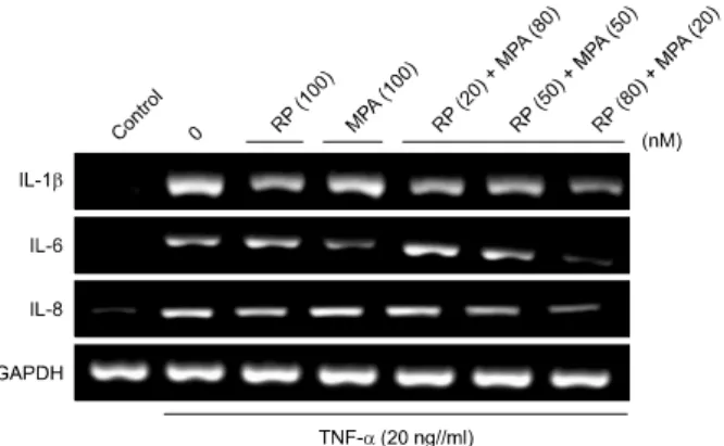
Fig. 2. Effects of a combination of rapamycin (RP) and mycop- henolic acid (MPA) on tumor necrosis factor (TNF)-α-induced expression of pro-inflammatory genes in HaCaT cells. HaCaT cells were pre-incubated for 24 h and then stimulated with TNF- α (20 ng/ml) after a 1 h pretreatment with RP, MPA, or a combination of the two drugs for 6 h. Pro-inflammatory gene expression was analyzed by reverse transcription-polymerase chain reaction (RT-PCR) using specific primers. The experiment was repeated independently at least three times. IL: interleukin, GAPDH: glyceraldehyde 3-phosphate dehydrogenase.
munoreactive bands was performed using a SuperSignal West Pico chemiluminescence substrate (PIERCE Biotechnology Inc., IL, USA). The values of the western blot were standardized with an internal control.
Statistical analysis
Data are presented as means±standard deviations. Data were evaluated using the Student’s t-test. A p-value of
<0.05 or <0.01 was considered statistically significant.
The analysis was performed with Statistical Package for Social Sciences (version 12.0; SPSS Inc., Chicago, IL, USA).
RESULTS
Cell viability after treatment with rapamycin, MPA or a combination of the two drugs in HaCaT cells
Cell viability was measured at different concentrations of rapamycin, MPA, or a combination of the two drugs by using MTT assay in HaCaT cells. HaCaT cells were in- cubated with various concentrations (10, 100, 1,000, or 5,000 nM) of rapamycin or MPA for 24 h (Fig. 1A).
Viability was not significantly altered by concentrations of rapamycin or MPA up to 1,000 nM. As shown Fig. 1B, the cells were also treated with a combination of different concentrations of rapamycin and MPA (Fig. 1C). The com- bination of these agents did not show cytotoxicity.
Inhibitory effects of rapamycin and MPA on TNF-α-in- duced expression of pro-inflammatory genes in HaCaT cells
To compare the effect of a combination of rapamycin and MPA to that of either agent alone, the expression levels of TNF-α-induced pro-inflammatory genes such as IL-1β, IL-6, and IL-8 were measured using reverse transcrip- tion-PCR (RT-PCR) in HaCaT cells. HaCaT cells were pre- treated with rapamycin, MPA, or a combination of rapa- mycin and MPA for 1 h, and then stimulated with TNF-α for 6 h. The expression of pro-inflammatory genes in- creased in TNF-α-stimulated HaCaT cells. The treatment of cells with rapamycin (100 nM) or MPA (100 nM) alone either slightly reduced or did not change the levels of IL-1β, IL-6, and IL-8 compared to the corresponding levels in the TNF-α-treated group. The treatment of cells with a combi- nation of rapamycin (20 nM) and MPA (80 nM), or rapa- mycin (50 nM) and MPA (50 nM) only slightly reduced the level of pro-inflammatory gene expression, whereas rapa- mycin at 80 nM in combination with MPA at 20 nM strongly inhibited the levels of IL-1β, IL-6, and IL-8. An effect was not seen with any other single or combination treatment. Use of equal amounts of cDNA was confirmed
on the basis of GAPDH expression (Fig. 2).
Inhibitory effects of rapamycin and MPA on the phos- phorylation of IκBα and the translocation of NF-κB p65 in HaCaT cells
NF-κB is a transcription factor for genes encoding inflam- matory cytokines, chemokines, and inflammation-related factors16. To verify the effects of combinations of rapamy- cin and MPA on TNF-α-induced NF-κB signaling cas- cades, the phosphorylation of IκBα and the translocation of NF-κB were measured by performing western blot.
Cells were pretreated with rapamycin (100 nM), MPA (100 nM), or a combination of both drugs for 1 h, before being stimulated with TNF-α. The activation of NF-κB molecules was analyzed using either nuclear extracts or whole-cell lysates. As shown in Fig. 3A, treatment with ra- pamycin or MPA slightly inhibited the phosphorylation of IκBα, and similar results were obtained from the combi- nation of rapamycin (20 nM) and MPA (80 nM) or rapamy- cin (50 nM) and MPA (50 nM). However, in the presence of rapamycin (80 nM) and MPA (20 nM), the inhibition of TNF-α-induced phosphorylation of IκBα was much higher than that with any other treatment. Moreover, a combination of rapamycin (80 nM) and MPA (20 nM) in- hibited the TNF-α-induced nuclear localization of NF-κB p65 in HaCaT cells when compared with individual drug treatments and other combinations of the two drugs.
These results suggest that the combination of rapamycin (80 nM) and MPA (20 nM) has an inhibitory effect on the
Fig. 3. Inhibitory effects of rapamy- cin (RP) and mycophenolic acid (MPA) on tumor necrosis factor (TNF)-α-induced activation of nu- clear factor-kappa B (NF-κB), extra- cellular signal-related kinases (ERK), p38, and c-Jun N-terminal kinases (JNK) mitogen activated protein ki- nases (MAPKs) in HaCaT cells. HaCaT cells were pre-incubated for 24 h.
The cells were then pre-treated with RP, MPA, or a combination of the two drugs for 1 h, and then sti- mulated by TNF-α (20 ng/ml) for 20 min. (A) The phosphorylation of IκB α and the translocation of NF-κB p65 in whole-cell protein lysates were determined by western blot analysis.
Cell lysates were prepared and sub- jected to western blot with the in- dicated antibodies. (B) The phospho- rylation of substrates in the MAPK family (ERK, p38, JNK) was detected by western blot analysis using specific antibodies. The densities of protein expression were expressed as per- centages of protein density in the TNF-α-only group. *p<0.05 and
**p<0.01 versus the TNF-α-treated group. The densitometric analysis of the western blot was standardized using glyceraldehyde 3-phosphate dehydrogenase (GAPDH) as an in- ternal control.
signaling pathway that leads to the activation of NF-κB in TNF-α-treated HaCaT cells.
Inhibitory effects of rapamycin and MPA on TNF-α- induced activation of ERK, p38, and JNK MAPKs in HaCaT cells
In order to assess the effects of a combination of these drugs on TNF-α-induced MAPK activation in HaCaT cells, we performed western blot analysis. We compared the in- hibitory effects of a single drug or a combination of both drugs on the MAPK signaling pathway. As shown in Fig.
3B, in the presence of a combination of 80 nM rapamycin and 20 nM MPA, the inhibition of TNF-α-induced ERK
activation was markedly higher than that in the presence of a combination of 50 nM rapamycin and 50 nM MPA.
Single-drug treatment and the combination of 20 nM rapa- mycin and 80 nM MPA did not inhibit TNF-α-induced ERK activation. TNF-α-induced p38 activation decreased in the MPA-only treatment group and in the combination groups but not in the rapamycin-only treatment group.
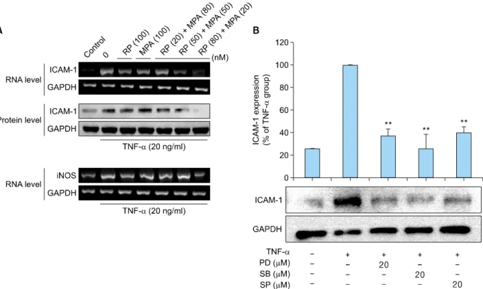
Inhibitory effects of rapamycin and MPA on TNF-α- induced ICAM-1 and iNOS expression in HaCaT cells To investigate the synergistic inhibitory effects of a combi- nation of rapamycin and MPA on TNF-α-induced ICAM-1 and iNOS expression, RT-PCR and western blot analysis
Fig. 4. Inhibitory effects of rapamycin (RP) and mycophenolic acid (MPA) on tumor necrosis factor (TNF)-α-induced intercellular adhesion molecule 1 (ICAM-1) and inducible nitric oxide synthase (iNOS) expression in HaCaT cells. (A) HaCaT cells were pre-incubated for 24 h, and then stimulated with TNF-α (20 ng/ml) in the presence of RP, MPA, or a combination of the two drugs for 3 h (for RNA) or 12 h (for protein). ICAM-1 and iNOS expression were determined after treatment with 100 nM RP, 100 nM MPA, and different combinations of the two drugs (20 nM RP and 80 nM MPA, 50 nM RP and 50 nM MPA, and 80 nM RP and 20 nM MPA) by reverse transcription-polymerase chain reaction (RT-PCR) and western blot. (B) HaCaT cells were pretreated with an extracellular signal-related kinases (ERK) inhibitor (PD98059), p38 inhibitor (SB203580), or c-Jun N-terminal kinases (JNK) inhibitor (SP600125) for 1 h, and then stimulated with TNF-α for 12 h. The band densities are expressed as percentages of the band density of the TNF-α-only group. **p<0.01 compared to treatment with TNF-α alone. The experiment was repeated independently at least three times. The densitometric analysis of the western blot was standardized using glyceraldehyde 3-phosphate dehydrogenase (GAPDH) as an internal control.
were performed. After TNF-α treatment in HaCaT cells, the HaCaT cells were treated with rapamycin and MPA alone, as well as with a combination of the two drugs.
Following treatment, the levels of ICAM-1 and iNOS were measured by RT-PCR and western blot. TNF-α-induced ICAM-1 and iNOS expression in HaCaT cells was slightly suppressed by rapamycin and MPA. The co-treatment of rapamycin and MPA resulted in significantly greater in- hibition compared to that with single-drug treatments. The greatest inhibitory effect was achieved with a combination of 80 nM rapamycin and 20 nM MPA (Fig. 4A).
In addition, we used pharmacological inhibitors of MAPK to investigate the functional relationship between MAPK activation and ICAM-1 expression. TNF-α-induced ICAM-1 expression was slightly suppressed in HaCaT cells with ERK, p38, and JNK inhibitors. These results suggest that the activation of ERK, p38, and JNK is involved in TNF-α
-induced ICAM-1 expression, and the combination of ra- pamycin (80 nM) and MPA (20 nM) modulates signaling cascades that involve TNF-α-induced activation of ERK and p38 but not JNK (Fig. 4B).
DISCUSSION
Many immunosuppressive drugs are currently being used clinically, including cyclosporine, FK-506, rapamycin, and MPA25,26. Rapamycin has been used in the treatment of various pathophysiological conditions including nephro- toxicity, hypertension, ischemia, glutamate neurotoxicity, autoimmune diseases, and inflammatory diseases, by reg- ulating the expression of nitric oxide and prostaglandin E227-30. MPA can induce the apoptosis of activated T-lym- phocytes by depleting guanosine nucleotides, and sup- press the expression and function of adhesion molecules
required for the recruitment of lymphocytes and mono- cytes to sites of inflammation31,32.
Recently, synergistic or additive effects of combined treat- ments with rapamycin and MPA in various pathological conditions have been reported in several studies. Various studies suggest possible synergistic effects of a combina- tion of rapamycin and imatinib on tuberous sclerosis com- plex neoplasia33, rapamycin and LY294002 in melanoma cells34, and rapamycin and cyclosporine A in an auto- immune disease model35.
In the present study, we found that the combination of ra- pamycin and MPA can synergistically inhibit pro-in- flammatory reactions and molecular regulatory mecha- nisms in TNF-α-stimulated HaCaT cells.
There are many reports that an increase in the expression of nitric oxide and adhesion molecules such as ICAM-1 in keratinocytes plays an important role in diseases involving skin inflammation6,36,37. Our results indicate that a combi- nation of rapamycin and MPA can inhibit TNF-α-induced ICAM-1 and iNOS expression in HaCaT cells. Interesting- ly, the combination of 80 nM rapamycin and 20 nM MPA resulted in significantly greater suppression than that ach- ieved with single-agent treatments and combinations with different doses. We also observed a decrease in the ex- pression of pro-inflammatory cytokines and chemokines in TNF-α-induced HaCaT cells.
This suggests that the anti-inflammatory effect of the com- bination of rapamycin and MPA is mediated by blocking the activation of NF-κB and the phosphorylation of ERK and p38 MAPK, which inhibits the expression of pro-in- flammatory genes induced by TNF-α.
In conclusion, the findings of the present study demon- strate that a combination of rapamycin and MPA synerg- istically inhibits the expression of pro-inflammatory factors in TNF-α-induced HaCaT cells. These findings indicate that the inhibition of ICAM-1 expression might cause the blockade of IL-6 or IL-8 expression through inhibition of ERK, P38, and NF-κB signaling pathways by rapamycin and MPA. The use of lower concentrations of rapamycin and MPA are expected to reduce the occurrence of side effects and toxicity. The different anti-inflammatory mech- anisms of rapamycin and MPA appear to result in a syner- gistic effect when the two drugs are used in combination.
However, the interactions of these two immunosuppressive drugs are still being examined, and additional evaluations with other types of immune cells and in vivo trials are needed.
ACKNOWLEDGMENT
This study was supported by a grant from the Korean
Health Technology R&D Project, Ministry for Health &
Welfare, Republic of Korea (A092258).
REFERENCES
1. Bos JD, Kapsenberg ML. The skin immune system: progress in cutaneous biology. Immunol Today 1993;14:75-78.
2. Kupper TS. Mechanisms of cutaneous inflammation. Inte- ractions between epidermal cytokines, adhesion molecules, and leukocytes. Arch Dermatol 1989;125:1406-1412.
3. Barker JN, Mitra RS, Griffiths CE, Dixit VM, Nickoloff BJ.
Keratinocytes as initiators of inflammation. Lancet 1991;
337:211-214.
4. Cho JW, Lee KS, Kim CW. Curcumin attenuates the ex- pression of IL-1beta, IL-6, and TNF-alpha as well as cyclin E in TNF-alpha-treated HaCaT cells; NF-kappaB and MAPKs as potential upstream targets. Int J Mol Med 2007;19:
469-474.
5. Albanesi C, Cavani A, Girolomoni G. Interferon-gamma- stimulated human keratinocytes express the genes nece- ssary for the production of peptide-loaded MHC class II molecules. J Invest Dermatol 1998;110:138-142.
6. Trefzer U, Brockhaus M, Loetscher H, Parlow F, Kapp A, Schöpf E, et al. 55-kd tumor necrosis factor receptor is expressed by human keratinocytes and plays a pivotal role in regulation of human keratinocyte ICAM-1 expression. J Invest Dermatol 1991;97:911-916.
7. Arnott CH, Scott KA, Moore RJ, Robinson SC, Thompson RG, Balkwill FR. Expression of both TNF-alpha receptor subtypes is essential for optimal skin tumour development.
Oncogene 2004;23:1902-1910.
8. Bachelez H. Immunopathogenesis of psoriasis: recent in- sights on the role of adaptive and innate immunity. J Autoimmun 2005;25(Suppl):69-73.
9. Bahar-Shany K, Ravid A, Koren R. Upregulation of MMP-9 production by TNFalpha in keratinocytes and its attenuation by vitamin D. J Cell Physiol 2010;222:729-737.
10. Banno T, Gazel A, Blumenberg M. Effects of tumor necrosis factor-alpha (TNF alpha) in epidermal keratinocytes revealed using global transcriptional profiling. J Biol Chem 2004;
279:32633-32642.
11. Chen G, Goeddel DV. TNF-R1 signaling: a beautiful path- way. Science 2002;296:1634-1635.
12. Mercurio F, Young DB, Manning AM. Detection and puri- fication of a multiprotein kinase complex from mammalian cells. IKK signalsome. Methods Mol Biol 2000;99:109-125.
13. Lawrence T, Gilroy DW, Colville-Nash PR, Willoughby DA.
Possible new role for NF-kappaB in the resolution of inflam- mation. Nat Med 2001;7:1291-1297.
14. Baldwin AS Jr. The NF-kappa B and I kappa B proteins: new discoveries and insights. Annu Rev Immunol 1996;14:649- 683.
15. Baeuerle PA, Baltimore D. NF-kappa B: ten years after. Cell 1996;87:13-20.
16. Young CN, Koepke JI, Terlecky LJ, Borkin MS, Boyd Savoy L, Terlecky SR. Reactive oxygen species in tumor necrosis factor-alpha-activated primary human keratinocytes: impli-
cations for psoriasis and inflammatory skin disease. J Invest Dermatol 2008;128:2606-2614.
17. Mason J. Pharmacology of cyclosporine (sandimmune). VII.
Pathophysiology and toxicology of cyclosporine in humans and animals. Pharmacol Rev 1990;41:423-434.
18. Lipsky JJ. Mycophenolate mofetil. Lancet 1996;348:1357- 1359.
19. Huang S, Houghton PJ. Inhibitors of mammalian target of rapamycin as novel antitumor agents: from bench to clinic.
Curr Opin Investig Drugs 2002;3:295-304.
20. Sabatini DM, Pierchala BA, Barrow RK, Schell MJ, Snyder SH. The rapamycin and FKBP12 target (RAFT) displays pho- sphatidylinositol 4-kinase activity. J Biol Chem 1995;270:
20875-20878.
21. Lynch WS, Roenigk HH Jr. Mycophenolic acid for psoriasis.
Arch Dermatol 1977;113:1203-1208.
22. Epinette WW, Parker CM, Jones EL, Greist MC. Myco- phenolic acid for psoriasis. A review of pharmacology, long-term efficacy, and safety. J Am Acad Dermatol 1987;
17:962-971.
23. Ransom JT. Mechanism of action of mycophenolate mofetil.
Ther Drug Monit 1995;17:681-684.
24. Allison AC, Eugui EM. Immunosuppressive and other effects of mycophenolic acid and an ester prodrug, mycophenolate mofetil. Immunol Rev 1993;136:5-28.
25. Langford CA, Klippel JH, Balow JE, James SP, Sneller MC.
Use of cytotoxic agents and cyclosporine in the treatment of autoimmune disease. Part 2: Inflammatory bowel disease, systemic vasculitis, and therapeutic toxicity. Ann Intern Med 1998;129:49-58.
26. Taylor AC, Connell WR, Elliott R, d'Apice AJ. Oral cyclo- sporin in refractory inflammatory bowel disease. Aust N Z J Med 1998;28:179-183.
27. Bobadilla NA, Gamba G, Tapia E, García-Torres R, Bolio A, López-Zetina P, et al. Role of NO in cyclosporin nephro- toxicity: effects of chronic NO inhibition and NO synthases gene expression. Am J Physiol 1998;274:F791-F798.
28. Gerber DA, Bonham CA, Thomson AW. Immunosuppres- sive agents: recent developments in molecular action and clinical application. Transplant Proc 1998;30:1573-1579.
29. Hutcheson AE, Rao MR, Olinde KD, Markov AK. Myocar- dial toxicity of cyclosporin A: inhibition of calcium ATPase
and nitric oxide synthase activities and attenuation by fructose-1,6-diphosphate in vitro. Res Commun Mol Pathol Pharmacol 1995;89:17-26.
30. Stein CM, Pincus T, Yocum D, Tugwell P, Wells G, Gluck O, et al. Combination treatment of severe rheumatoid arthritis with cyclosporine and methotrexate for forty-eight weeks: an open-label extension study. The Methotrexate- Cyclosporine Combination Study Group. Arthritis Rheum 1997;40:1843-1851.
31. Cohn RG, Mirkovich A, Dunlap B, Burton P, Chiu SH, Eugui E, et al. Mycophenolic acid increases apoptosis, lysosomes and lipid droplets in human lymphoid and monocytic cell lines. Transplantation 1999;68:411-418.
32. Blaheta RA, Leckel K, Wittig B, Zenker D, Oppermann E, Harder S, et al. Mycophenolate mofetil impairs transen- dothelial migration of allogeneic CD4 and CD8 T-cells.
Transplant Proc 1999;31:1250-1252.
33. Govindarajan B, Willoughby L, Band H, Curatolo AS, Veledar E, Chen S, et al. Cooperative benefit for the com- bination of rapamycin and imatinib in tuberous sclerosis complex neoplasia. Vasc Cell 2012;4:11.
34. Werzowa J, Cejka D, Fuereder T, Dekrout B, Thallinger C, Pehamberger H, et al. Suppression of mTOR complex 2-dependent AKT phosphorylation in melanoma cells by combined treatment with rapamycin and LY294002. Br J Dermatol 2009;160:955-964.
35. Martin DF, DeBarge LR, Nussenblatt RB, Chan CC, Roberge FG. Synergistic effect of rapamycin and cyclosporin A in the treatment of experimental autoimmune uveoretinitis. J Immunol 1995;154:922-927.
36. Krutmann J, Czech W, Parlow F, Trefzer U, Kapp A, Schöpf E, et al. Ultraviolet radiation effects on human keratinocyte ICAM-1 expression: UV-induced inhibition of cytokine-in- duced ICAM-1 mRNA expression is transient, differentially restored for IFN gamma versus TNF alpha, and followed by ICAM-1 induction via a TNF alpha-like pathway. J Invest Dermatol 1992;98:923-928.
37. Winiski AP, Foster CA. ICAM-1 expression in a spon- taneously transformed human keratinocyte cell line: charac- terization by a simple cell-ELISA assay. J Invest Dermatol 1992;99:48-52.