ISSN 2234-3806 • eISSN 2234-3814
http://dx.doi.org/10.3343/alm.2016.36.3.263 www.annlabmed.org 263
Ann Lab Med 2016;36:263-265
http://dx.doi.org/10.3343/alm.2016.36.3.263
Letter to the Editor
Diagnostic Hematology
Minor BCR-ABL1-Positive Acute Myeloid Leukemia
Associated With the NPM1 Mutation and FLT3 Internal Tandem Duplication
Moon Jung Kim, M.D.1, Sunhyun Ahn, M.D.2,3, Seong-hyun Jeong, M.D.4, Ja-Hyun Jang, M.D.5, Jae Ho Han, M.D.6, Jong Rak Choi, M.D.7, and Sung Ran Cho, M.D.2
Department of Laboratory Medicine1, Seonam University College of Medicine, Goyang; Departments of Laboratory Medicine2 and Hematology-Oncology4, Ajou University School of Medicine, Suwon; SQ Laboratory3, Yongin; Green Cross Laboratories5, Yongin; Department of Pathology6, Ajou University School of Medicine, Suwon; Department of Laboratory Medicine7, Yonsei University College of Medicine, Seoul, Korea
Dear Editor,
Philadelphia chromosome-positive (Ph+) or BCR-ABL1-positive AML accounts for <1% of newly diagnosed AML cases [1]. Al- though the role of Ph in CML pathogenesis has been elucidated, Ph+ AML pathogenesis remains controversial. Ph+ AML is be- lieved to represent the de novo myeloid blast phase (BP) of CML and, therefore, was not recognized in the 2008 WHO clas- sification of myeloid neoplasms and acute leukemia [2].
Konoplev et al. [3] presented molecular evidence suggesting that Ph+ AML is a clinicopathological entity distinct from CML- BP, in a study involving nine patients with Ph+ AML and five pa- tients with CML-BP at their initial presentations. They showed that patients with Ph+ AML carried the nucleophosmin (NPM1) mutation at a similar frequency as that seen in all cases of AML, and that they lacked ABL1 mutations that occur in a proportion of patients with CML-BP. Internal tandem duplication (ITD) of the Fms-like receptor tyrosine kinase 3 (FLT3) was detected in a Ph+ AML patient, but no Ph+ AML patients harbored both the NPM1 mutation and the FLT3 ITD. We present a rare case of BCR-ABL1-positive AML with both the NPM1 mutation and the
FLT3 ITD.
A 79-yr-old woman was referred to our hospital after several months of general weakness and weight loss. She had under- gone surgery for acute appendicitis one year earlier. Her initial white blood cell count was 157.5×10⁹/L (80% blasts and 1%
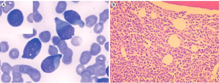
basophils), hemoglobin was 11.8 g/dL, and platelet count was 87×109/L. She did not show organomegaly or lymphadenopa- thy. Her bone marrow (BM) was packed with blasts (92.3% of all nucleated cells) with cup-like nuclear indentation (Fig. 1) and showed no significant dyshematopoietic features. Flow cy- tometry revealed that the blasts were positive for CD13 (94%), CD33 (98%), CD34 (72%), HLA-DR (46%), and cytoplasmic myeloperoxidase (86%). Chromosome analysis revealed t(2;5) (p12;p15) in 20 metaphases, which was considered a constitu- tional chromosomal abnormality. Multiplex reverse transcription (RT)-PCR using HemaVision kit (DNA Diagnostic A/S, Risskov, Denmark) revealed the presence of minor BCR-ABL1 (e1a2) fu- sion transcripts. Interphase FISH analysis of formalin-fixed par- affin-embedded tissue from BM trephine biopsy section showed no fusion signals by using locus specific identifier BCR/ABL
Received: October 1, 2015
Revision received: December 21, 2015 Accepted: January 29, 2016
Corresponding author: Sung Ran Cho
Department of Laboratory Medicine, Ajou University School of Medicine, 164 Worldcup-ro, Yeongtong-gu, Suwon 16499, Korea
Tel: +82-31-219-5780, Fax: +82-31-219-5778 E-mail: [email protected]
© The Korean Society for Laboratory Medicine.
This is an Open Access article distributed under the terms of the Creative Commons Attribution Non-Commercial License (http://creativecommons.org/licenses/by-nc/3.0) which permits unrestricted non-commercial use, distribution, and reproduction in any medium, provided the original work is properly cited.
Kim MJ, et al.
BCR-ABL1 (+) AML with NPM1 mutation and FLT3 ITD
264 www.annlabmed.org http://dx.doi.org/10.3343/alm.2016.36.3.263 dual-color, dual-fusion translocation probe set (Abbott Molecu-
lar/Vysis, Des Plaines, IL, USA).
Genomic DNA was amplified to detect the FLT3 ITD and
NPM1 exon 12 mutations. Total RNA was extracted and RT- PCR was carried out to amplify the BCR/ABL kinase domain.
Direct sequencing of the amplified products revealed that our Fig. 1. Bone marrow findings of the patient. (A) Aspirate smear showing increased blasts (Wright-Giemsa, ×1,000). The blasts with cup- like nuclei and fish mouth nuclei (inlet) are observed. (B) Biopsy section showing a diffuse infiltration by the blasts (Hematoxylin and eosin,
×200).
Fig. 2. Sequencing analyses show the presence of type A mutation (c.863_864insTCTG) in the nucleophosmin gene (NPM1) (A) and an FLT3 internal tandem duplication (ITD) with a 45 bp in-frame duplication (c.1788_1832dup) (B). Arrows indicate the insertion sites.
A B
A
B
Kim MJ, et al.
BCR-ABL1 (+) AML with NPM1 mutation and FLT3 ITD
http://dx.doi.org/10.3343/alm.2016.36.3.263 www.annlabmed.org 265
patient had type A mutations of NPM1 (Fig. 2A) and FLT3 ITD (Fig. 2B), but no ABL1 mutations.
The patient was diagnosed as having AML without matura- tion, according to the 2008 WHO classification of haematopoi- etic and lymphoid tumors [4], and was considered to have a cryptic e1a2 BCR-ABL1 fusion. Although cytarabine and decitabine therapy was started, the patient died one week after AML diagnosis.
The clinicopathological features that suggested Ph+ AML rather than CML-BP included the absence of a clinical history of hematologic disorders (including CML and MDS); no dwarf megakaryocytes, splenomegaly, or basophilia; and the absence of an extra Ph, trisomy 8, trisomy 19, or isochromosome 17q [3, 5]. This case showed clinical and laboratory features compatible with Ph+ AML, as mentioned above.
Considering the findings of Konoplev’s cases [3] and this case, NPM1 mutations and FLT3 ITD may be concurrent genetic ab- normalities in patients with Ph+ AML. The coexistence of the FLT3 ITD and the NPM1 mutations, as observed in our patient, is reported in about 40% of patients with AML and mutated NPM1 [6]. This case supports the idea that Ph+ AML is a dis- tinct entity. Another study also showed that NPM1 mutations oc- cur only in patients with AML and not in those with CML, whereas ABL1 mutations are common in patients with CML [7].
Furthermore, a high-resolution array-based comparative ge- nomic hybridization study indicated that a unique loss within im- munoglobulin genes provides a simple test to differentiate clini- cally similar de novo Ph+ AML cases from CML-BP cases [1].
To our knowledge, our patient is the first case of Ph+ AML with both FLT3 ITD and NPM1 mutations. Ph+ AML cases seem to have different molecular or genomic features from those with CML-BP [1, 3, 8]. However, to describe Ph+ AML as a discrete entity in the WHO classification, it is important to doc- ument more cases and perform more aggressive diagnostic workups, including FLT3 ITD and NPM1 mutational analyses and analysis of immunoglobulin genes. Especially, a hemato- pathologist should keep in mind that cup-like nuclei and fish mouth nuclei may represent an important morphologic clue to
provoke testing for both the FLT3 ITD and the NPM1 mutations [9, 10].
Authors’ Disclosures of Potential Conflicts of Interest
No potential conflicts of interest relevant to this article were re- ported.
REFERENCES
1. Nacheva EP, Grace CD, Brazma D, Gancheva K, Howard-Reeves J, Rai L, et al. Does BCR/ABL1 positive acute myeloid leukaemia exist? Br J Haematol 2013;161:541-50.
2. Vardiman JW, Thiele J, Arber DA, Brunning RD, Borowitz MJ, Porwit A, et al. The 2008 revision of the World Health Organization (WHO) classi- fication of myeloid neoplasms and acute leukemia: rationale and impor- tant changes. Blood 2009;114:937-51.
3. Konoplev S, Yin CC, Kornblau SM, Kantarjian HM, Konopleva M, An- dreeff M, et al. Molecular characterization of de novo Philadelphia chro- mosome-positive acute myeloid leukemia. Leuk Lymphoma 2013;
54:138-44.
4. Swerdlow SH, Campo E, et al. eds. WHO classification of tumours of haematopoietic and lymphoid tissues. 4th ed. Lyon: IARC, 2008:130-9.
5. Soupir CP, Vergilio JA, Dal Cin P, Muzikansky A, Kantarjian H, Jones D, et al. Philadelphia chromosome-positive acute myeloid leukemia: a rare aggressive leukemia with clinicopathologic features distinct from chron- ic myeloid leukemia in myeloid blast crisis. Am J Clin Pathol 2007;
127:642-50.
6. Falini B, Mecucci C, Tiacci E, Alcalay M, Rosati R, Pasqualucci L, et al.
Cytoplasmic nucleophosmin in acute myelogenous leukemia with a normal karyotype. N Engl J Med 2005;352:254-66.
7. Watkins DB, Hughes TP, White DL, D’Andrea RJ. NPM1 mutations oc- cur rarely or not at all in chronic myeloid leukaemia patients in chronic phase or blast crisis. Leukemia 2013;27:489-90.
8. Bacher U, Haferlach T, Alpermann T, Zenger M, Hochhaus A, Beelen DW, et al. Subclones with the t(9;22)/BCR-ABL1 rearrangement occur in AML and seem to cooperate with distinct genetic alterations. Br J Haematol 2011;152:713-20.
9. Park BG, Chi HS, Jang S, Park CJ, Kim DY, Lee JH, et al. Association of cup-like nuclei in blasts with FLT3 and NPM1 mutations in acute my- eloid leukemia. Ann Hematol 2013;92:451-7.
10. Jain P, Vega-Vazquez F, Faderl S. “Cup-like” blasts and NPM1 and FLT3 (ITD) mutations in acute myeloid leukemia (AML). Int J Hematol 2013;98:3.