Copyright © 2019. Anatomy & Cell Biology
Introduction
Tongue muscles change in shape and position during speaking and swallowing [1]. Reductions in tongue muscle strengths trigger problems in both the pharyngeal and oral phases, particularly in terms of poor bolus formation during chewing and swallowing [2]. Tongue strength and accuracy must be improved if such problems develop; the TPS-100 device measures the maximum tongue isometric strength [3, 4]. The buccinator muscle presses the cheek against the teeth, sending food to tooth occlusal surfaces to aid mastication [5].
Muscle strength and co-ordination may improve with train- ing; normal functioning may resume. The strengths and ac- tivities of the tongue and buccinator muscles have been stud- ied. However, the relationships between the muscles remain unclear.
Postural techniques improve swallowing safety by control- ling food and liquid flow and reducing the aspiration risk. For example, in patients who have suffered cerebrovascular ac- cidents, the head should be turned toward the more-involved side to close off the weaker pharyngeal wall, which makes swallowing safer [6]. In patients with pharyngeal disorders, head rotation toward the paralyzed side reduces the volume of the pyriform sinus, after which the bolus descends down the non-affected side [7], reducing pressure on the upper esophageal sphincter and pressurizing the thyroid cartilage.
This in turn promotes the closure of the vocal cords [8]. Oral cancer patients who have undergone partial tongue resection can move a bolus from the mouth to the pharynx by extend-
Corresponding author:
Da-Hye Kim
Department of Dental Hygiene, Division of Health Sciences, Dongseo University, 47 Jurye-ro, Sasang-gu, Busan 47011, Korea
Tel: +82-51-320-2660, Fax: +82-51-320-2732, E-mail: dahye1124@dongseo.
ac.kr
The effects of head rotation and tilt on oral pressure and muscle activity
Tae-Hoon Kim
1, Da-Hye Kim
21Department of Occupational Therapy, Division of Health Science, Dongseo University, Busan, 2Department of Dental Hygiene, Division of Health Sciences, Dongseo University, Busan, Korea
Abstract: We present basic data on head positions that can serve as compensatory interventions for patients with weak tongue and buccinator muscles. We studied 30 Korean adults (15 males, 15 females; mean age, 23 years; range, 20–30 years). A TPS- 100 instrument was used to measure tongue and cheek pressures and suprahyoid and buccinator muscle activities at various head rotations and tilts, as independent variables. The data were subjected to one-way analysis of variance and post-hoc (linear contrast) testing. Tongue elevation pressures differed significantly when the head was flexed or extended compared to the neutral position (P<0.01). Suprahyoid muscle activity varied significantly when the head was rotated left or right compared to neutral, or tilted with the tongue elevated (P<0.01). Cheek pressure varied significantly when the head was rotated left or right compared to neutral, or tilted (P<0.01). Both tongue and cheek pressures increased significantly when the head was extended or rotated contralaterally compared to the neutral position. Suprahyoid muscle activity increased when the head was flexed or extended, or contralaterally or ipsilaterally rotated compared to the neutral position. Therefore, we suggest that head rotation or tilting could be used to vary oral pressure and muscle activity.
Key words: Head position, Suprahyoid muscles, Buccinator, Oral pressure, Muscle activity Received September 3, 2019; Revised November 22, 2019; Accepted December 2, 2019
ing the head [6]. Most stroke patients are at risk of food aspi- ration because the swallowing reflex is delayed. Head flexion reduces this risk by holding the bolus temporarily in the val- lecula [9]. Thus, head rotations and tilts are often prescribed by occupational therapists. However, most prior studies have not explored individual swallowing physiologies. Few Korean studies have biomechanically analyzed oral pressures or su- prahyoid/buccinator muscle activities.
Electromyography (EMG) is commonly used to explore movements of the upper and lower limbs. Commencing in 2017, oral pressure-measuring equipment has been increas- ingly used in clinics. However, no study has yet explored how head position affects oral pressure and associated muscle ac- tivities. Preliminary work with healthy adults is essential prior to studying patients with weak tongue or buccinator muscles.
We thus gathered basic data on how head positioning might aid such patients, exploring how head rotation and tilt affect oral pressure and muscle activities.
Materials and Methods
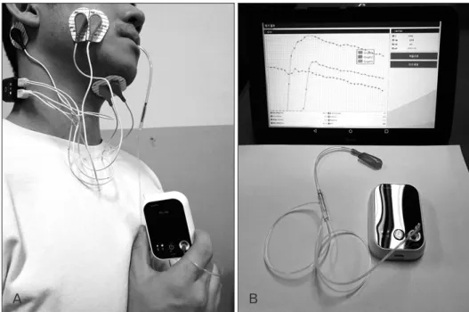
Oral pressure measurementThe TPS-100 device (Cybermedic, Iksan, Korea) is used to analyze and strengthen tongue (front and rear) movements in patients with swallowing disorders. Tongue coordination enhances bolus movement and oral pressurization during de- glutition. The device features an air bulb, a tube, a sensor, and a pressure-measuring device. We measured tongue and cheek
pressures three times, and calculated averages in hPa (Fig. 1).
Surface EMG
We used a surface EMG device (2EM 4D-MT, Relive, Gim- hae, Korea) to measure suprahyoid and buccinator muscle activities. The signals were bandpass-filtered, preserving only those of 25–300 Hz. Skin resistance was minimized by removing hair and wiping the skin with an ethanol swab. The interelectrode distance was 1 cm. To measure suprahyoid activity, two electrodes were attached to the skin over the midline of the submental triangle; the ground electrode was placed on the right mastoid process. To assess right buccina- tor muscle activity, the first electrode was attached lateral to the mouth and the second just lateral to the first (Fig. 1) [10, 11]. To measure the percentage reference voluntary contrac- tion (%RVC), each subject was asked to swallow saliva three times at intervals of 3 minutes.
Subjects
We briefed recruited subjects on oral anatomical struc- tures, our experimental plan, and the clinical significance of the study. We enrolled only volunteers. We excluded those with surgical injuries to the tongue and cheek. The subjects included 30 Korean adults (15 males, 15 females; mean age, 23 years; range, 20–30 years). The study was approved by the Dongseo University Institutional Review Board (No. 1041493- A-2019-004) and we obtained informed consent from the subjects.
A B
Fig. 1. Measurement of oral pressure and muscle activity using TPS-100 and 2EM 4D-MT devices. (A) The air bulb of TPS-100 is located in the oral cavity.
The electrodes of 2EM 4D-MT are attached to the suprahyoid and bucci- nator muscles. (B) The TPS-100 device is consist of an air bulb, a tube, a sensor, and a pressure-measuring device. The patients provided written informed consent for the publication and the use of their images.
Experimental procedures
Head rotation and tilt served as independent variables. The tongue pressure applied to the front of the palate when the tongue was elevated was measured, and the activities of the suprahyoid and buccinator muscles were recorded. Next, the air bulb was positioned between the right upper and lower first molars and the right buccal mucosa, and cheek pressure and suprahyoid and buccinator muscle activities were record- ed during cheek contraction. All tests were run three times at 3-minute intervals. The order of the head positions tested was randomized.
Statistical analysis
The SPSS software ver. 24 (IBM Corp., Armonk, NY, USA) was used to compare all data. The level of statistical signifi- cance (α value) was set to 0.05. The following items were ana- lyzed via one-way analysis of variance accompanied by post- hoc (linear contrast) testing: (1) oral pressures of the tongue tip and cheek in the neutral position, and after left and right head rotation; (2) oral pressures of the tongue tip and cheek in the neutral position, and after head flexion and extension;
(3) buccinator and suprahyoid muscle activities in the neutral position, and after left and right head rotation; (4) buccinator and suprahyoid muscle activities in the neutral position, and after head flexion and extension.
Results
Oral pressure in relation to head position and tongue elevation
The tongue pressure after tongue elevation did not differ when the head was in the neutral position or rotated to the left or right (F=2.95, P>0.05). When the tongue was elevated, the pressure differed significantly when the head was in the neutral position versus when it was flexed or extended (F=8.25, P<0.05). On post-hoc analysis, the difference when the head was extended compared to the neutral position re- mained significant (P<0.01), but the difference when the head was flexed did not (P>0.05) (Table 1, Fig. 2).
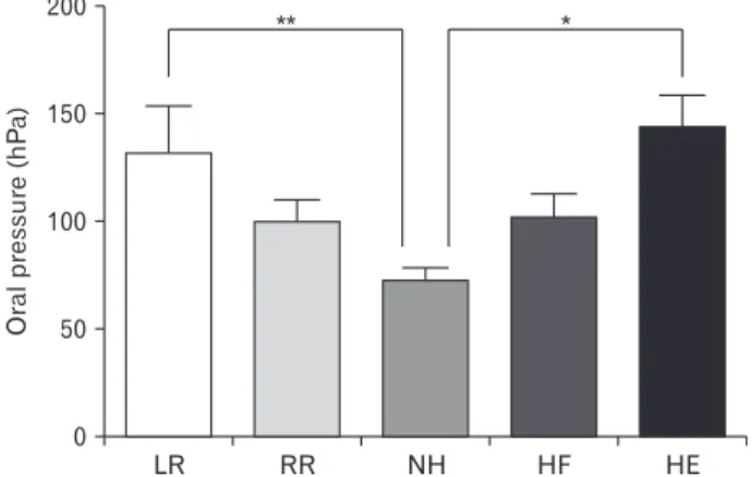
Oral pressure in relation to head position and cheek constriction
In terms of head rotation with cheek constriction, the cheek pressures differed significantly between the neutral position and upon left or right rotation (F=3.48, P<0.05). On post-hoc analysis, the difference between the neutral position and left rotation was maintained (P<0.01), but the effect of right rotation (compared to no or left rotation) was not (both P>0.05). The cheek pressure differed significantly when the head was in the neutral position versus flexed or extended (F=10.34, P<0.01). Post-hoc analysis showed that the differ-
Table 1. Oral pressure according to head position and oral movement (n=30)
LR RR NH HF HE
Tongue elevation 216.98±143.62 182.07±94.39 139.46±57.26 213.44±118.86 331.22±233.63
Cheek compression 130.55±110.84 98.59±50.77 72.84±24.37 102.11±55.61 144.65±63.79
Values are presented as mean±SD (hPa). LR, left rotation; RR, right rotation; NH, neutral head; HF, head flexion; HE, head extension.
LR RR NH HF HE
500
400
300
200
100
Oralpressure(hPa)
0
**
Fig. 2. Oral pressure according to head position with tongue elevation (**P<0.01). LR, left rotation; RR, right rotation; NH, neutral head;
HF, head flexion; HE, head extension.
LR RR NH HF HE
200
150
100
pressureOral(hPa) 50
0
** *
Fig. 3. Oral pressure according to head position with tongue elevation (*P<0.05, **P<0.01). LR, left rotation; RR, right rotation; NH, neutral head; HF, head flexion; HE, head extension.
ence upon extension (compared to neutral/flexed) (P<0.05) was maintained, but the difference upon flexion was not (both P>0.05) (Table 1, Fig. 3).
Buccinator muscle activity in relation to head position and tongue elevation
When the head was rotated with the tongue elevated, no significant difference in buccinator muscle activity between the neutral and left- or right-rotated positions was evident (F=0.33, P>0.05). In terms head flexion or extension with the tongue elevated, the buccinator muscle activity did not vary (F=0.74, P>0.05) (Table 2, Fig. 4).
Buccinator muscle activity in relation to head position and cheek constriction
No significant difference in buccinator muscle activity was noted when the head was in the neutral position versus rotated left or right (F=3.02, P>0.05). No significant differ- ence in buccinator muscle activity was noted when the head was in the neutral position versus extended or flexed (F=1.27, P>0.05) (Table 2, Fig. 5).
Suprahyoid muscle activity in relation to head position and tongue elevation
In terms head rotation with the tongue elevated, the supra-
hyoid muscle activity varied significantly (F=255.65, P<0.01) when the head was in the neutral position versus rotated to the left or right (post-hoc analysis; both P<0.01), but the activ- ities upon left and right rotation were equivalent (P>0.05). In terms of head flexion or extension with the tongue elevated, the suprahyoid muscle activity varied significantly (F=100.49, P<0.01) when the head was in the neutral position versus flexed or extended (post-hoc analysis; both P<0.01); the effects of flexion and extension differed (P<0.01) (Table 3, Fig. 6).
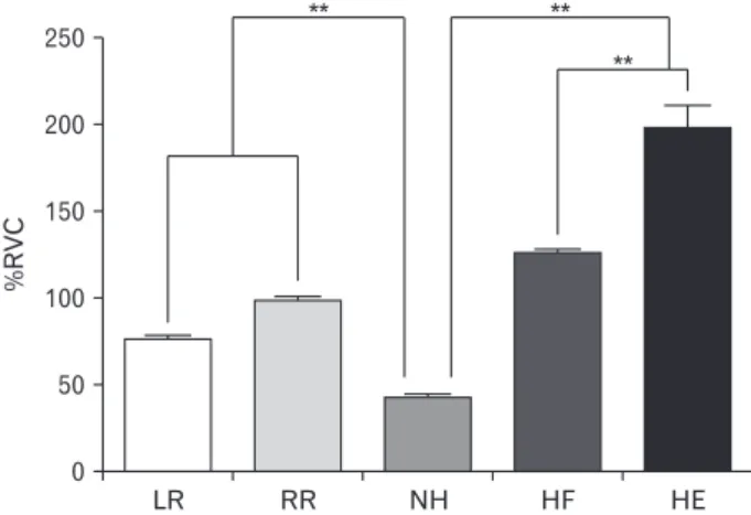
Suprahyoid muscle activity in relation to head position and cheek constriction
The suprahyoid muscle activity differed significantly (F=4.29, P<0.05) when the head was in the neutral position versus rotated left or right (P<0.05). The suprahyoid muscle activity differed significantly (F=4.30, P<0.05) when the head was in the neutral position versus flexed or extended (P<0.05) (Table 3, Fig. 7).
Discussion
The tongue is critical in terms of food movement, and the buccinator muscles of the cheek serve as lateral retainers that prevent food particles from falling into the sulcus between the jaw and cheek [12]. Postural interventions for those ex-
Table 2. Muscle activity of the buccinator according to head position and oral movement (n=30)
LR RR NH HF HE
Tongue elevation 123.86±133.66 110.46±78.25 100.48±52.92 119.88±56.64 127.02±101.78
Cheek compression 308.70±213.86 206.54±128.67 204.69±128.42 286.29±196.92 252.27±174.74
Values are presented as mean±SD (%RVC). LR, left rotation; RR, right rotation; NH, neutral head; HF, head flexion; HE, head extension.
LR RR NH HF HE
200
150
100
50
%RVC
0
Fig. 4. Muscle activity of the buccinator according to head position with tongue elevation. %RVC, percentage reference voluntary con trac- tion; LR, left rotation; RR, right rotation; NH, neutral head; HF, head flexion; HE, head extension.
LR RR NH HF HE
400
300
200
100
%RVC
0
Fig. 5. Muscle activity of the buccinator according to head position with cheek constriction. %RVC, percentage reference voluntary con- trac tion; LR, left rotation; RR, right rotation; NH, neutral head; HF, head flexion; HE, head extension.
hibiting swallowing impairments have traditionally sought to functionally modify the tongue and cheek biomechanics [13]. EMG has been used to aid patients with chronic dyspha- gia, affording useful biofeedback on oral muscle tone, thus improving swallowing [14]. We found that the tongue and cheek pressures increased significantly when the head was extended; specifically, the cheek pressure increased on contra- lateral (left) head rotation. When the muscle fibers and soft tissues are stretched, the tongue and cheek pressures change;
passive pressure develops when the muscle and elastic com- ponents are stretched beyond their resting lengths [15]. If a patient exhibits low tongue or cheek tension, head extension or contralateral rotation is helpful. Passive tension induced by stretching increases internal pressure, and active tensioning on contraction enhances EMG activity. Suprahyoid muscle activity increases on ipsilateral (right) rotation because the pyriform sinus volume is reduced and the bolus descends on the opposite side; the suprahyoid muscles contract [16].
These muscles elevate the hyoid either anteriorly or posteri- orly. When the swallowing response is triggered, the tongue base rises to direct the bolus into the pharynx and the hyoid becomes elevated and moves anteriorly [17]. We found that the suprahyoid muscle activity was higher when the head was flexed or extended, rather than neutral. Head flexion directly
moves the hyoid upward and forward because of the length- tension relationship; the force produced by the contractile elements (lying parallel to the elastic components) of the su- prahyoid tendons increases [18]. Head extension widens the laryngeal vestibule and narrows the vallecular space; physi- ological difficulties may follow [17]. However, if oral transfer is impaired in patients who have undergone supraglottic resection, head extension is a useful postural remedy [19]. In dysphagic patients, head flexion expands the vallecular space, pushes the tongue base toward the pharynx, and protects the epiglottis. Therefore, head flexion is frequently used during deglutition training [20].
We found that the suprahyoid muscle activity increased upon contralateral (left) head rotation. Such rotation would be less favored than the neutral position in healthy adults, who use the suprahyoid muscles symmetrically. However, many stroke patients use the perioral muscles asymmetrically [21]. In left hemiplegic patients, left rotation would increase right-side muscle activity and decrease left-side muscle activ- ity. Thus, head rotation would compensate for the suprahy- oid muscle weakness of the involved side. Turning the head toward that side eliminates that region of the pharynx from muscle activation, rendering the non-impaired side more ac- tive [22].
The buccinator muscle is used to position food for chew-
LR RR NH HF HE
250
200
150
100
50
%RVC
0
**
**
**
Fig. 6. Muscle activity of the suprahyoid according to head position with tongue elevation (**P<0.01). %RVC, percentage reference volun- tary contraction; LR, left rotation; RR, right rotation; NH, neutral head; HF, head flexion; HE, head extension.
LR RR NH HF HE
250
200
150
100
50
%RVC
0
Fig. 7. Muscle activity of the suprahyoid according to head position with cheek constriction. %RVC, percentage reference voluntary con- trac tion; LR, left rotation; RR, right rotation; NH, neutral head; HF, head flexion; HE, head extension.
Table 3. Muscle activity of the suprahyoid according to head position and oral movement (n=30)
LR RR NH HF HE
Tongue elevation 75.25±7.85 97.71±6.69 41.05±10.28 124.71±9.24 197.45±61.69
Cheek compression 127.47±116.85 119.95±104.71 51.91±29.75 157.91±161.55 164.62±178.27
Values are presented as mean±SD (%RVC). LR, left rotation; RR, right rotation; NH, neutral head; HF, head flexion; HE, head extension.
ing and to control the bolus. We found that the cheek pressure increased significantly on contralateral head rotation and ex- tension, compensating for buccinator weakness. Cheek pres- sure is influenced by cheek space between fixed part (teeth, alveolar bone) and unfixed part (buccinator muscle). Head position (contralateral rotation, extension) reduced the space and pressure was increased consequently. However, there were no significant changes in the buccinator muscle activ- ity. We assumed the origin and insertion of the muscle was hardly affected by head position [5]. The change in head posi- tion significantly increases the pressure in the cheek area even though it does not affect the buccinator muscle activity, which may be helpful for patients with buccinator weakness.
We measured oral pressure and suprahyoid and buccinator muscle activities related to head position. Both the tongue and cheek pressure increased on head extension or contralateral rotation. The suprahyoid muscle activity increased upon head flexion and extension, and contralateral and ipsilateral rota- tion. In conclusion, head extension or contralateral rotation would aid those with poor tongue or cheek pressure. Head flexion/extension, or contralateral/ipsilateral rotation, in- crease suprahyoid muscle activity; the optimal head position will vary individually in patients with functional dysphagia or who have undergone supraglottic resection.
ORCID
Tae-Hoon Kim: https://orcid.org/0000-0002-5747-2856 Da-Hye Kim: https://orcid.org/0000-0002-6684-7023
Author Contributions
Conceptualization: THK. Data acquisition: THK, DHK.
Data analysis or interpretation: THK. Drafting of the manu- script: THK, DHK. Critical revision of the manuscript: THK, DHK. Approval of the final version of the manuscript: all au- thors.
Conflicts of Interest
No potential conflict of interest relevant to this article was reported.
Acknowledgements
This work was supported by Dongseo University, "Dong-
seo Cluster Project" Research Fund of 2019 (DSU-20190002).
References
1. Fregosi RF, Ludlow CL. Activation of upper airway muscles dur- ing breathing and swallowing. J Appl Physiol (1985) 2014;116:
291-301.
2. Youmans SR, Youmans GL, Stierwalt JA. Differences in tongue strength across age and gender: is there a diminished strength reserve? Dysphagia 2009;24:57-65.
3. Yeates EM, Molfenter SM, Steele CM. Improvements in tongue strength and pressure-generation precision following a tongue- pressure training protocol in older individuals with dysphagia:
three case reports. Clin Interv Aging 2008;3:735-47.
4. Moon JH, Kim HJ, Kang MK, Won YS. Effects of tongue strength and accuracy training on tongue strength, swallowing function, and quality of life in chronic stroke patients with dysphagia. J Korea Contents Assoc 2016;16:605-13.
5. Drake RL, Vogl AW, Mitchell AW. Gray's anatomy for students.
4th ed. Philadelphia, PA: Elsevier/Churchill Livingstone; 2019.
6. Pendleton HM, Schultz-Krohn W. Pedretti's occupational therapy: practice skills for physical dysfunction. 8th ed. St. Louis, MO.: Mosby/Elsevier; 2018.
7. Logemann JA, Kahrilas PJ, Kobara M, Vakil NB. The benefit of head rotation on pharyngoesophageal dysphagia. Arch Phys Med Rehabil 1989;70:767-71.
8. Lee H, Rho H, Cheon HJ, Oh SM, Kim YH, Chang WH. Selec- tion of head turn side on pharyngeal dysphagia in hemiplegic stroke patients: a preliminary study. Brain Neurorehabil 2018;11:
e19.
9. Kumai Y, Miyamoto T, Matsubara K, Samejima Y, Yoshida N, Baba H, Orita Y. Determining the efficacy of the chin-down maneuver following esophagectomy with fiberoptic endoscopic evaluation of swallowing. Arch Phys Med Rehabil 2019;100:
1076-84.
10. Criswell E. Cram’s introduction to surface electromyography.
2nd ed. Sudbury, MA: Jones and Bartlett Publishers; 2011.
11. Taniguchi H, Tsukada T, Ootaki S, Yamada Y, Inoue M. Cor- respondence between food consistency and suprahyoid muscle activity, tongue pressure, and bolus transit times during the oro- pharyngeal phase of swallowing. J Appl Physiol (1985) 2008;105:
791-9.
12. Logemann JA, Curro FA, Pauloski B, Gensler G. Aging effects on oropharyngeal swallow and the role of dental care in oropharyn- geal dysphagia. Oral Dis 2013;19:733-7.
13. Gonzalez-Fernandez M, Ottenstein L, Atanelov L, Christian AB.
Dysphagia after stroke: an overview. Curr Phys Med Rehabil Rep 2013;1:187-96.
14. Huckabee ML, Cannito MP. Outcomes of swallowing rehabilita- tion in chronic brainstem dysphagia: A retrospective evaluation.
Dysphagia 1999;14:93-109.
15. Langenbach GE, Hannam AG. The role of passive muscle ten- sions in a three-dimensional dynamic model of the human jaw.
Arch Oral Biol 1999;44:557-73.
16. Burbidge AS, Cichero JA, Engmann J, Steele CM. “A day in the life of the fluid bolus”: an introduction to fluid mechanics of the oropharyngeal phase of swallowing with particular focus on dys- phagia. Appl Rheol 2016;26:64525.
17. Walton J, Silva P. Physiology of swallowing. Surgery 2018;36:529- 34.
18. Okeson JP. Management of temporomandibular disorders and occlusion: e-book. 7th ed. St. Louis, MO: Elsevier Health Sci- ences; 2014.
19. Cohen EE, LaMonte SJ, Erb NL, Beckman KL, Sadeghi N, Hutcheson KA, Stubblefield MD, Abbott DM, Fisher PS, Stein
KD, Lyman GH, Pratt-Chapman ML. American Cancer Society head and neck cancer survivorship care guideline. CA Cancer J Clin 2016;66:203-39.
20. Ertekin C, Keskin A, Kiylioglu N, Kirazli Y, On AY, Tarlaci S, Aydogdu I. The effect of head and neck positions on oropharyn- geal swallowing: a clinical and electrophysiologic study. Arch Phys Med Rehabil 2001;82:1255-60.
21. Schimmel M, Leemann B, Christou P, Kiliaridis S, Herrmann FR, Muller F. Quantitative assessment of facial muscle impairment in patients with hemispheric stroke. J Oral Rehabil 2011;38:800-9.
22. Logemann JA. Treatment of oral and pharyngeal dysphagia.
Phys Med Rehabil Clin N Am 2008;19:803-16.