경추부 연부조직 계측에 있어 자기공명영상과 초음파의 비교
Comparison of Ultrasonography and MRI in Measuring of Cervical Soft Tissue Structure
심대무 • 김태균 • 이석중 • 송승엽
원광대학교 의과대학 정형외과학교실
접수일 2010년 8월 17일 게재확정일 2011년 4월 27일 교신저자 김태균
전북 익산시 신용동 344-2, 원광대학교 의과대학 정형외과학교실 TEL 063-859-1360, FAX 063-852-9329
E-mail [email protected]
*본 논문은 2008년도 원광대학 병원의 임상 연구비의 지원을 받아 이루어졌음.
Copyright © 2011 by The Korean Orthopaedic Association
“This is an Open Access article distributed under the terms of the Creative Commons Attribution Non-Commercial License (http://creativecommons.org/licenses/by-nc/3.0/) which permits unrestricted non-commercial use, distribution, and reproduction in any medium, provided the original work is properly cited.”
대한정형외과학회지:제 46권 제 4호 2011
서 론
초음파 기구의 해상도 개선은 초음파를 이용한 연부조직 질환의 진단을 가능하게 하였고 이는 정형외과 영역과 스포츠 의학 영역 에서 진단적 기구로써의 비중을 점진적으로 증가시키고 있다.1)
초음파는 근육 두께, 근섬유속 길이의 변화 등을 평가하는데 있 어 인정받고 있는 진단 방법일 뿐만 아니라 가격면에서 가장 효 율적이고 비침습적이며 주변 근육들과 혼잡을 일으키지 않는다.
또한 실시간, 역동적인 진단 방법이라는 장점을 갖는다.2-4) 그러 나 그동안 요추부 구조를 보여주는 데에는 많이 이용되어 온 반 면에 경추부 구조를 평가하는데 이용된 연구는 매우 드물고 이제 시작 단계에 있다.5,6) 하지만 초음파는 연부조직의 병변이 주인 견 관절의 경우 진단의 민감도가 자기공명영상에 필적하며,7,8) 척추 영역에서도 외래를 기반으로 후관절차단술이나 선택적 신경근 차단술이 빈번하게 시행되고 있다.9-12)
이에 저자들은 외래에서 경추부 자기공명영상을 촬영하고 초 목적: 초음파를 이용한 경추부 해부학적 구조를 탐색하고 이를 자기공명영상 결과와 비교하여 경추부 구조 탐색에 있어 초음파 이용의 유 용성을 알아보고자 하였다.
대상 및 방법: 2008년 5월에서 2009년 1월까지 경추부 통증을 주소로 본원 외래 내원하여 경추부 자기공명영상 검사를 시행하고 추시 진 료 중인 16명(남 8명, 여 8명, 평균 55.5세)을 대상으로 시행하였다. 초음파를 이용하여 윤상연골 부위, 기관의 중심을 기준으로 내경동맥 (Internal carotid artery), 내경정맥(Internal jugular vein), 흉쇄유돌근(sternocleidomastoid muscle), 경장근(logus colli muscle)까지의 최단 거리를 측정하였으며, 경장근(longus colli)에 대해 두께를 측정하였다. 또한 자기공명영상의 축상면상(axial view) 제6경추에 해당하는 높이 에서 각각의 최단거리 및 경장근의 두께를 측정하여 평균 값을 구하고 이를 비교하였다.
결과: 초음파를 이용하여 측정한 거리는 내경동맥까지 평균 2.12 cm, 내경정맥까지는 평균 3.04 cm, 흉쇄유돌근까지는 평균 4.34 cm, 경 장근까지는 평균 0.68 cm이었다. 경추부 자기공명영상 검사를 이용하여 측정한 거리는 내경동맥까지 평균 2.23 cm, 내경정맥까지는 평 균 3.14 cm, 흉쇄유돌근까지는 평균 4.39 cm, 경장근까지는 평균 0.70 cm이었다. 또한 경장근에 대해 측정한 두께는 초음파에서 평균 0.77 cm이였고, 자기공명영상 검사에서는 평균 0.76 cm를 보였다. 초음파를 이용한 측정과 자기공명영상 검사를 이용한 측정에서의 거리는 통계학적으로 의미있는 결과를 보이지는 않았으나 경장근의 두께에 대해 측정한 결과는 통계학적으로 의미있는 결과를 보였다.
결론: 초음파를 이용한 경추부 주요 구조물들의 계측은 자기공명영상을 이용한 결과와 비교했을 때 경장근 두께를 측정에서 유의한 결과를 보여, 초음파가 향후 경추부 진단 및 치료에 유용한 정보를 제공해 줄 수 있는 방법으로 생각된다.
색인단어: 경추, 초음파, 자기공명영상
음파로 후관절차단술이나 선택적 신경근 차단술을 시행받는 환 자들은 대상으로 하여 초음파를 이용하여 경추부 주요 해부학적 구조물을 확인하고, 이를 자기공명영상검사 소견과 비교하여 그 결과를 분석하고자 하였다.
대상 및 방법
2008년 5월에서 2009년 1월까지 경추부 통증을 주소로 본원 외래 내원하여 경추부 자기공명영상 검사를 시행하고 추시진료 중에 초음파를 이용한 후관절 차단술이나 선택적 신경차단술을 시행 받은 16명을 대상으로 하였고 이 중 남자가 8명, 여자는 8명이었 으며 평균 연령은 55.5세(33-85세)였다. 윤상연골부위 기관의 중 심을 기준으로 내경동맥(Internal carotid artery)의 내측벽, 내경정 맥(Internal jugular vein)의 내측벽, 흉쇄유돌근(sternocleidomastoid muscle)의 내측 경계선, 경장근(logus colli muscle) 정중면까지의 거리를 측정하여, 이에 대해 자기공명영상에서 측정한 값과 비교 하였다(Fig. 1, 2).
환자들은 무릎을 굽히고 앙와위 자세로 누워있고 팔은 가슴에 교차한 자세를 취한 후에 검사를 시행하였으며 제4-5경추가 갑 상연골(thyroid cartilage), 제6경추가 윤상연골(cricoids cartilage)임 에 유의하고, 제7경추의 횡돌기에 전방결절이 없음을 이용하여 제6경추임을 확인하고 수치를 측정하였다.13) 또한, 수치를 측정하 는 데에 있어서 인공음영이 발생하지 않도록 매질에 수직인 상태 에서 수치를 계측하고자 노력하였다.14) 또한 피검자 간의 오차를 줄이기 위하여 연부조직을 압박하는 강도를 일정하게 유지하려 고 노력하였으며 이를 위하여 cervical fascia의 superficial layer와 prove 간의 거리를 환자간, 시술 범위간 최대한 일정하게 유지하 Figure 1. MRI measurements of cervical soft tissue structure at C6 level.
Figure 2. Ultrasonographic measurements of cervical soft tissue structure. (A) Distance between the center of trachea and internal carotid artery. (B) Distance between the center of trachea and internal jugular vein. (C) Distance between the center of trachea and longus colli. (D) Distance between the center of trachea and SCM. (E) Thickness of longus colli. SCM, sternocleidomastoid muscle.
도록 노력하였다.
저자들이 사용한 MRI는 Philips(Gyroscan), 1.5 Tesla scanner를 이용하였으며, 초음파는 ALOKA 9-5 MHz linear transducer를 이 용하였고, 검사자간의 오차를 줄이기 위하여 한 명의 진단 방사 선과 전문의에 의해서 시행되었다. 영상은 2-D를 얻어서 측정하 였다.
결과에 관한 통계 처리는 SPSS version 17.0 (SPSS Inc., Chicago, IL, USA)을 이용한 Intraclass correlation coefficient test를 이용하 여 p-value가 0.05 이하인 것을 유의한 것으로 평가하였다.
결 과
초음파를 이용하여 측정한 거리는 내경동맥(Internal carotid ar- tery)까지 평균 2.12 cm (2.04-2.24 cm), 내경정맥(Internal jugular vein)까지는 3.04 cm (2.92-3.14 cm), 흉쇄유돌근(sternocleido- mastoid muscle)까지는 4.34 cm (4.22-4.44 cm), 경장근(logus colli muscle)까지는 0.68 cm (0.66-0.74 cm)이였고 경장근의 두께는 0.77 cm (0.73-0.84 cm)이었다.
자기공명영상에서 측정한 거리는 내경동맥(Internal carotid artery)까지 평균 2.32 cm (2.24-2.44 cm), 내경정맥(Internal jugular vein)까지는 3.18 cm (3.02-3.26 cm), 흉쇄유돌근(sternocleido- mastoid muscle)까지는 4.42 cm (4.14-4.54 cm), 경장근(logus colli muscle)까지는 0.64 cm (0.62-0.72 cm)이었고 경장근의 두께는 0.76 cm (0.71-0.82 cm)이었다.
초음파를 이용한 측정 결과와 자기공명영상에서의 측정 결과 를 비교하였고 통계 처리 결과 척추체의 중심에서 경추부 구조물 까지의 거리는 p-value 0.07으로 확인되어 유의하지 않은 것으로 확인되었고 경장근의 두께에서는 p-value 0.03로 확인되어 유의 한 것으로 나타났다(Table 1, 2).
고 찰
근골격계의 초음파 검사는, 1979년 Seltzer가 처음으로 의학계에 초음파 검사를 보고한 이후 1980년대 중반에 이르러 활성화되었 으며 견관절의 검사부터 시작되었다.15)
초음파 검사는 비침습적으로 통증이 없으며 자기공명영상에
비해 비교적 저렴한 비용으로, 실시간에 관심부위에 대해 동적 검사를 할 수 있을 뿐 아니라 도플러 검사를 파노라마로 관찰할 수 있고 환자의 치료 경과 관찰, 초음파 유도 주사 등에 활용할 수 있는 장점을 갖는다. 하지만, 검사자의 경험에 의존적이고 주관적 이며 습득하는데 오랜 시간이 걸린다는 단점이 있으며 근골격계 구조의 단면에 대한 해부학적 지식이 있어야 한다는 어려움이 있 다. 또한 인공음영이 많은 것이 역시 단점으로 꼽히나 인공음영 중 상당 부분이 진단에 매우 중요한 단서를 제공하므로 반드시 단점이라 할 수는 없다.7,16)
견관절의 경우 골성 구조보다는 연부 조직의 이상에 의해 통증 이 발생하는 경우가 대부분이므로 고해상도 변환기의 사용 이후 견관절에서의 초음파는 자기공명영상에 필적할 만한 민감도와 특이도를 나타내고 있으나, 경추에서 초음파는 연부조직의 구조 적 변화나 골, 연골 표면에 생긴 변화는 관찰 가능하나 척추관 내 의 척추나 디스크 등 구조물을 볼 수 없어 진단에 한계가 있고 지 금까지 자기공명영상으로 많은 정보를 얻을 수 있어왔기 때문에 보다 큰 역할을 할 수 없을 것으로 생각하기 쉽다. 그러나 척추관 내 병변 외 구조물의 상태를 실시간 및 동적으로 파악할 수 있다 는 점에서 초음파의 용도는 많다.8,17)
후관절차단술이나 선택적 신경근 차단술에 있어서 초음파는, 기존의 방사선 투시기 하에서 시행하는 것보다 척추 옆 구조물에 대한 실시간적 파악이 가능하고 방사선 투시기의 이차원적인 접 근에 비해 삼차원적 접근을 가능하게 하여 보다 안전하고 정확하 게 실시할 수 있게 해준다.9-12)
초음파를 이용한 계측에 있어서는 근육의 넓이나 면적, 면적비 측정에서는 의미가 없으나 근육의 두께 측정에서는 의미가 있는 것으로 알려졌다.18) 본 논문에서도 경추부 구조물 사이의 거리 측 정에서는 통계학적으로 의미가 없는 것으로 나타났고 경장근의 두께 측정에서는 의미가 있는 것으로 나타났다. 이밖에도 초음파 를 이용하여, 경추부 전방 도달법을 이용한 수술 후 합병으로 발 Table 2. Compare US and MRI Measurements of the Thickness of Longus Colli
US MRI
Longus colli 0.77 cm (0.73-0.84 cm) 0.76 cm (0.71-0.82 cm) p>0.05.
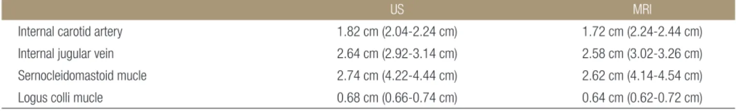
Table 1. Comparison of Ultrasound and MRI Measurements of the Width of the Cervical Structures
US MRI
Internal carotid artery 1.82 cm (2.04-2.24 cm) 1.72 cm (2.24-2.44 cm) Internal jugular vein 2.64 cm (2.92-3.14 cm) 2.58 cm (3.02-3.26 cm) Sernocleidomastoid mucle 2.74 cm (4.22-4.44 cm) 2.62 cm (4.14-4.54 cm) Logus colli mucle 0.68 cm (0.66-0.74 cm) 0.64 cm (0.62-0.72 cm) p<0.05.
생가능한 혈종 형성과 내고정물로 인한 연하곤란 여부를 파악 할 수 있으며 골격과골증에 의한 연하곤란을 동적으로 파악 할 수 있다고 한다.17,19,20)
또한, Lee 등18)은 만성경부통을 않는 환자에서 경추다열근의 수 축 시 면적 변화를 초음파와 자기공명영상 사이의 비교를 통하여 이해하려 하였다. 이러한 초음파를 사용한 길이 및 면적의 계측 은 구조물이 많은 경부의 해부학적 관찰을 용이하게 하며, 실시 간 관찰하여 침습적인 시술을 돕는데 다른 영역에서보다 기타 구 조물의 손상을 줄일 수 있는 계기가 될 수 있다.
본 연구에서는 Lee 등18)의 연구에서와는 달리 부피에서는 자기 공명영상의 결과와 일치하지 않았는데 이는 초음파의 이차원적 인 특성 때문인 것으로 생각 되었다. 반면 계측된 경부 구조물 중 경장근의 두께 측정만이 유의하게 자기공명영상과 유의한 수준 의 일치를 보였다.
이는 초음파를 이용한 경추부 신경 차단술이나 다른 침습적 시 술 시 경장근이 비교적 고정된 구조물로서 지표(landmark)가 될 수 있으며, 계측치를 고려하여 자기공명영상과 차이를 인지할 수 있다는 점에서 임상적 효용이 있다 하겠다.
자기공명영상이 대상을 수 밀리미터(mm) 간격으로 임의적으 로 절단하여 영상을 얻은 후 이에 대한 파악이 이루어지며 신호 강도의 차이로 높은 민감도와 특이도를 가지는데 비하여 초음파 는 실시간, 동적으로 도플러를 이용하여 연부조직에 대한 파악이 가능하다는 점과 비교적 가격이 싸며, 비침습적이라는 점을 생각 한다면 진단이나 치료 목적에서의 활용도는 보다 증가할 것으로 기대된다.18,21)
결 론
초음파를 이용하여 경추부 주요 구조물들의 위치를 파악할 수 있 으나, 자기공명영상에서의 결과와 비교했을 때 일부 차이가 있었 으며, 이는 초음파 측정 시에 발생한 오차로 생각된다. 이에 초음 파를 이용하여 경추부 구조물의 두께 측정이 유용할 것으로 생각 되고 발생 가능한 오차를 줄이기 위한 노력과 함께 술기의 보완 이 이루어진다면 초음파를 이용한 경추부 구조 탐색은 향후 진단 및 치료에 있어 많은 도움이 될 것으로 생각된다.
참고문헌
1. Nofsinger C, Konin JG. Diagnostic ultrasound in sports medi- cine: current concepts and advances. Sports Med Arthrosc.
2009;17:25-30.
2. Hodges PW, Pengel LH, Herbert RD, Gandevia SC. Measure- ment of muscle contraction with ultrasound imaging. Muscle Nerve. 2003;27:682-92.
3. McMeeken JM, Beith ID, Newham DJ, Milligan P, Critchley DJ. The relationship between EMG and change in thickness of transversus abdominis. Clin Biomech (Bristol, Avon).
2004;19:337-42.
4. Bunce SM, Moore AP, Hough AD. M-mode ultrasound: a reliable measure of transversus abdominis thickness? Clin Biomech (Bristol, Avon). 2002;17:315-7.
5. Emshoff R, Bertram S, Strobl H. Ultrasonographic cross- sectional characteristics of muscles of the head and neck. Oral Surg Oral Med Oral Pathol Oral Radiol Endod. 1999;87:93- 106.
6. Kristjansson E. Reliability of ultrasonography for the cervical multifi dus muscle in asymptomatic and symptomatic subjects.
Man Th er. 2004;9:83-8.
7. Jacobson JA. Musculoskeletal ultrasound and MRI: which do I choose? Semin Musculoskelet Radiol. 2005;9:135-49.
8. Iannotti JP, Ciccone J, Buss DD, et al. Accuracy of offi ce-based ultrasonography of the shoulder for the diagnosis of rotator cuff tears. J Bone Joint Surg Am. 2005;87:1305-11.
9. Shim DM, Kim TK, Oh SK, Choi YH, Lee SJ. Eff ectiveness of selective nerve root block on the need for surgical treatment of lumbar spine: a minimum 5 years follow up. J Korean Or- thop Assoc. 2009;44:118-22.
10. Davies MJ, Silbert BS, Scott DA, Cook RJ, Mooney PH, Blyth C. Superficial and deep cervical plexus block for carotid ar- tery surgery: a prospective study of 1000 blocks. Reg Anesth.
1997;22:442-6.
11. Dhonneur G, Saidi NE, Merle JC, Asfazadourian H, Ndoko SK, Bloc S. Demonstration of the spread of injectate with deep cervical plexus block: a case series. Reg Anesth Pain Med.
2007;32:116-9.
12. Saranteas T, Paraskeuopoulos T, Anagnostopoulou S, Kanel- lopoulos I, Mastoris M, Kostopanagiotou G. Ultrasound anat- omy of the cervical paravertebral space: a preliminary study.
Surg Radiol Anat. 2010;32:617-22.
13. Suk SI. Textbook of spinal surgery. 2nd ed. Korea; 2004.
14. Martinoli C, Bianchi S, Santacroce E, Pugliese F, Graif M, Der- chi LE. Brachial plexus sonography: a technique for assessing the root level. AJR Am J Roentgenol. 2002;179:699-702.
15. Hashimoto BE, Kramer DJ, Wiitala L. Applications of muscu- loskeletal sonography. J Clin Ultrasound. 1999;27:293-318.
16. Iannotti JP, Ciccone J, Buss DD, et al. Accuracy of offi ce-based ultrasonography of the shoulder for the diagnosis of rotator cuff tears. J Bone Joint Surg Am. 2005;87:1305-11.
17. Zhao J, Quan Z, Ou Y, Jiang D. Prevention and treatment of early postoperative complications of anterior cervical spi- nal surgery. Zhongguo Xiu Fu Chong Jian Wai Ke Za Zhi.
2008;22:901-4.
18. Lee JP, Tseng WY, Shau YW, Wang CL, Wang HK, Wang SF. Measurement of segmental cervical multifidus contrac- tion by ultrasonography in asymptomatic adults. Man Ther.
2007;12:286-94.
19. Urrutia J, Bono CM. Long-term results of surgical treatment
of dysphagia secondary to cervical diff use idiopathic skeletal hyperostosis. Spine J. 2009;9:e13-7.
20. Miyamoto K, Sugiyama S, Hosoe H, Iinuma N, Suzuki Y, Shimizu K. Postsurgical recurrence of osteophytes causing dysphagia in patients with diff use idiopathic skeletal hyperos- tosis. Eur Spine J. 2009;18:1652-8.
21. Sinnatamby CS. Head and neck and spine. In: Sinnatamby CS, ed. Last’s anatomy, 11th ed. London: Churchill Livingstone;
2006. 341-72.
Comparison of Ultrasonography and MRI in Measuring of Cervical Soft Tissue Structure
Dae Moo Shim, M.D., Ph.D., Tae Kyun Kim, M.D., Ph.D., Seok Jung Lee, M.D., Ph.D., and Seung Yeop Song, M.D.
Department of Orthopaedic Surgery, School of Medicine, Wonkwang University, Iksan, Korea
Purpose: We wanted to determine the usefullness of ultrasonography for exploring cervical anatomical structures and to compare
its results with those of magnetic resonance imaging.Materials and Methods: Between May, 2008 and January, 2009, 16 patient (8 male patients and 8 female patients, average
age: 55.5 years old) with neck pain following in out patient clinic and who had cervical MRI performed were selected. By using ultrasonography, we measured the shortest distance of the internal carotid artery, internal jugular vein, sternocleidomastoid muscle and longus colli muscle based on the vertebral body of cricoid cartilage. We also measured the thickness of the longus colli muscle.We measured the average length of the shortest distance and thickness of C6 on MRI axial view.
Results: The length using ultrasonography showed that the average length was 2.12 cm for the internal carotid artery, 3.04 cm for
the internal jugular vein, 4.34 cm for the sternocleidomastoid muscle and 0.68 cm for the longus colli muscle. Cervical MRI was used to measure the same structures. Its results were 2.23 cm for the internal carotid artery, 3.14 cm for the internal jugular vein, 4.39 cm for the sternocleidomastoid muscle and 0.70 cm for the longus colli muscle. We also measured the thickness of the longus colli muscle with ultrasonography and cervical MRI , and the results were 0.77 cm and 0.76 cm, respectively. There was no statistically significant difference between ultrasonography and MRI for the measurement of length, but there was a statistically significant difference for the measurement of thickness.Conclusion: There was a signifi cant difference for the measurement of cervical anatomical stuctures using ultrasonography, and
especially the longus coli thickness, as compared with that of MRI. Therefore, we suggest that ultrasonography can give more useful information for the diagnosis and treatment of problems in the cervical spinal fi eld.Key words: cervical spine, ultrasonography, MRI
Received August 17, 2010 Accepted April 27, 2011 Correspondence to: Tae Kyun Kim, M.D.
Department of Orthopaedic Surgery, School of Medicine, Wonkwang University, 344-2, Shinyong-dong, Iksan 570-749, Korea