INTRODUCTION
Muscle weakness is a common impairment among patients
with cerebral palsy (CP) and is associated with difficulties per- forming functional activities.1,2 It is known that the weakness found in CP is attributable to both altered neural mechanisms and muscle tissue changes.3 Lower limb muscle strength is re- duced by 6–59% compared with peers of typical development, depending on the muscle group.4
Many clinicians have regarded spasticity as a major factor that contributes to functional limitations and abnormal move- ment patterns.5,6 However, recent studies now recognize weak- ness as a prominent impairment observed in patients with CP.1,7,8 Therefore, many researchers have reported a relationship between muscle strength and motor function, and there is in- creasing interest in measuring muscle strength among patients with CP.2,4,7,9-11 A number of studies have shown that muscle
Relationships between Isometric Muscle Strength, Gait Parameters, and Gross Motor Function Measure in Patients with Cerebral Palsy
Hyung-Ik Shin1*, Ki Hyuk Sung2*, Chin Youb Chung2, Kyoung Min Lee2, Seung Yeol Lee3, In Hyeok Lee4, and Moon Seok Park2
1Department of Rehabilitation Medicine, Seoul National University Hospital, Seoul;
2Department of Orthopaedic Surgery, Seoul National University Bundang Hospital, Seongnam;
3Department of Orthopaedic Surgery, Ewha Womans University Mokdong Hospital, Seoul;
4Department of Orthopaedic Surgery, Sungkyunkwan University Samsung Changwon Hospital, Changwon, Korea.
Purpose: This study investigated the correlation between isometric muscle strength, gross motor function, and gait parameters in patients with spastic cerebral palsy and to find which muscle groups play an important role for gait pattern in a flexed knee gait.
Materials and Methods: Twenty-four ambulatory patients (mean age, 10.0 years) with spastic cerebral palsy who were scheduled for single event multilevel surgery, including distal hamstring lengthening, were included. Preoperatively, peak isometric muscle strength was measured for the hip flexor, hip extensor, knee flexor, and knee extensor muscle groups using a handheld dyna- mometer, and three-dimensional (3D) gait analysis and gross motor function measure (GMFM) scoring were also performed.
Correlations between peak isometric strength and GMFM, gait kinematics, and gait kinetics were analyzed.
Results: Peak isometric muscle strength of all muscle groups was not related to the GMFM score and the gross motor function classification system level. Peak isometric strength of the hip extensor and knee extensor was significantly correlated with the mean pelvic tilt (r=-0.588, p=0.003 and r=-0.436, p=0.033) and maximum pelvic obliquity (r=-0.450, p=0.031 and r=-0.419, p=0.041). There were significant correlations between peak isometric strength of the knee extensor and peak knee extensor mo- ment in early stance (r=0.467, p=0.021) and in terminal stance (r=0.416, p=0.043).
Conclusion: There is no correlation between muscle strength and gross motor function. However, this study showed that muscle strength, especially of the extensor muscle group of the hip and knee joints, might play a critical role in gait by stabilizing pelvic motion and decreasing energy consumption in a flexed knee gait.
Key Words: Muscle strength, gross motor function, gait parameter, cerebral palsy Yonsei Med J 2016 Jan;57(1):217-224
http://dx.doi.org/10.3349/ymj.2016.57.1.217 pISSN: 0513-5796 · eISSN: 1976-2437
Received: December 17, 2014 Revised: April 7, 2015 Accepted: May 28, 2015
Corresponding author: Dr. Moon Seok Park, Department of Orthopaedic Surgery, Seoul National University Bundang Hospital, 82 Gumi-ro 173beon-gil, Bundang-gu, Seongnam 13620, Korea.
Tel: 82-31-787-7203, Fax: 82-31-787-4056, E-mail: [email protected]
*Hyung-Ik Shin and Ki Hyuk Sung contributed equally to this work.
•The authors have no financial conflicts of interest.
© Copyright: Yonsei University College of Medicine 2016
This is an Open Access article distributed under the terms of the Creative Com- mons Attribution Non-Commercial License (http://creativecommons.org/ licenses/
by-nc/3.0) which permits unrestricted non-commercial use, distribution, and repro- duction in any medium, provided the original work is properly cited.
strength is significantly associated with gross motor function and that strength is more highly related to function than spas- ticity.7 In addition, various studies have reported that muscle- strengthening exercises may result in functional improve- ments.2,8,12,13 On the other hand, some studies reported that muscle-strengthening training did not improve functional ac- tivity,14,15 and other components, such as balance and coordi- nation, may influence the functional ability to a greater degree than muscle strength alone. Therefore, it is questionable wheth- er muscle strength is actually related to motor function.
This study investigated the correlations between isometric muscle strength and gross motor function, gait kinematics, and gait kinetics among ambulatory patients with spastic CP. In ad- dition, we aimed to identify the muscle groups that play an im- portant role in gait pattern in a flexed knee gait.
MATERIALS AND METHODS
This study was approved by the Institutional Review Board at our institution (a tertiary referral center for CP). Informed con- sent was obtained from the patients and relevant guardians.
The inclusion criteria were as follows: 1) consecutive ambula-
tory patients with spastic CP [gross motor function classifica- tion system (GMFCS) level I–III], 2) patients with flexed knee gait, who were scheduled for a single event multilevel surgery, including distal hamstring lengthening, 3) patients who had gross motor function measure (GMFM) scores and preopera- tive three-dimensional (3D) gait analysis, which are the primary and secondary outcome measures, respectively. Patients who had a history of gait correcting surgery and who were unable to accept and follow verbal instructions were excluded. Demo- graphic data, such as gender, age, height, weight, and GMFCS, were obtained from a review of medical records.
Measuring the isometric muscle strength
Peak isometric muscle strength was measured in all patients using a digital force dynamometer (Compact Force Gauge®, Mecmesin Corporation, West Sussex, UK) with a range of 0–500 N. Test position was standardized for each muscle group in such a way that gravitational forces minimally influenced the assessment.16 Investigated muscle groups were knee extensor, knee flexor, hip extensor, and hip flexor, and these were evalu- ated bilaterally. Dynamometer was anchored to a rigid frame and was connected to a patient’s limb with a strap. Peak iso- metric strength tests of the hip flexor and hip extensor muscle
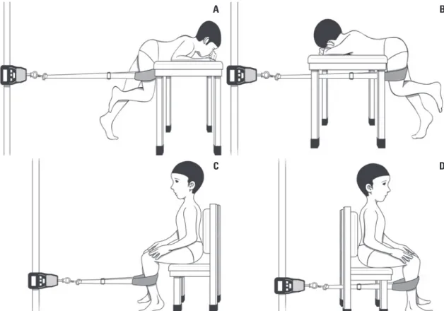
Fig. 1. Measurement of peak isometric strength for the hip flexor (A), hip extensor (B), knee flexor (C), and knee extensor (D) muscle group. (A and B) The leg to be measured is flexed at 90º at the hip and knee joint. The patient may hold the examination table and presses toward the ground with the other leg as depicted. The dynamometer is fixed to a rigid frame. The patient presses the knee slowly forward (A) or backward (B) against the band and the pelvis stays rigid. (C and D) Knee joint to be measured is flexed at 90º and hands are relaxed or folded. The band is placed on the lower leg, with its distal border as close as possible to the ankle joint and the dynamometer is fixed to a rigid frame. The patient presses the lower legs slowly backward (C) or forward (D) against the band. The trunk remains upright and no weight shifting is allowed.
A
C
B
D
group were carried out with patients in a prone standing posi- tion with the hip and knee in 90º flexion. In this position, the patients’ trunk was supported by the examination table and the standing leg, while the other leg was tested (Fig. 1A and B). Peak isometric strength tests of the knee flexor and extensor muscle group were performed with patients positioned in a supported sitting position with the hip and knee in 90º flexion (Fig. 1C and D). Children were given practice trials for each test until the in- vestigator was confident that they understood the task. They were instructed to increase their isometric muscle strength continuously and gradually by pushing or pulling the strap until the maximum strength plateau was reached. After practicing, each child performed three trials with an interval of approxi- mately 30 seconds for each muscle group, and the peak force values from the dynamometer were recorded. During the inter- val of each trial, we made efforts to relax the patients’ muscle due to the presence of spasticity.
All strength tests were conducted, and scores were recorded
a day before surgery by a researcher. The researcher was experi- enced in the measurements used in the study to assure stan- dardization of the procedures and accuracy of the measures.
The values for each muscle group were normalized by the indi- vidual’s body mass (N/kg) for statistical analysis because weight has been shown to be related to force in patients with CP.17 Gross motor function measure (GMFM)
The GMFM is a validated instrument designed to assess motor function in CP. It consists of 88 items within five dimensions: A, lying and rolling; B, sitting; C, crawling and kneeling; D, stand- ing; and E, walking, running, and jumping.18 A percentage score of each dimension and a total percentage score are provided.
The authors deemed dimensions D and E as the most relevant to this study in terms of functional abilities. Therefore, the pa- tients included in this study were scored by their physiothera- pist using GMFM dimensions D and E a few days before the surgery.
Fig. 2. Scatter plots of isometric muscle strength and gait kinematics.
30.00
20.00
10.00
0.00
-10.00
Mean pelvic tilt (degree)
Hip extensor (N/kg)
0.00 0.50 1.00 1.50 2.00
15.00
10.00
5.00
0.00
-5.00
Maximum pelvic obliquity (degree)
Hip extensor (N/kg)
0.00 0.50 1.00 1.50 2.00
30.00
20.00
10.00
0.00
-10.00
Mean pelvic tilt (degree)
Knee extensor (N/kg)
0.00 0.20 0.40 0.60 0.80 1.00
15.00
10.00
5.00
0.00
-5.00
Maximum pelvic obliquity (degree)
Knee extensor (N/kg)
0.00 0.20 0.40 0.60 0.80 1.00
3D gait analysis
3D Gait analysis was performed a few days before surgery using a motion analysis system (Motion Analysis, Santa Rosa, CA, USA) equipped with seven charge-coupled device cameras and two force plates.19 Markers were placed for the Helen Meyer marker set20 by an experienced operator. Patients were asked to walk three times barefooted on a 9 m walkway. Average kine- matic and kinetic data and temporal parameters were archived.
Temporal parameters, such as stride length, cadence, and walk- ing speed were normalized according to the patients’ height for analysis.
Statistical analysis
The prior sample size for the reliability testing was calculated to determine the minimum number of patients required. The in- traclass correlation coefficients (ICCs) were used for reliability testing at a target value of 0.85 and a 95% confidence interval of 0.2 for three trials. The minimal sample size needed was 20 us- ing Bonett’s approximation.21 For the purpose of statistical in- dependence, one limb from patients with diplegia or quadri- plegia and the involved limb from patients with hemiplegia were included for statistical analysis.22
Descriptive statistics were used to summarize the patients’
demographics. The ICCs and their 95% confidence intervals were used to summarize the intra-session reliability of the peak isometric muscle strength of each muscle group and were cal- culated in the setting using a two-way random effect model, as- suming a single measurement and absolute agreement.23
Differences in peak muscle strength values according to walking ability (GMFCS level) were analyzed using the Kruscal- Wallis test. Correlations between peak isometric strength in each muscle group and GMFM, gait kinematics, and gait kinet- ics were determined using Spearman correlation coefficients.
Statistical analysis was performed using SPSS version 20.0 for Windows (IBM Co., Chicago, IL, USA), and null hypotheses of no difference were rejected if the p values were <0.05.
Source of funding
There was no external funding source for this investigation.
RESULTS
Thirty-three consecutive patients were initially enrolled. After testing against the exclusion criteria, 24 patients were ultimately included in this study. There were 13 male and 11 female pa- tients. Six patients had hemiplegia and 18 had diplegia. The mean patient age was 10.0±5.2 years (range, 5.3 to 19.6 years).
There were 6 with GMFCS level I, 13 with GMFCS level II, and 5 with GMFCS level III. The mean height of the patients was 129.6±19.7 cm and the mean weight was 31.8±15.6 kg (Table 1).
All patients underwent distal hamstring lengthening for flexed knee deformity and tendo-Achilles lengthening for equinus
foot deformity. Of these, 18 patients (75%) underwent rectus femoris transfer for a stiff knee gait due to quadriceps spasticity.
In term of intra-session reliability of the peak isometric strength measurements, the ICC values were 0.975 (95% confi- dence interval, 0.951 to 0.988), 0.973 (95% confidence interval, 0.948 to 0.987), 0.986 (95% confidence interval, 0.972 to 0.993), and 0.974 (95% confidence interval, 0.949 to 0.988) for the hip flexor, hip extensor, knee flexor, and knee extensor group, re- spectively.
There was no correlation between the GMFM score and nor- malized isometric strength of all muscle groups (Table 2). Peak isometric muscle strength of all muscle groups was not related to the GMFCS level (Table 3).
The mean knee flexion contracture was 1.7±4.6 degrees Table 1. Demographics, GMFM, and Peak Isometric Strength of Patients
Age (yr) 10.0±5.2 (range 5.3 to 19.6)
Gender (M/F) 13/11
Type (hemi/diplegia) 6/18
GMFCS level (I/II/III) 6/13/5
Height (cm) 129.6±19.7
Weight (kg) 31.8±15.6
GMFM-D (%) 80.6±14.6
GMFM-E (%) 72.8±20.4
Muscle group Peak isometric strength
Hip flexor (N) 8.61±6.65
Hip flexor (N/kg) 0.27±0.12
Hip extensor (N) 15.67±12.54
Hip extensor (N/kg) 0.52±0.35
Knee flexor (N) 6.54±5.58
Knee flexor (N/kg) 0.21±0.15
Knee extensor (N) 10.15±5.47
Knee extensor (N/kg) 0.34±0.20
GMFM, gross motor function measure; GMFCS, gross motor function classifi- cation system.
Data are presented as mean±SD.
Table 2. Correlations between Peak Isometric Muscle Strength and GMFM
Muscle groups GMFM-D GMFM-E
r p value r p value
Hip flexor 0.147 0.503 0.326 0.130
Hip extensor 0.121 0.582 0.324 0.132
Knee flexor 0.178 0.406 0.360 0.084
Knee extensor 0.057 0.792 0.219 0.304
GMFM, gross motor function measure.
Table 3. Peak Isometric Muscle Strength According to GMFCS Levels Muscle groups GMFCS I GMFCS II GMFCS III p value Hip flexor (N/kg) 0.29±0.03 0.29±0.04 0.18±0.03 0.292 Hip extensor (N/kg) 0.59±0.09 0.59±0.12 0.21±0.05 0.147 Knee flexor (N/kg) 0.27±0.06 0.23±0.05 0.11±0.05 0.265 Knee extensor (N/kg) 0.39±0.08 0.36±0.07 0.23±0.03 0.321 GMFCS, gross motor function classification system.
Data are presented as mean±SD.
(range, 0 to 20 degrees) and was significantly related to knee flexion at initial contact (r=0.527, p=0.008). Peak isometric strength of hip extensor and knee extensor was significantly cor- related with the mean pelvic tilt (r=-0.588, p=0.003 and r=-0.436, p=0.033) and maximum pelvic obliquity (r=-0.450, p=0.031 and r=-0.419, p=0.041) (Fig. 2). There was a significant correlation between the peak isometric strength of hip extensor and maxi- mum hip flexion in swing (r=-0.542, p=0.008). Peak isometric strength of the knee extensor was related to the knee flexion at initial contact (r=0.477, p=0.018), minimum knee flexion in stance (r=0.527, p=0.008), and knee flexion at terminal swing (r=0.467, p=0.012) (Table 4).
There were significant correlations between peak isometric strength of the knee extensor and peak knee extensor moment
in early stance (r=0.467, p=0.021) and in terminal stance (r=0.416, p=0.043). Peak knee absorption power in terminal swing was related to isometric strength of all included muscle groups (r=0.469, 0.481, 0.494, and 0.468) (Table 4).
DISCUSSION
We investigated the association between maximal isometric muscle strength and gross motor function, gait kinematics, and gait kinetics in patients with spastic CP. Our study showed no correlation between isometric muscle strength and gross motor function. However, we found that higher muscle strength, espe- cially the hip extensor and knee extensor, decreased the pelvic Table 4. Correlations between Peak Isometric Muscle Strength and Gait Kinetics
Gait parameters Hip flexor Hip extensor Knee flexor Knee extensor
r p value r p value r p value r p value
Stride length -0.078 0.725 -0.080 0.718 0.123 0.568 -0.020 0.927
Cadence -0.181 0.408 -0.068 0.757 -0.131 0.541 -0.100 0.642
Walking speed -0.138 0.530 -0.117 0.595 -0.032 0.882 -0.195 0.362
Minimum pelvic tilt -0.201 0.357 -0.581 0.004 -0.167 0.436 -0.482 0.017
Maximum pelvic tilt -0.178 0.416 -0.558 0.006 -0.100 0.642 -0.392 0.058
Mean pelvic tilt -0.203 0.354 -0.588 0.003 -0.136 0.526 -0.436 0.033
Minimum pelvic obliquity 0.267 0.218 0.128 0.562 0.180 0.401 -0.051 0.812
Maximum pelvic obliquity -0.197 0.368 -0.450 0.031 -0.202 0.344 -0.419 0.041
Mean pelvic obliquity 0.053 0.811 -0.194 0.375 -0.008 0.970 -0.270 0.202
Minimum hip flexion -0.066 0.764 -0.286 0.186 0.023 0.917 -0.050 0.818
Maximum hip flexion in stance -0.189 0.387 -0.362 0.089 -0.013 0.950 -0.096 0.655
Maximum hip flexion in swing -0.349 0.103 -0.542 0.008 -0.235 0.268 -0.282 0.182
Knee flexion at initial contact -0.001 0.997 0.301 0.163 0.108 0.617 0.477 0.018
Minimum knee flexion in stance 0.164 0.456 0.469 0.024 0.163 0.447 0.527 0.008
Peak knee flexion in swing -0.331 0.123 -0.176 0.421 -0.277 0.191 -0.095 0.659
Knee flexion at terminal swing 0.028 0.900 0.301 0.163 0.113 0.598 0.467 0.021
Hip flexion/extension moment
Peak extensor in early stance 0.226 0.229 -0.076 0.730 0.208 0.330 -0.128 0.551
Peak flexor in stance -0.059 0.791 0.064 0.772 -0.009 0.968 0.155 0.470
Peak extensor in swing 0.202 0.356 -0.089 0.687 0.043 0.842 -0.263 0.215
Hip flexion/extension power
Peak generation in early stance 0.168 0.444 -0.092 0.677 0.187 0.381 -0.045 0.835
Peak absorption in mid-stance 0.312 0.147 0.267 0.218 0.186 0.384 0.229 0.281
Peak generation in terminal stance -0.062 0.778 -0.195 0.373 -0.078 0.715 -0.213 0.318
Knee flexion/extension moment
Peak extensor in early stance 0.192 0.381 0.414 0.049 0.322 0.125 0.467 0.021
Peak Flexor in mid-stance -0.087 0.685 0.256 0.239 -0.031 0.887 0.312 0.138
Peak extensor in terminal stance 0.090 0.684 0.283 0.190 0.180 0.400 0.416 0.043
Peak flexor in swing 0.120 0.586 0.135 0.540 0.094 0.661 0.255 0.230
Knee flexion/extension power
Peak absorption in early stance 0.362 0.090 0.083 0.707 0.196 0.359 0.028 0.896
Peak generation during stance 0.144 0.512 0.297 0.169 0.281 0.184 0.310 0.141
Peak absorption in terminal stance -0.074 0.738 -0.072 0.743 -0.046 0.830 -0.069 0.750
Peak absorption in terminal swing 0.469 0.024 0.481 0.020 0.494 0.014 0.468 0.021
tilt and the pelvic obliquity during walking, and this makes walking more energy-efficient in a flexed knee gait.
There are several limitations with our study. First, the results are valid for patients with GMFCS level I to III spastic CP. These results cannot be generalized to children with lower functional levels or with other clinical sub-types of CP. However, children at GMFCS level IV and V are unable to participate in muscle strength measurements because of muscle weakness and cog- nitive difficulties. Second, in measuring muscle strength, the other muscle groups that also play an important role in gait and gross motor function, such as the ankle dorsiflexor and ankle plantarflexor,7,24 were not included. However, all patients in this study had fixed equinus foot deformity; for these patients, it is difficult to objectively measure ankle plantar flexor and dorsi- flexor strength using a digital force dynamometer. Therefore, we excluded the ankle plantar flexors and dorsiflexors and fo- cused on the flexor and extensor muscle groups of the hip and knee joints. Third, the isometric strength assessments used do not contain any information about dynamic muscle function, and walking may differ from the isometric test results at fixed joint angles. Isokinetic strength, which involves joint move- ments rather than isometric muscle strength, may be more re- lated to motor function.17 Fourth, no control was recruited for the comparison of muscle strength for typically developing per- sons. We hypothesized that the muscle strength of patients with CP is lower than of normally developing persons as demon- strated in previous studies.1,4,25 Fifth, gait performance varies according to the geographical type of CP. This study included patients with hemiplegia and diplegia. However, we believe that muscle strength is more closely related to patients’ func- tional levels than to the geographical type of CP. Therefore, the
current study included ambulatory patients (GMFCS I, II, and III) with a homogenous gait pattern consisting of a flexed knee gait. Consequently, we think that investigating the relationship in this cohort is meaningful. Sixth, spasticity might be an im- portant factor in the evaluation of gross motor function and gait pattern. However, recent studies reported that muscle strength may have a greater influence than spasticity on gross motor function and gait pattern. Therefore, we did not evaluate spas- ticity but instead focused on the muscle strength when investi- gating gait pattern.
Measuring isometric muscle strength with a dynamometer is an easy method to measure muscle strength in clinical practice.
Recently, several authors have shown that it can be measured with sufficient reliability in patients with CP,11,25-27 including our study, which demonstrated excellent intra-session reliability with ICC values ranging from 0.973 to 0.984.
Previous studies have investigated the relationship between muscle strength and gross motor function in patients with CP (Table 5). They reported a significant correlation between mus- cle strength and the GMFM, indicating that muscle strength/
weakness affects walking ability.2,4,7,9-11 They have also found a statistically significant difference in muscle strength between the GMFCS levels.4,28 Of these studies, Damiano and Abel2 used absolute strength values without normalization and the mean values of both extremities for the analysis. They found that the mean strength had significant correlation with total GMFM.
Ross and Engsberg7 measured the isokinetic muscle strength, which may be related more to Dimensions D and E of GMFM than the isometric muscle strength. They found that aggregate strength was highly related to the total GMFM. Goh, et al.10 re- cruited the school-aged (range, 7 to 12 years) children with
Table 5. Previous Studies on Correlation between Muscle Strength and Gross Motor Function
Study No. of subjects Muscle group GMFM total GMFM-D GMFM-E
r p value r p value r p value
Damiano and Abel2 11 - 0.59 0.05 - - - -
Eek and Beckung4 55
Hip extensor 0.59
<0.01
0.43
<0.01
0.54
<0.01
Hip flexor 0.73 0.59 0.61
Hip abductor 0.71 0.61 0.66
Hip adductor 0.64 0.56 0.63
Knee extensor 0.52 0.48 0.53
Knee flexor 0.76 0.69 0.77
Ankle dorsiflexor 0.68 0.68 0.67
Ankle plantarflexor 0.80 0.72 0.76
Damiano, et al.9 10 Knee extensor 0.57 <0.05 - - - -
Goh, et al.10 27 Knee extensor 0.78
<0.01 0.75
<0.01 0.77
<0.01
Knee flexor 0.84 0.82 0.81
Ross and Engsberg7 97 - 0.83 <0.01 0.81 <0.01 - -
Berry, et al.11 15
Hip abductor
- -
0.64 0.014 0.64 0.014
Knee extensor 0.38 0.16 0.38 0.16
Knee flexor 0.48 0.08 0.60 0.023
GMFM, gross motor function measure.
spastic diplegia, and reported that the strength of quadriceps and hamstring had significant relationships with total GMFM.
However, our present study did not show a correlation between GMFM score and isometric muscle strength of the included muscle groups. In addition, there was no difference in the peak isometric muscle strength based on the patients’ GMFCS levels.
We believe that if the muscle strength of patients exceeds a cer- tain level, it may not provide any additional advantage in their functional abilities. In addition, other components such as bal- ance and coordination may influence functional ability to a greater degree than muscle strength alone.
Muscle strength has previously been found to correlate with kinematic gait variables, walking velocity, and stride length.2,7,29 In this study, there was no correlation between isometric mus- cle strength and normalized temporal parameters, such as stride length, cadence, and walking speed, contrary to the re- sults of other studies. A significant correlation was found be- tween strength of the hip extensor and the pelvic mean anterior tilt (r=-0.14),29 and between aggregate strength and the pelvic tilt (r=-0.50).7 Our present study revealed a significant correla- tion between peak isometric strength of the hip extensor and knee extensor, and the pelvic tilt and pelvic obliquity. These re- sults infer that the extensor muscles of the hip and knee joint might play a critical role in gait by stabilizing the pelvic motion during walking.
There have been several reports investigating the relation- ships between muscle strength and kinetic gait parame- ters.24,29,30 Eek, et al.24 reported that there was a significant corre- lation between muscle strength in almost all muscle groups and plantarflexing gait moments and ankle generating power in children with CP, and that ankle plantarflexors were impor- tant muscles with weakness that affects the kinetic gait pattern.
Desloovere, et al.29 showed weak correlations between lower limb strength and joint kinetics in children with CP. Dallmeijer, et al.30 found a significant difference between isometric hip flexor strength and the peak hip flexor moment during gait in patients with diplegic CP. No significant relationships between the isometric strength of the other lower limb muscle groups and corresponding peak moment during gait were found. In this study, the knee extensor moments in the early and terminal stances were significantly related to the isometric strength of the knee extensor. This study included only patients with a flexed knee gait. The flexed knee locates the ground reaction force posterior to the knee joint. If the extensor muscle of the hip or knee does not produce sufficient extension moments, the flexed knees become further flexed and the energy con- sumption is increased. Our findings demonstrated that greater strength of the knee extensor muscle produces sufficient knee extensor moments in the stance and might decrease energy consumption during walking. There is a significant correlation between isometric muscle strength in all of the measured mus- cle groups and the peak knee absorption power in the terminal swing. The power curves of the joint kinetics estimate the work
performed at the joint during a single gait cycle.31 Therefore, these results indicate that the greater the muscle strength, the less eccentric work is performed, and that greater muscle strength makes walking more energy efficient.
In conclusion, there is no evidence of a correlation between muscle strength and gross motor function. However, this study showed that muscle strength, especially of the extensor muscle group of the hip and knee joints, might play a critical role in gait by stabilizing pelvic motion and decreasing energy consump- tion in a flexed knee gait. Further studies regarding changes in the muscle strength after a single event multilevel surgery and their relationships with gait kinematics and kinetics are needed.
REFERENCES
1. Wiley ME, Damiano DL. Lower-extremity strength profiles in spastic cerebral palsy. Dev Med Child Neurol 1998;40:100-7.
2. Damiano DL, Abel MF. Functional outcomes of strength training in spastic cerebral palsy. Arch Phys Med Rehabil 1998;79:119-25.
3. Mockford M, Caulton JM. The pathophysiological basis of weak- ness in children with cerebral palsy. Pediatr Phys Ther 2010;22:222- 33.
4. Eek MN, Beckung E. Walking ability is related to muscle strength in children with cerebral palsy. Gait Posture 2008;28:366-71.
5. Fasano VA, Broggi G, Barolat-Romana G, Sguazzi A. Surgical treat- ment of spasticity in cerebral palsy. Childs Brain 1978;4:289-305.
6. Bobath K. Neurophysiological basis for the treatment of cerebral palsy. 2nd ed. London: William Heimenamm Medical Books Ltd;
1980.
7. Ross SA, Engsberg JR. Relationships between spasticity, strength, gait, and the GMFM-66 in persons with spastic diplegia cerebral palsy. Arch Phys Med Rehabil 2007;88:1114-20.
8. Eek MN, Tranberg R, Zügner R, Alkema K, Beckung E. Muscle strength training to improve gait function in children with cere- bral palsy. Dev Med Child Neurol 2008;50:759-64.
9. Damiano DL, Martellotta TL, Sullivan DJ, Granata KP, Abel MF.
Muscle force production and functional performance in spastic cerebral palsy: relationship of cocontraction. Arch Phys Med Reha- bil 2000;81:895-900.
10. Goh HT, Thompson M, Huang WB, Schafer S. Relationships among measures of knee musculoskeletal impairments, gross motor function, and walking efficiency in children with cerebral palsy.
Pediatr Phys Ther 2006;18:253-61.
11. Berry ET, Giuliani CA, Damiano DL. Intrasession and intersession reliability of handheld dynamometry in children with cerebral pal- sy. Pediatr Phys Ther 2004;16:191-8.
12. Engsberg JR, Ross SA, Collins DR. Increasing ankle strength to im- prove gait and function in children with cerebral palsy: a pilot study. Pediatr Phys Ther 2006;18:266-75.
13. Lee JH, Sung IY, Yoo JY. Therapeutic effects of strengthening exercise on gait function of cerebral palsy. Disabil Rehabil 2008;30:1439-44.
14. Scholtes VA, Dallmeijer AJ, Rameckers EA, Verschuren O, Tempe- laars E, Hensen M, et al. Lower limb strength training in children with cerebral palsy--a randomized controlled trial protocol for functional strength training based on progressive resistance exer- cise principles. BMC Pediatr 2008;8:41.
15. Scianni A, Butler JM, Ada L, Teixeira-Salmela LF. Muscle strength- ening is not effective in children and adolescents with cerebral palsy: a systematic review. Aust J Physiother 2009;55:81-7.
16. Stoll T, Huber E, Seifert B, Stucki G, Michel BA. Isometric muscle
strength measurement. New York: Thieme Stuttgart; 2002.
17. MacPhail HE, Kramer JF. Effect of isokinetic strength-training on functional ability and walking efficiency in adolescents with cere- bral palsy. Dev Med Child Neurol 1995;37:763-75.
18. Russell DJ, Rosenbaum PL, Cadman DT, Gowland C, Hardy S, Jar- vis S. The gross motor function measure: a means to evaluate the effects of physical therapy. Dev Med Child Neurol 1989;31:341-52.
19. Chesnin KJ, Selby-Silverstein L, Besser MP. Comparison of an in- shoe pressure measurement device to a force plate: concurrent valid- ity of center of pressure measurements. Gait Posture 2000;12:128-33.
20. Kadaba MP, Ramakrishnan HK, Wootten ME. Measurement of lower extremity kinematics during level walking. J Orthop Res 1990;
8:383-92.
21. Bonett DG. Sample size requirements for estimating intraclass correlations with desired precision. Stat Med 2002;21:1331-5.
22. Park MS, Kim SJ, Chung CY, Choi IH, Lee SH, Lee KM. Statistical consideration for bilateral cases in orthopaedic research. J Bone Joint Surg Am 2010;92:1732-7.
23. Lee KM, Lee J, Chung CY, Ahn S, Sung KH, Kim TW, et al. Pitfalls and important issues in testing reliability using intraclass correla- tion coefficients in orthopaedic research. Clin Orthop Surg 2012;
4:149-55.
24. Eek MN, Tranberg R, Beckung E. Muscle strength and kinetic gait pattern in children with bilateral spastic CP. Gait Posture 2011;33:
333-7.
25. van der Linden ML, Aitchison AM, Hazlewood ME, Hillman SJ, Robb JE. Test-retest repeatability of gluteus maximus strength test- ing using a fixed digital dynamometer in children with cerebral palsy. Arch Phys Med Rehabil 2004;85:2058-63.
26. Crompton J, Galea MP, Phillips B. Hand-held dynamometry for muscle strength measurement in children with cerebral palsy. Dev Med Child Neurol 2007;49:106-11.
27. Taylor NF, Dodd KJ, Graham HK. Test-retest reliability of hand- held dynamometric strength testing in young people with cere- bral palsy. Arch Phys Med Rehabil 2004;85:77-80.
28. Thompson N, Stebbins J, Seniorou M, Newham D. Muscle strength and walking ability in diplegic cerebral palsy: implications for as- sessment and management. Gait Posture 2011;33:321-5.
29. Desloovere K, Molenaers G, Feys H, Huenaerts C, Callewaert B, Van de Walle P. Do dynamic and static clinical measurements cor- relate with gait analysis parameters in children with cerebral pal- sy? Gait Posture 2006;24:302-13.
30. Dallmeijer AJ, Baker R, Dodd KJ, Taylor NF. Association between isometric muscle strength and gait joint kinetics in adolescents and young adults with cerebral palsy. Gait Posture 2011;33:326-32.
31. Gage JR. The clinical use of kinetics for evaluation of pathologic gait in cerebral palsy. Instr Course Lect 1995;44:507-15.