http://e-nrp.org
Protective effect of Cordyceps militaris against hydrogen peroxide-induced oxidative stress in vitro
Mei Tong He
1, Ah Young Lee
2, Chan Hum Park
3and Eun Ju Cho
1§1Department of Food Science and Nutrition, Pusan National University, Busandaehak-ro 63 beon-gil 2, geumjeong-gu, Busan 46241, Korea
2Department of Food Science, Gyeongnam National University of Science and Technology, Jinju 52725, Korea
3Department of Medicinal Crop Research, National Institute of Horticultural and Herbal Science, Rural Development Administration, Eumseong 27709, Korea
BACKGROUND/OBJECTIVES: Excessive production of reactive oxygen species (ROS) such as hydroxyl (·OH), nitric oxide (NO), and hydrogen peroxide (H
2O
2) is reported to induce oxidative stress. ROS generated by oxidative stress can potentially damage glial cells in the nervous system. Cordyceps militaris (CM), a kind of natural herb widely found in East Asia. In this study, we investigated the free radical scavenging activity of the CM extract and its neuroprotective effects in H
2O
2-induced C6 glial cells.
MATERIALS/METHODS: The ethanol extract of CM (100-1,000 μg/mL) was used to measure DPPH, ·OH, and NO radical scavenging activities. In addition, hydrogen peroxide (H
2O
2)-induced C6 glial cells were treated with CM at 0.5-2.5 μg/mL for measurement of cell viability, ROS production, and protein expression resulting from oxidative stress.
RESULTS: The CM extract showed high scavenging activities against DPPH, ·OH, and NO radicals at concentration of 1,000 μg/mL. Treatment of CM with H
2O
2-induced oxidative stress in C6 glial cells significantly increased cell viability, and decreased ROS production. Cyclooxygenase-2 and inducible nitric oxide synthase protein expression was down-regulated in CM-treated groups. In addition, the protein expression level of phospho-p38 mitogen-activated protein kinase (p-p38 MAPK), phospho-c-Jun N-terminal kinase (p-JNK), and phospho-extracellular regulated protein kinases (p-ERK) in H
2O
2-induced C6 glial cells was down-regulated upon CM administration.
CONCLUSION: CM exhibited radical scavenging activity and protective effect against H
2O
2as indicated by the increased cell viability, decreased ROS production, down-regulation of inflammation-related proteins as well as p-p38, p-JNK, and p-ERK protein levels. Therefore, we suggest that CM could play the protective role from oxidative stress in glial cells.
Nutrition Research and Practice 2019;13(4):279-285; https://doi.org/10.4162/nrp.2019.13.4.279; pISSN 1976-1457 eISSN 2005-6168
Keywords: Cordyceps militaris, free radicals, neuroglia, hydrogen peroxide, oxidative stress
INTRODUCTION
1)The occurrence of oxidative stress is relevant to both free radicals and reactive oxygen species (ROS) [1,2]. Free radicals, such as superoxide (·O
2-), hydroxyl radical (·OH), nitric oxide (NO), and the non-radical oxidizing agents like hydrogen peroxide (H
2O
2) can generate ROS. Actually, under homeostasis, ROS participates in a series of cellular functional reactions.
However, in an excessively exposed situation, ROS becomes toxic and increases oxidative damage, especially being harmful for cells [3-5]. Cellular damage by oxidative stress disrupts the normal mechanisms of cellular signaling [6,7]. According to Gille and Joenje [8], disturbing the prooxidant-antioxidant balance is a way to induce oxidative stress. Generally, H
2O
2itself is stable in culture media; however, it can generate toxic ·OH in the cell through the transition metal-catalyzed reaction. Although cellular catalase has been reported to decompose H
2O
2to water and oxygen to prevent ·OH formation, the function of catalase
might loss balance when treated with high doses of H
2O
2and the catalase activity is inversely correlated with the cytotoxic effect of H
2O
2[9]. The exposure of cells to H
2O
2results in DNA damage and eventually causes cellular apoptosis or/and induces inflammatory responses [10]. These alterations of homeostasis cause cellular damage by H
2O
2-induced oxidative stress. In the central nervous system, glial cells play a crucial role in protec- ting neurons [11,12]. Oxidative stress in glial cells is closely related to neurodegenerative dysfunctions such as Alzheimer’s disease (AD) and Parkinson’s disease [13,14]. Therefore, research on the regulation of glial cell function and protection from oxidative stress has gained interest.
Oxidative stress occurs with inflammation and activates apoptosis pathways [12,15]. Inflammatory cells release cyclooxy- genase-2 (COX-2) and inducible nitric oxide synthase (iNOS) mediators, which are associated with age-related degenerative diseases [16,17]. Inflammatory cytokines activate mitogen- activated protein kinases (MAPKs) pathways, including c-Jun
§Corresponding Author: Eun Ju Cho, Tel. 82-51-510-2779, Fax. 82-51-583-3648, Email: [email protected] Received: September 5, 2018, Revised: November 8, 2018, Accepted: May 14, 2019
This is an Open Access article distributed under the terms of the Creative Commons Attribution Non-Commercial License (http://creativecommons.org/licenses/by-nc/3.0/) which permits unrestricted non-commercial use, distribution, and reproduction in any medium, provided the original work is properly cited.
NH
2-terminal kinase (JNK) and p38 MAPK, which are important for cellular proliferation and apoptosis. Moreover, the phos- phorylated JNK and p38 are known to regulate apoptotic cell death [18].
The edible mushroom Cordyceps militaris (CM) is a kind of traditional medical herb widely used for hundred years in Asia.
Several studies have shown that CM has exerted functions on immunomodulatory, neuroprotection, anti-bacterial and anti- tumor properties [19-21]. A previous research indicated the protective effect of CM against oxidative damage under high glucose induction conditions [22]. According to Ng and Wang [23], the Cordyceps species were reported to contain several active compounds, including cordycepin, ergosterol, and poly- saccharides. Of these, the antioxidant property of cordycepin isolated from CM (3’-deoxyadenosine, C
10H
13N
5O
3) has been studied recently [24,25]. Ramesh et al. [24] have reported that cordycepin attenuates oxidative stress and exerts antioxidant capacity in vivo. Moreover, polysaccharides isolated from CM have also demonstrated to inhibit mitochondrial injury and improve immune responses by scavenging ROS [26,27]. In addition, CM has been reported to inhibit ROS generation and protect against oxidative stress induced in human dermal fibroblast cells [28]. The present study aimed to investigate the protective effect of CM against H
2O
2-induced oxidative stress and explore the related molecular mechanisms against oxidative stress and apoptosis in C6 glial cells.
MATERIALS AND METHODS Sample and experimental groups
The ethanol extract of CM was obtained from the National Institute of Horticultural and Herbal Science (RDA, Jeolabuk-do, Korea). CM was dissolved in dimethyl sulfoxide (DMSO) to prepare a stock solution and further diluted with Dulbecco’s modified Eagle’s medium (DMEM) before the experiment. The experimental groups of C6 glial cells were categorized as follows:
(1) Normal: no treatment with CM or H
2O
2; (2) Control: treatment with 300 μM H
2O
2;
(3) 0.5 μg/mL: treatment with CM (0.5 μg/mL) + H
2O
2(300 μM);
(4) 1 μg/mL: treatment with CM (1 μg/mL) + H
2O
2(300 μM);
(5) 1.25 μg/mL: treatment with CM (1.25 μg/mL) + H
2O
2(300 μM);
(6) 2.5 μg/mL: treatment with CM (2.5 μg/mL) + H
2O
2(300 μM).
Reagents
Griess reagent, 1,1-Diphenyl-2-picrylhydrazyl (DPPH), 2-deoxyri- bose, 3-(4,5-Dimethylthiazol-2-yl)-2,5-diphenyltetrazolium bromide (MTT), dichlorofluorescin diacetate (DCF-DA), and DMSO were purchased from Sigma Aldrich Co. (St. Louis, MO, USA).
Thiobarbituric acid (TBA) and trichloroactetic acid (TCA) were obtained from Acros Organics (Kangnam-gu, Seoul, Korea) and Kanto Chemical Co. (Tokyo, Japan), respectively. H
2O
2and sodium pentacyanonitrosylferrate (III) dihydrate (SNP) were purchased from Junsei Chemical Co. (Tokyo, Japan). DMEM, fetal bovine serum (FBS), and penicillin-streptomycin were acquired from Welgene (Daegu, Korea). Radioimmunoprecipitation assay (RIPA) buffer and the protein marker were obtained from Elpis
Biotech (Daejeon, Korea). The polyvinylidene fluoride (PVDF) membrane was purchased from Millipore (Billerica, MA, USA).
Primary and secondary antibodies were purchased from Cell Signaling Technology (Beverly, MA, USA) and Santa Cruz Biotechnology (Santa Cruz, MA, USA), respectively.
DPPH assay
The DPPH radical scavenging assay was performed as described previously by Hatano et al. [29]. Sixty micromoles of the DPPH solution were added following treatment of the sample (100, 250, 500, 1,000 μg/mL). The absorbance was measured at 540 nm following incubation for 30 min at room temperature.
DPPH radical scavenging activity (%)
= (Abs
control- Abs
sample/Abs
control) × 100
·OH assay
The scavenging activity of the ·OH radical was measured according to the method described by Chung et al. [30]. The CM extract was mixed with a solution of 10 mM FeSO
4· H
2O-EDTA, 10 mM 2-deoxyribose, and 10 mM H
2O
2. The sample mixture was incubated at 37ºC for 4 h and was added to a solution containing 1.0% TBA and 2.8% TCA. The solution was boiled for 20 min and cooled in ice bath. The absorbance was measured at 490 nm.
·OH radical scavenging activity (%)
= [1-(Abs
sample-Abs
blank)/(Abs
control-Abs
blank)] × 100 NO assay
The scavenging activity of the NO radical was measured according to the method described by Marcocci et al. [31]. The sample (100, 250, 500, 1,000 μg/mL) was mixed with 5 mM SNP solution and incubated for 150 min [31], after which it was mixed with Griess reagent in a ratio of 1:1 and incubated at room temperature for 30 min. The absorbance was measured at 540 nm.
Cell culture
C6 glial cells were obtained from the Korea Cell Line Bank (Seoul, Korea) and were cultured in T75-flasks using DMEM, 10%
FBS, and 100 U/mL penicillin- streptomycin at 37ºC under an atmosphere of 5% CO
2. For subculture, the cell culture medium was replaced daily by using 0.05% trypsin-EDTA.
Measurement of cell viability
Cell viability was determined using the MTT assay as described by Mosmann [32]. C6 glial cells were seeded in a 96-well plate at a density of 5 × 10
4per well and incubated for 24 h. Different concentrations of CM (0.5, 1, 1.25, 2.5 μg/mL) was administered to the cells for 1 h after which H
2O
2(300 μM) was added. Following overnight incubation, the cell culture media was replaced with MTT solution and the plate was incubated for 30 min. Finally, cell viability was determined by measuring absorbance at 540 nm.
Measurement of ROS production
DCF-DA solution was used as a fluorochrome in ROS
Treatment (μg/mL)
Scavenging activity (%)
DPPH ·OH NO
0 2.75 ± 5.09d 0.95 ± 0.73e 1.62 ± 0.41e 100 3.88 ± 2.29d 90.65 ± 0.18d 5.14 ± 1.39d 250 25.89 ± 2.76c 95.31 ± 0.56c 13.62 ± 2.23c 500 43.52 ± 2.25b 102.47 ± 0.31b 23.68 ± 0.65b 1,000 59.16 ± 4.09a 104.34 ± 0.23a 29.68 ± 2.99a Values are mean ± SD.
a-eThe different letters indicate significantly differences in the mean (P< 0.05) using the Duncan’s multiple-range test.
Table 1. DPPH, ·OH, and NO radical scavenging activity of Cordyceps militaris (CM)
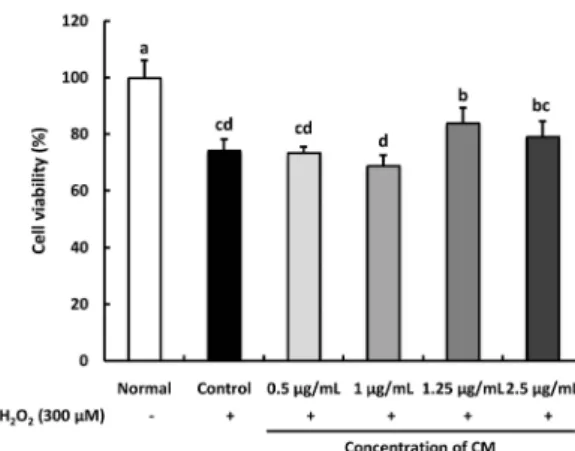
Fig. 1. Effect of Cordyceps militaris (CM) on H2O2-induced C6 glial cell viability.
Cells were pre-treated with CM extract (0.5, 1, 1.25, 2.5 μg/mL) for 1 h, followed by the addition of 300 μM H2O2 for 24 h. Values are presented as the mean ± SD. The different letters (a-d) indicate significant differences (P< 0.05) using the Duncan’s multiple-range test. Normal: no treatment with CM or H2O2; Control: treatment with 300 μM H2O2; 0.5 μg/mL: treatment with CM (0.5 μg/mL) + H2O2 (300 μM); 1 μg/mL: treatment with CM (1 μg/mL) + H2O2 (300 μM); 1.25 μg/mL: treatment with CM (1.25 μg/mL) + H2O2 (300 μM);
2.5 μg/mL: treatment with CM (2.5 μg/mL) + H2O2 (300 μM).
Fig. 2. Effect of Cordyceps militaris (CM) on H2O2-induced C6 glial cell viability.
Cells were pre-treated with the CM extract (0.1, 0.25, 0.5, 1, 1.25, 2.5, and 5 μg/mL) for 1 h, followed by the addition of 300 μM H2O2 for 24 h. Values are presented as the mean ± SD. The different letters (a-d) indicate significant differences (P< 0.05) using the Duncan’s multiple-range test.
generation according to the method from Ma et al. [33]. At a concentration of 5 × 10
4cells/mL, C6 glial cells were seeded into a 96-well plate and incubated for 24 h. The cells were treated with CM (0.5, 1, 1.25, 2.5 μg/mL) for 1 h, following which 300 μM H
2O
2was added. Finally, DCF-DA (80 μM) was added to the cells followed by incubation for 30 min. ROS production in cells was monitored by FLUO star OPTIMA (BMG labtech, Ortenberg, Germany) at excitation and emission wavelengths of 480 nm and 535 nm, respectively.
Western blotting
The cells were washed with phosphate-buffered saline (PBS) after aspiration of the culture medium. Thereafter, the cells were lysed in lysis buffer containing a mixture of RIPA buffer and 1× protease inhibitor cocktail. For western blotting, an equal amount of proteins was loaded in each lane. Fifteen micrograms of total protein were separated using a 10% or 13% SDS- polyacrylamide gel and were transferred to PVDF membranes.
The membranes were blocked with 5% non-fat milk and incubated overnight at 4ºC with primary antibodies [COX-2 and iNOS (1:200, Calbiochem); p-p38 (1:1,000, Cell Signaling); p-JNK (1:200, Santa Cruz); p-ERK (1:1,000, Cell Signaling); and β-actin (1:1,000, Cell Signaling)]. Finally, the membranes were probed with HRP-linked secondary antibodies. Protein expression was measured using the enhanced chemiluminescence detection system (Davinci Chemi, Seoul, Korea).
Statistical analysis
The data are represented as the mean ± standard deviation (SD). Statistical significance was determined using one-way analysis of variance (ANOVA), followed by the Duncan’s multiple- range test. Significance was considered at P < 0.05.
RESULTS
DPPH radical scavenging activity
DPPH is widely used to measure the ability of various biological substances to scavenge free radicals [34]. As shown in Table 1, the treatment with CM exact of the concentrations from 100 to 1,000 μg/mL showed a significant scavenging effect of DPPH radical. Moreover, the capacity on DPPH radical scavenging increased concentration-dependently.
·OH radical scavenging activity
The ·OH radical generated by the H
2O
2/iron system is highly reactive and unstable [35]. Table 1 showed that the CM extract
exhibited strong scavenging activity for the ·OH radical. The treatment of CM at the concentration of 100 μg/mL showed 90.65% ·OH scavenging activity.
NO radical scavenging activity
The NO radical is supposed to be related to the formation of peroxynitrite anion with high toxicity and oxidative damage [36]. Table 1 illustrated the NO radical scavenging activity of CM extract. It indicated that CM extract significantly increased NO scavenging ability in a concentration-dependent manner.
Effect of CM on cell viability in H
2O
2-induced C6 glial cells
We performed the MTT cytotoxicity assay to determine the
effect of CM on cell viability (Fig. 1). We assessed the toxicity
of various concentrations (0.1, 0.25, 0.5, 1, 1.5, 2.5, and 5 μg/mL)
of the CM extract in C6 glial cells. At 5 μg/mL, the cell viability
decreased to below 80%, indicating that CM might produce a
toxic effect on glial cells at high doses (Fig. 2). When C6 glial
cells were treated with 300 μM of H
2O
2, the cell viability was
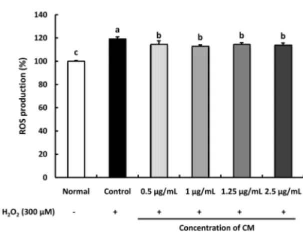
Fig. 3. Effect of Cordyceps militaris (CM) on the level of reactive oxygen species (ROS) in H2O2-induced C6 glial cell. Cells were pre-treated with the CM extract (0.5, 1, 1.25, and 2.5 μg/mL) for 1 h, followed by the addition of 300 μM H2O2 for 24 h. Values are presented as the mean ± SD. The different letters (a-d) indicate significant differences (P< 0.05) using the Duncan’s multiple-range test. Normal: no treatment with CM or H2O2
in the C6 glial cell; Control: treatment with 300 μM H2O2; 0.5 μg/mL: treatment with CM (0.5 μg/mL) + H2O2 (300 μM); 1 μg/mL: treatment with CM (1 μg/mL) + H2O2 (300 μM);
1.25 μg/mL: treatment with CM (1.25 μg/mL) + H2O2 (300 μM); 2.5 μg/mL: treatment with CM (2.5 μg/mL) + H2O2 (300 μM).
Fig. 4. Effect of Cordyceps militaris (CM) on COX-2 and iNOS protein expression in H2O2-induced C6 glial cells. Cells were pre-treated with the CM extract (0.5 and 1.25 μg/mL) for 1 h, followed by the addition of 300 μM H2O2 for 24 h. Cellular protein was isolated and subjected to western blot analysis. The membrane was probed with anti-COX-2 and anti-iNOS antibodies. Beta-actin was used as an internal control. Values are presented as the mean ± SD. The different letters (a-d) indicate significant differences (P< 0.05) using the Duncan’s multiple-range test. Normal: no treatment with CM or H2O2 in the C6 glial cell; Control: treatment with 300 μM H2O2; 0.5 μg/mL: treatment with CM (0.5 μg/mL) + H2O2
(300 μM); 1.25 μg/mL: treatment with CM (1.25 μg/mL) + H2O2 (300 μM).
decreased to 74.13%, whereas the normal group was observed to be 100%. However, treatment of H
2O
2-induced cells with CM showed a significant increase in cell viability to 83.84% and 79.09% at the concentration of 1.25 and 2.5 μg/mL, respectively.
Effect of CM on ROS production in H
2O
2-induced C6 glial cells The ability of CM to inhibit ROS production over a time course of 1 h was determined (Fig. 3). ROS levels in the H
2O
2-induced
control group increased significantly than that in the normal group. Conversely, CM-treated groups significantly inhibited ROS production.
Effect of CM on COX-2 and iNOS protein expression in H
2O
2-induced C6 glial cells
The protein expression of COX-2 and iNOS was up-regulated in the H
2O
2-induced control group relative to that in the normal group (Fig. 4). However, the CM-treated groups significantly inhibited COX-2 and iNOS protein expressions in the C6 glial cells. These results indicate that CM regulates the inflammatory pathway in H
2O
2-induced C6 glial cells by down-regulating of COX-2 and iNOS protein expressions.
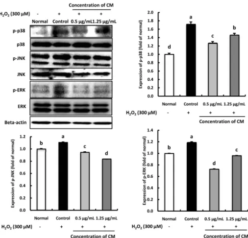
Effect of CM on p-p38, p-JNK, and p-ERK protein expression in H
2O
2-induced C6 glial cells
The inhibitory effect of CM on p-p38, p-JNK, and p-ERK protein expression in H
2O
2-induced C6 glial cells are shown in Fig. 5. The protein levels of p-p38 and p-JNK were increased when C6 cells were treated with H
2O
2compared with the normal group. Moreover, compared with the normal group, p-ERK protein expression was up-regulated in the H
2O
2-treated control group, while the groups treated with CM significantly decreased the level of p-ERK, indicating that under H
2O
2- induced oxidative stress, CM might inhibit the p-ERK expression.
Furthermore, p-p38, p-JNK, and p-ERK protein expression was
significantly low in the 1.25 μg/mL CM-treated group. These
results suggest that CM might inhibit p-p38, p-JNK, and p-ERK
protein expressions under H
2O
2-induced oxidative stress.
Fig. 5. Effect of Cordyceps militaris (CM) on phospho-p38 (p-p38), phospho-JNK (p-JNK), and phospho-ERK (p-ERK) protein expression in H2O2-induced C6 glial cells.
Cells were pre-treated with the CM extract (0.5 and 1.25 μg/mL) for 1 h, followed by the addition of 300 μM H2O2 for 24 h. Cellular protein was isolated and subjected to western blot analysis. The membrane was probed with anti-p-p38, anti-p-JNK and anti-p-ERK antibodies. Beta-actin was used as an internal control. Values are presented as the mean ± SD.
The different letters (a-d) indicate significant differences (P< 0.05) using the Duncan’s multiple-range test. Normal: no treatment with CM or H2O2 in the C6 glial cell; Control: treatment with 300 μM H2O2; 0.5 μg/mL: treatment with CM (0.5 μg/mL) + H2O2 (300 μM); 1.25 μg/mL: treatment with CM (1.25 μg/mL) + H2O2 (300 μM).
DISCUSSION
Free radicals are involved in neural disorders that closely related to oxidative damage [37,38]. In addition, researchers have demonstrated the correlation between oxidative stress and ROS generation, which synergistically cause degenerative diseases by inflammation and cell death [39]. Synthetic drugs are widely used as a treatment regimen for oxidative stress- related degenerative diseases, but it can cause irreversible side effects. Therefore, the use of natural medicines becomes hot topic of discussion. Dong et al. [40] has demonstrated antioxi- dant and cytotoxic properties of the methanol extract of CM against tumor cells. In addition, CM exhibits a protective effect in human dermal fibroblasts, suggesting the promising potential of CM treatment for degenerative diseases [28]. However, the neuroprotective effect of CM against oxidative stress has not been studied yet. The objective of this study was to investigate the neuroprotective effect of CM on oxidative stress-induced C6 glial cells.
Glial cells are closely related to the progression of degene- rative diseases in the nervous system, including AD [41,42]. Choi et al. [43] has reported on the C6 glial cell as a well-established cellular model for studying ROS generation and apoptotic
pathways in neurodegenerative diseases. Impairment of neurons and glial cells by free radicals causes inflammation, which in turn promotes damage of the central nervous system [44,45].
Furthermore, inflammatory cytokines released from glial cells and cellular apoptosis are important components of neurode- generative pathology [46]. A recent study by Yu et al. [47]
showed that COX-2 and iNOS protein expression is up-regulated under oxidative stress by H
2O
2in PC12 cells - a model for neurotoxicological study - indicating that H
2O
2treatment induces neuroinflammation. Furthermore, rapid expression of COX-2 in glial cells may lead to neuroinflammation and affect brain function; there, COX-2 is gaining interest as a target in neurological diseases [48]. Additionally, iNOS has been shown to be involved in COX-2 regulation [49]. The present study demonstrates the role of CM in H
2O
2-induced inflammation in C6 glial cells by determining COX-2 and iNOS protein expression levels. The results indicate that treatment with the CM extract down-regulates COX-2 and iNOS protein expression in H
2O
2- induced C6 glial cells, whereas H
2O
2up-regulates COX-2 and iNOS expression.
To investigate the protective effect of CM against oxidative
stress and inflammatory in C6 glial cells, the expressions of JNK,
p-38 and ERK were observed. Treatment of glial cells with H
2O
2led to neuron death by up-regulating p38 and JNK protein expression. Xie et al. [50] have demonstrated evidence that the signaling pathways of the phosphorylated forms of JNK and p38 contribute to neuronal dysfunction and death. Kwon et al.
[51] have also indicated the role of JNK and p38 signaling pathways in neurodegenerative diseases. Furthermore, ERK has been shown to participate in the regulation of cell growth and differentiation, response to cellular stress, and cytokines. Choi et al. [52] have demonstrated the inhibitory role of cordycepin on ERK phosphorylation in LPS-stimulated RAW 264.7 cells. In this study, we observed a significant up-regulation in the protein expression levels of p-JNK, p-p38, and p-ERK in H
2O
2- treated glial cells; however, treatment of H
2O
2-induced cells with CM showed a suppressive effect on the p-JNK and p-p38 signaling pathways, suggestive of the neuroprotective ability of CM.
The present study revealed that among the various concentrations of CM (0.5, 1, 1.25, 2.5 μg/mL), 1.25 μg/mL showed protective effect against oxidative stress in C6 glial cells. In our previous study, we investigated that oral administration of the CM extract (100 and 200 mg/kg/day) attenuates amyloid beta- induced cognitive impairment in mice models. We have shown that a dose of 200 mg/kg of the CM extract significantly improves learning and memory ability without demonstrating any side effects or hepatotoxicity (data not shown). According to Shin et al. [53], a dose of 200 mg/kg for animals could be translated into approximately 16 mg/kg for humans (60 kg) by using km factor (human: 37, mouse: 3). Therefore, the daily intake dose of CM extract for humans is approximately 960 mg [53].
In conclusion, this study demonstrates that H
2O
2-induced cell injury is related to ROS overproduction. However, treatment with CM significantly attenuates the damage in C6 glial cells by up-regulation of COX-2 and iNOS protein expression, and down-regulation of p-JNK and p-p38 protein expression. These results show the neuroprotective effects of CM against oxidative stress. It suggested that Cordyceps militaris could play the protective role against oxidative stress in glial cells.
CONFLICT OF INTEREST
The authors declare no potential conflicts of interests.
ORCID
Mei Tong He: https://orcid.org/0000-0001-5766-6178 Ah Young Lee: https://orcid.org/0000-0002-3489-7798 Chan Hum Park: https://orcid.org/0000-0003-1183-7159 Eun Ju Cho: https://orcid.org/0000-0003-4282-3219
REFERENCES
1. Liochev SI. Reactive oxygen species and the free radical theory of aging. Free Radic Biol Med 2013;60:1-4.
2. Juránek I, Bezek S. Controversy of free radical hypothesis: reactive oxygen species--cause or consequence of tissue injury? Gen Physiol Biophys 2005;24:263-78.
3. Bayir H. Reactive oxygen species. Crit Care Med 2005;33:S498-501.
4. Ray PD, Huang BW, Tsuji Y. Reactive oxygen species (ROS) homeostasis and redox regulation in cellular signaling. Cell Signal 2012;24:981-90.
5. Ott M, Gogvadze V, Orrenius S, Zhivotovsky B. Mitochondria, oxidative stress and cell death. Apoptosis 2007;12:913-22.
6. Siddique YH, Beg T, Afzal M. Protective effect of ascorbic acid against oxidative damage induced by hydrogen peroxide in cultured human peripheral blood lymphocytes. Indian J Clin Biochem 2009;24:294-300.
7. Chandra K, Salman AS, Mohd A, Sweety R, Ali KN. Protection against FCA induced oxidative stress induced DNA damage as a model of arthritis and in vitro anti-arthritic potential of Costus speciosus rhizome extract. Inter J Pharm Phytochem Res 2015;7:383-9.
8. Gille JJ, Joenje H. Cell culture models for oxidative stress: superoxide and hydrogen peroxide versus normobaric hyperoxia. Mutat Res 1992;275:405-14.
9. Spitz DR, Li GC, McCormick ML, Sun Y, Oberley LW. Stable H2O2-resistant variants of Chinese hamster fibroblasts demonstrate increases in catalase activity. Radiat Res 1988;114:114-24.
10. Rojkind M, Domínguez-Rosales JA, Nieto N, Greenwel P. Role of hydrogen peroxide and oxidative stress in healing responses. Cell Mol Life Sci 2002;59:1872-91.
11. Minghetti L, Polazzi E, Nicolini A, Levi G. Opposite regulation of prostaglandin E2 synthesis by transforming growth factor-β1 and interleukin 10 in activated microglial cultures. J Neuroimmunol 1998;82:31-9.
12. Prajeeth CK, Huehn J, Stangel M. Regulation of neuroinflammatory properties of glial cells by T cell effector molecules. Neural Regen Res 2018;13:234-6.
13. Barnham KJ, Masters CL, Bush AI. Neurodegenerative diseases and oxidative stress. Nat Rev Drug Discov 2004;3:205-14.
14. Beal MF. Mitochondria take center stage in aging and neurodegeneration. Ann Neurol 2005;58:495-505.
15. Loh KP, Huang SH, De Silva R, Tan BK, Zhu YZ. Oxidative stress:
apoptosis in neuronal injury. Curr Alzheimer Res 2006;3:327-37.
16. Li YZ, Ren S, Yan XT, Li HP, Li W, Zheng B, Wang Z, Liu YY.
Improvement of Cisplatin-induced renal dysfunction by Schisandra chinensis stems via anti-inflammation and anti-apoptosis effects. J Ethnopharmacol 2018;217:228-37.
17. Choi YJ, Kim HS, Lee J, Chung J, Lee JS, Choi JS, Yoon TR, Kim HK, Chung HY. Down-regulation of oxidative stress and COX-2 and iNOS expressions by dimethyl lithospermate in aged rat kidney.
Arch Pharm Res 2014;37:1032-8.
18. Kumamoto H, Ooya K. Immunohistochemical detection of phosphorylated JNK, p38 MAPK, and ERK5 in ameloblastic tumors.
J Oral Pathol Med 2007;36:543-9.
19. Wu XF, Zhang M, Bhandari B, Li Z. Effects of microwave-assisted pulse-spouted bed freeze-drying (MPSFD) on volatile compounds and structural aspects of Cordyceps militaris. J Sci Food Agric 2018;98:4634-43.
20. Huang SJ, Huang FK, Li YS, Tsai SY. The quality improvement of solid-state fermentation with Cordyceps militaris by UVB irradiation.
Food Technol Biotechnol 2017;55:445-53.
21. Das SK, Masuda M, Sakurai A, Sakakibara M. Medicinal uses of the mushroom Cordyceps militaris: current state and prospects.
Fitoterapia 2010;81:961-8.
22. Chu HL, Chien JC, Duh PD. Protective effect of Cordyceps militaris against high glucose-induced oxidative stress in human umbilical
vein endothelial cells. Food Chem 2011;129:871-6.
23. Ng TB, Wang HX. Pharmacological actions of Cordyceps, a prized folk medicine. J Pharm Pharmacol 2005;57:1509-19.
24. Ramesh T, Yoo SK, Kim SW, Hwang SY, Sohn SH, Kim IW, Kim SK.
Cordycepin (3'-deoxyadenosine) attenuates age-related oxidative stress and ameliorates antioxidant capacity in rats. Exp Gerontol 2012;47:979-87.
25. Bawadekji A, Al Ali K, Al Ali M. A review of the bioactive compound and medicinal value of Cordyceps militaris. J North Basic Appl Sci 2016;347:1-3.
26. Li XT, Li HC, Li CB, Dou DQ, Gao MB. Protective effects on mitochondria and anti-aging activity of polysaccharides from cultivated fruiting bodies of Cordyceps militaris. Am J Chin Med 2010;38:1093-106.
27. Lin R, Liu H, Wu S, Pang L, Jia M, Fan K, Jia S, Jia L. Production and in vitro antioxidant activity of exopolysaccharide by a mutant, Cordyceps militaris SU5-08. Int J Biol Macromol 2012;51:153-7.
28. Park JM, Lee JS, Lee KR, Ha SJ, Hong EK. Cordyceps militaris extract protects human dermal fibroblasts against oxidative stress-induced apoptosis and premature senescence. Nutrients 2014;6:3711-26.
29. Hatano T, Edamatsu R, Hiramatsu M, Mori A, Fujita Y, Yasuhara T, Yoshida T, Okuda T. Effects of the interaction of tannins with co-existing substances. VI.: effects of tannins and related polyphenols on superoxide anion radical, and on 1, 1-Diphenyl-2-picrylhydrazyl radical. Chem Pharm Bull (Tokyo) 1989;37:2016-21.
30. Chung SK, Osawa T, Kawakishi S. Hydroxyl radical-scavenging effects of spices and scavengers from brown mustard (Brassica nigra). Biosci Biotechnol Biochem 1997;61:118-23.
31. Marcocci L, Maguire JJ, Droy-Lefaix MT, Packer L. The nitric oxide-scavenging properties of Ginkgo biloba extract EGb 761.
Biochem Biophys Res Commun 1994;201:748-55.
32. Mosmann T. Rapid colorimetric assay for cellular growth and survival:
application to proliferation and cytotoxicity assays. J Immunol Methods 1983;65:55-63.
33. Ma WW, Hou CC, Zhou X, Yu HL, Xi YD, Ding J, Zhao X, Xiao R.
Genistein alleviates the mitochondria-targeted DNA damage induced by β-amyloid peptides 25-35 in C6 glioma cells. Neurochem Res 2013;38:1315-23.
34. Blois MS. Antioxidant determinations by the use of a stable free radical. Nature 1958;181:1199-200.
35. Hayyan M, Hashim MA, AlNashef IM. Superoxide ion: generation and chemical implications. Chem Rev 2016;116:3029-85.
36. Patel Rajesh M, Patel Natvar J. In vitro antioxidant activity of coumarin compounds by DPPH, Superoxide and nitric oxide free radical scavenging methods. J Adv Pharm Technol Res 2011;1:52-68.
37. Aruoma OI. Free radicals, oxidative stress, and antioxidants in human health and disease. J Am Oil Chem Soc 1998;75:199-212.
38. Valko M, Leibfritz D, Moncol J, Cronin MT, Mazur M, Telser J. Free radicals and antioxidants in normal physiological functions and human disease. Int J Biochem Cell Biol 2007;39:44-84.
39. Apel K, Hirt H. Reactive oxygen species: metabolism, oxidative
stress, and signal transduction. Annu Rev Plant Biol 2004;55:373-99.
40. Dong CH, Yang T, Lian T. A comparative study of the antimicrobial, antioxidant, and cytotoxic activities of methanol extracts from fruit bodies and fermented mycelia of caterpillar medicinal mushroom Cordyceps militaris (Ascomycetes). Int J Med Mushrooms 2014;16:485-95.
41. Jessen KR, Mirsky R. Glial cells in the enteric nervous system contain glial fibrillary acidic protein. Nature 1980;286:736-7.
42. Sadigh-Eteghad S, Majdi A, Mahmoudi J, Golzari SE, Talebi M.
Astrocytic and microglial nicotinic acetylcholine receptors: an overlooked issue in Alzheimer's disease. J Neural Transm (Vienna) 2016;123:1359-67.
43. Choi EO, Jeong JW, Park C, Hong SH, Kim GY, Hwang HJ, Cho EJ, Choi YH. Baicalein protects C6 glial cells against hydrogen peroxide-induced oxidative stress and apoptosis through regulation of the Nrf2 signaling pathway. Int J Mol Med 2016;37:798-806.
44. Valori CF, Brambilla L, Martorana F, Rossi D. The multifaceted role of glial cells in amyotrophic lateral sclerosis. Cell Mol Life Sci 2014;71:287-97.
45. Puves D, Augustine GJ, Fitzpatrick D, Hall WC, LaMantia AS, White LE. Neuroscience. 5th ed. Sunderland (MA): Sinauer Associates, Inc.;2012. p.560-80.
46. McGeer PL, McGeer EG. Glial cell reactions in neurodegenerative diseases: pathophysiology and therapeutic interventions. Alzheimer Dis Assoc Disord 1998;12 Suppl 2:S1-6.
47. Yu H, Liu Z, Zhou H, Dai W, Chen S, Shu Y, Feng J. JAK-STAT pathway modulates the roles of iNOS and COX-2 in the cytoprotection of early phase of hydrogen peroxide preconditioning against apoptosis induced by oxidative stress. Neurosci Lett 2012;529:166-71.
48. Minghetti L. Cyclooxygenase-2 (COX-2) in inflammatory and dege- nerative brain diseases. J Neuropathol Exp Neurol 2004;63:901-10.
49. Landino LM, Crews BC, Timmons MD, Morrow JD, Marnett LJ.
Peroxynitrite, the coupling product of nitric oxide and superoxide, activates prostaglandin biosynthesis. Proc Natl Acad Sci U S A 1996;93:15069-74.
50. Xie Z, Smith CJ, Van Eldik LJ. Activated glia induce neuron death via MAP kinase signaling pathways involving JNK and p38. Glia 2004;45:170-9.
51. Kwon SH, Kim JA, Hong SI, Jung YH, Kim HC, Lee SY, Jang CG.
Loganin protects against hydrogen peroxide-induced apoptosis by inhibiting phosphorylation of JNK, p38, and ERK 1/2 MAPKs in SH-SY5Y cells. Neurochem Int 2011;58:533-41.
52. Choi YH, Kim GY, Lee HH. Anti-inflammatory effects of cordycepin in lipopolysaccharide-stimulated RAW 264.7 macrophages through Toll-like receptor 4-mediated suppression of mitogen-activated protein kinases and NF-κB signaling pathways. Drug Des Devel Ther 2014;8:1941-53.
53. Shin JW, Seol IC, Son CG. Interpretation of animal dose and human equivalent dose for drug development. J Korean Orient Med 2010;31:1-7.