A Doctoral Dissertation
Protective effect of esculetin against
oxidative stress induced cell damage via
scavenging reactive oxygen species
So Hyung Kim
Department of Medicine
Graduate School
Cheju National University
산화적인
스트레스에 의해 유도된 세포손상에서
활성산소
제거를 통한 Esculetin 의 세포보호 효과
지도교수 강 현 욱김
소 형
이
논문을 의학 박사학위 논문으로 제출함
2009 년 2 월김소형의
의학 박사학위 논문을 인준함
심사위원장
印
위
원 印
위
원 印
위
원 印
위
원 印
제주대학교
대학원
2009년 2월Protective effect of esculetin against oxidative
stress induced cell damage via scavenging
reactive oxygen species
So Hyung Kim
(Supervised by Professor Hyun Wook Kang)
A thesis submitted in partial fulfillment of the requirement for the
degree of doctor of philosophy in medicine
Date Approved : Department of Medicine Graduate School Cheju National University
CONTENTS
CONTENTS ..............................ⅰ
LIST of FIGURES ............................ii 1. ABSTRACT .............................1 2. INTRODUCTION .........................3 3. MATERIALS AND METHODs......................5
3-1. Cell culture and reagents
3-2. DPPH radical scavenging activity 3-3. Detection of hydroxyl radical 3-4. Intracellular ROS measurement 3-5. Lipid Peroxidation Inhibitory Activity 3-6. Protein carbonyl formation
3-7. Western blot
3-8. Immunocytochemistry 3-9. Cell viability
3-10. Statistical analysis
4. Results ........................................................ 13
4-1. Radical scavenging activity of esculetin in a cell-free system 4- 2. Intracellular ROS scavenging activity of esculetin
4-3. The effect of esculetin against the damage of cellular components induced by H2O2
treatment
4-4. Protective effect of esculetin on cell damage induced by H2O2
5. Discussion ..............................25 6. Reference ..............................29
LIST OF FIGURES
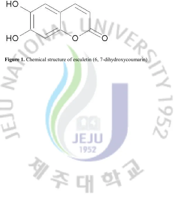
Figure 1. Chemical structure of esculetin (6, 7-dihydroxycoumarin). ...... . 6 Figure 2. Effects of esculetin on the scavenging of DPPH radicals and hydroxyl radicals. ................................13
Figure 3. Effect of esculetin on the scavenging of intracellular ROS. ......15 Figure 4. The effect of esculetin on lipid peroxidation. .. . . .. . . . . .19
Figure 5. The effect of esculetin on protein carbonyl formation.........20
Figure 6. The effect of esculetin on DNA damage ..............21
1. Abstract
Aim: To investigate the antioxidant properties of esculetin (6, 7-dihydroxycoumarin) against H2O2-induced Chinese hamster lung fibroblast (V79-4)
damage. Methods: The radical scavenging activity was assessed by 1, 1-diphenyl-2-picrylhydrazyl (DPPH) radical, hydroxyl radical, and intracellular reactive oxygen species (ROS). In addition, lipid peroxidation was assayed by the measure of related substances which react with thiobarbituric acid. The amount of carbonyl formation in protein was determined using a protein carbonyl ELISA kit. As well, cellular DNA damage was detected by western blot and immuno-fluorescence image. Cell viability was assessed by 3-(4, 5-dimethylthiazole-2-yl)-2, 5-diphenyltetrazolium bromide assay. Results: Esculetin exhibited DPPH radical scavenging, hydroxyl radical scavenging, and intracellular ROS scavenging activities. The radical scavenging activity of esculetin resulted in the protection of cells from lipid peroxidation, protein carbonyl, and DNA damage induced by H2O2. Therefore, esculetin recovered cell
viability exposed to H2O2. Conclusion: Esculetin efficiently attenuated the oxidative
stress induced cell damage via its antioxidant properties. As a result, esculetin may be useful in the development of functional food and raw materials of medicine.
Key words
antioxidant activity; esculetin; oxidative stress; cytoprotective activity; reactive oxygen species
2. INTRODUCTION
A redox imbalance in a healthy living system leads to the malfunctioning of cells which ultimately results in various reactive oxygen species (ROS) related diseases, including cancer, neurological degeneration, arthritis, and aging process[1, 2]. The
consequences of this redox imbalance become even more harmful when genetic variation impairs the normal degradation of these altered proteins. Therefore, therapeutic strategies should be focused on for the reduction of free radical formation and scavenging of free radicals[3].
Plants, including fruits, vegetables, and medicinal herbs, may contain a wide variety of free radical scavenging molecules such as polyphenols. Natural polyphenols can be divided into several different classes depending on their basic chemical structure ranging from simple molecules to highly polymerized compounds. Coumarin, which is known as 1, 2-benzopyrone, consists of fused benzene and a-pyrone rings, and is an important low-molecular weight phenolic group[4] widely used for the prevention and treatment of venous thromboembolism, myocardial infarction, and strokes[5]. Natural and synthetic coumarins possess antioxidant, anti-inflammatory, anticoagulation, and anticancer activities[6]. For the in vivo experiment, rats treated with coumarin ameliorated the hepatotoxicity induced by oxidative
stress[7]. Therefore, researchers are looking for natural antioxidants with strong
pharmacological action and less cytotoxic properties. The efficiency of phenolic compounds as antiradicals and antioxidants is variable and depends on many factors such as the number of hydroxyl groups bonded to the aromatic ring, the site of bonding, and the mutual position of hydroxyls in the aromatic ring. For example, previous studies have found that the advantageous effect of an o-dihydroxy substitution in the aromatic ring of phenol for radical scavenging activity is achieved by producing quinone via hydrogen donation[8, 9].
Here, we demonstrate the esculetin (6, 7-dihydroxycoumarin) was found to possess cytoprotective properties against oxidative stress induced cell damage.
3. Material and Methods 3.1 Cell culture and reagents
Chinese hamster lung fibroblast (V79-4) cells were obtained from the American type culture collection and cultured in Dulbecco’s modified Eagle’s medium which contained 10% heat-inactivated fetal calf serum, streptomycin (100 mg/mL) and penicillin (100 unit/mL) at 37 °C in a humidified atmosphere of 5% CO2. Esculetin
(Figure 1) was purchased from Wako Pure Chemical Ind., Ltd. (Tokyo, Japan) and dissolved in dimethylsulfoxide (DMSO) and final concentration of DMSO did not exceed 0.02%. The 1, 1-diphenyl-2-picrylhydrazyl (DPPH) radical, 5, 5-dimethyl-1-pyrroline-N-oxide (DMPO), and 2', 7'-dichlorodihydrofluorescein diacetate (DCF-DA) were purchased from Sigma (St. Louis, MO, USA). The thiobarbituric acid was purchased from BDH laboratories (Poole, Dorset, UK). Anti-phospho histone H2A.X antibody was purchased from Upstate Biotechnology (Lake Placid, NY, USA). All other chemicals and reagents used were of analytical grade.
3.2 DPPH radical scavenging activity
Esculetinat 0.1, 1, and 10 mg/mL was added to a 1 ´ 10-4 mol/L solution of DPPH
in methanol, and the reaction mixture was shaken vigorously. After 5 h, the remaining DPPH was determined by measuring its absorbance at 520 nm[10]. The
DPPH radical scavenging activity (%) was calculated as; [(optical density of DPPH radical treatment)-(optical density of esculetin with DPPH radical treatment)]/(optical density of DPPH radical treatment)×100.
3.3 Detection of hydroxyl radical
The hydroxyl radicals were generated by the Fenton reaction (H2O2 + FeSO4),
and then reacted with a nitrone spin trap, 5, 5-dimethyl-1-pyrroline-N-oxide (DMPO). The resultant DMPO-OH adducts were detected using an electron spin resonance (ESR) spectrometer. The ESR spectrum was recorded using a JES-FA ESR spectrometer (JEOL, Tokyo, Japan), at 2.5 min after mixing with phosphate buffer solution (pH 7.4) with 0.2 mL of 0.3 mol/L DMPO, 0.2 mL of 10 mmol/L FeSO4, 0.2
mL of 10 mmol/L H2O2, and esculetin at 10 mg/mL. The parameters of the ESR
spectrometer were set at a magnetic field of 336.5 mT, power of 1.00 mW, frequency of 9.4380 GHz, modulation amplitude of 0.2 mT, gain of 200, scan time of 0.5 min, scan width of 10 mT, time constant of 0.03 sec, and a temperature of 25 °C[11, 12].
3.4 Intracellular ROS measurement
]The DCF-DA method was used to detect the levels of intracellular ROS[13]. The
V79-4 cells were seeded in a 96 well plate at 2 ´ 104 cells/well. Sixteen hours after
plating, the cells were treated with esculetin at 0.1, 1, and 10 mg/mL. After 30 min, 1 mmol/L of H2O2 was added to the plate. The cells were incubated for an additional
30 min at 37 °C. After adding 25 mmol/L of DCF-DA solution for 10 min, the fluorescence of 2', 7'-dichlorofluorescein was detected using a Perkin Elmer LS-5B spectrofluorometer and using flow cytometry (Becton Dickinson, Mountain View, CA, USA), respectively. The intracellular ROS scavenging activity (%) was calculated as follows; [(optical density of H2O2 treatment) - (optical density of
esculetin with H2O2 treatment)]/(optical density of H2O2 treatment)×100. The image
analysis for the generation of intracellular ROS was performed by seeding the cells on a cover-slip loaded six well plate at 2 ´ 105 cells/well. Sixteen hours after plating,
the cells were treated with 10 mg/mL of esculetin. After 30 min, 1 mmol/L of H2O2
was added to the plate. After changing the media, 100 mmol/L of DCF-DA was added to each well and was incubated for an additional 30 min at 37 °C. After washing with PBS, the stained cells were mounted onto microscope slides in mounting medium (DAKO, Carpinteria, CA, USA). The microscopic images were collected using the
Laser Scanning Microscope 5 PASCAL program (Carl Zeiss, Jena, Germany) on a confocal microscope.
3.5 Lipid peroxidation detection
Lipid peroxidation was assayed by the measurement of related substances that reacts with thiobarbituric acid (TBARS)[14]. The V79-4 cells were seeded in a culture
dish at a concentration of 1 ´ 105 cells/mL, and 16 h after plating, were treated with
esculetin at 10 mg/mL. After 1 h, 1 mmol/L H2O2 was added to the plate, which was
incubated more for a further 1 h. The cells were then washed with cold PBS, scraped and homogenized in ice-cold 1.15% KCl. About 100 mL of cell lysates was combined with 0.2 mL of 8.1% SDS, 1.5 mL of 20% acetic acid (adjusted to pH 3.5) and 1.5 mL of 0.8% thiobarbituric acid (TBA). The mixture was adjusted to a final volume of 4 mL with distilled water and heated to 95 °C for 2 h. After cooling to room temperature, 5 mL of a mixture of n-butanol and pyridine (15:1) was added to each sample, and the mixture shaken vigorously. After centrifugation at 1000´g for 10 min, the supernatant fraction was isolated, and the absorbance measured at 532 nm.
3.6 Protein carbonyl formation
H2O2 was added to the plate, and the mixture was incubated for 24 h.The amount of
carbonyl formation in protein was determined using an OxiselectTM protein carbonyl
ELISA kit purchased from Cell Biolabs (San Diego, CA, USA) according to the manufacturer’s instructions. Cellular protein was isolated using protein lysis buffer (50 mmol/L Tris (pH 7.5), 10 mmol/L EDTA (pH 8), 1 mmol/L PMSF) and quantified using a spectrophotometer.
3.7 Western blot
The cells were treated with 10 mg/mL of esculetin, and after 1 h, 1 mmol/L of H2O2 was added to the plate, and the mixture was incubated for 24 h. The cells were
harvested, and washed twice with PBS. The harvested cells were then lysed on ice for 30 min in 100 mL of lysis buffer [120 mmol/L NaCl, 40 mmol/L Tris (pH 8), 0.1% NP 40] and centrifuged at 13,000´g for 15 min. Supernatants were collected from the lysates and protein concentrations were determined. Aliquots of the lysates (40 μg of protein) were boiled for 5 min and electrophoresed in 10% SDS-polyacrylamide gel. Blots in the gels were transferred onto nitrocellulose membranes (Bio-Rad, Hercules, CA, USA), which were then incubated with primary antibody. The membranes were further incubated with secondary immunoglobulin G-horseradish peroxidase conjugates (Pierce, Rockland, IL, USA), and then exposed to
X-ray film. The protein bands were detected using an enhanced chemiluminescence Western blotting detection kit (Amersham, Little Chalfont, Buckinghamshire, UK).
3.8 Immunocytochemistry
Cells plated on coverslips were fixed with 4% paraformaldehyde for 30 min and permeabilized with 0.1% Triton X-100 in PBS for 2.5 min. Cells were treated with blocking medium (3% bovine serum albumin in PBS) for 1 h and incubated with anti-phospho histone H2A.X antibody diluted in blocking medium for 2 h. Primary phospho histone H2A.X antibody was detected by a 1:500 dilution of FITC-conjugated secondary antibody (Jackson Immuno Research Laboratories, West Glove, PA, USA) for 1 h. After washing with PBS, the stained cells were mounted onto microscope slides in mounting medium with DAPI (Vector, Burlingame, CA, USA). The images were collected using the Laser Scanning Microscope 5 PASCAL program (Carl Zeiss, Jena, Germany) on a confocal microscope.
3.9 Cell viability
The effect of esculetin on the viability of cells was determined by the 3-(4, 5-dimethylthiazole-2-yl)-2, 5-diphenyltetrazolium bromide (MTT) assay, which is based on the cleavage of a tetrazolium salt by mitochondrial dehydrogenase in viable cells[15]. The cells were seeded in a 96 well plate at a concentration of 1 ´ 105
cells/mL. At sixteen hours after plating, the cells were treated with 10 g/mL of esculetin, and 1 h later 1 mmol/L H2O2 was added to the plate and incubated for an
additional 24 h at 37 °C. Fifty microliter of MTT stock solution (2 mg/mL) was then added to each well of a total reaction volume of 200 mL. After incubating for 4 h, the plate was centrifuged at 800´g for 5 min and the supernatants aspirated. The formazan crystals in each well were dissolved in 150 mL DMSO and the A540 was
read on a scanning multi-well spectrophotometer. 3.10 Statistical analysis
]The data were presented as the means ± standard error (SE). The results were subjected to an analysis of the variance (ANOVA) using the Tukey test to analyze the difference. p<0.05 were considered significantly.
4. Results
4.1 Radical scavenging activity of esculetin in a cell-free system
The radical scavenging effects of esculetin on DPPH radicals and hydroxyl radicals were measured. The DPPH radical scavenging activity of esculetin was 8% at 0.1 mg/mL, 28% at 1 mg/mL, and 77% at 10 mg/mL (Figure 2A). In addition, the hydroxyl radicals generated by the Fenton reaction (FeSO4 + H2O2) in a cell-free
system was detected by ESR spectrometry. The ESR results revealed that a specific signal was not clearly detected in the control and the 10 mg/mL of esculetin, however, the signal of the hydroxyl radical increased up to 4769 in the FeSO4 + H2O2 system.
Esculetin treatment was found to decrease the hydroxyl radical signal to 2769 (Figure 2B).
Figure 2. Effects of esculetin on the scavenging of DPPH radicals and hydroxyl radicals. (A) The amount of DPPH radicals was determined spectrophotometrically at 520 nm. *Significantly different from the control cells (p<0.05). (B) The hydroxyl radicals generated by the Fenton reaction (H2O2 + FeSO4) were reacted with DMPO,
4.2 Intracellular ROS scavenging activity of esculetin
The intracellular ROS scavenging activity of esculetin in V79-4 cells after H2O2
treatment was detected by the DCF-DA assay. The fluorescence spectrometric data revealed that the intracellular ROS scavenging activity of esculetin was 1% at 0.1 mg/mL, 40% at 1 mg/mL, and 75% at 10 mg/mL (Figure 3A). Moreover, the fluorescence intensity of DCF-DA staining was measured using a flow cytometer and a confocal microscope. The level of ROS was detected using a flow cytometer and revealed a fluorescence intensity value of 167 for the ROS stained by DCF-DA fluorescence dye in H2O2 treated cells with 10 mg/mL of esculetin, compared to a
Figure 3. Effect of esculetin on the scavenging of intracellular ROS. (A) The cells were treated with esculetin at 0.1, 1, and 10 mg/mL. After 30 min, 1 mmol/L of H2O2
was added to the plate. After an additional 30 min, the DCF-DA was added and the intracellular ROS generated were detected by spectrofluorometry. *Significantly different from control cells (p<0.05). (B) The cells were treated with esculetin at 10
30 min, the intracellular ROS generated were detected by flow cytometry after the DCF-DA treatment. *Significantly different from control cells (p<0.05). **Significantly different from H2O2-treated cells (p<0.05). (C) The representative
confocal images illustrate the increase in the red fluorescence intensity of DCF produced by ROS in H2O2-treated cells, compared to the control and the lowered
fluorescence intensity in the H2O2-treated cells with esculetin at 10 mg/mL (original
4.3 The effect of esculetin against the damage of cellular components induced by H2O2 treatment
The H2O2-induced damage of cellular components represents the primary cause
of cell viability loss. The effect of esculetin on the damage of membrane lipid, protein, and cellular DNA in H2O2-treated cells was investigated. V79-4 cells
exposed to H2O2 were found to increase lipid peroxidation, as evidenced by the
generation of TBARS. However, esculetin treatment was found to prevent the H2O2
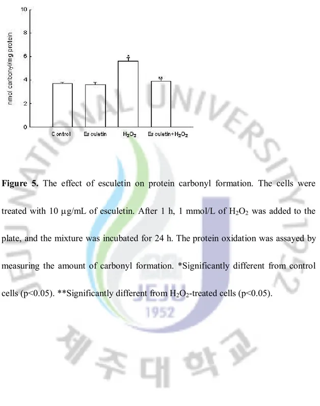
-induced peroxidation of lipids (Figure 4). The protein carbonyl formation serves as a biomarker for cellular oxidative protein damage[16]. Moreover, the accumulation of oxidatively modified protein carbonyls may disrupt cellular function either by the loss of catalytic and structural integrity or by the interruption of regulatory pathways[17]. Moreover, the protein carbonyl content in cells increased significantly after H2O2 treatment, whereas esculetin prevented the H2O2-induced protein carbonyl
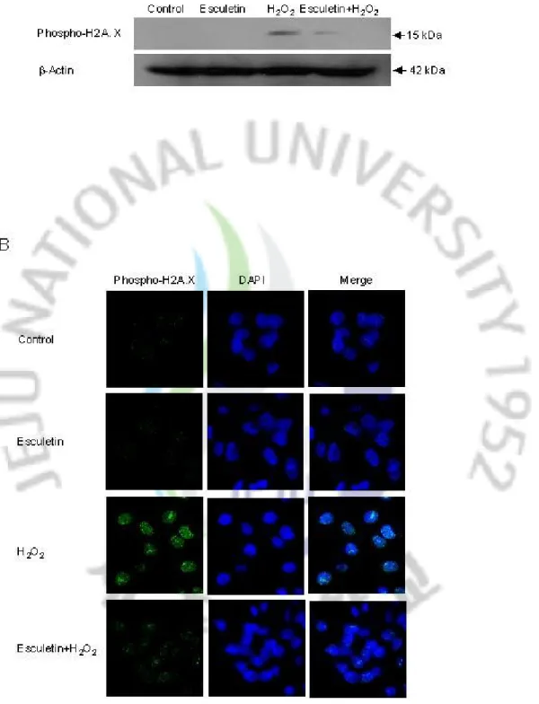
formation (Figure 5). Further, damage to cellular DNA induced by H2O2 exposure
was detected by phospho histone-H2A.X expression in nucleus. The phosphorylation of the nuclear histone H2A.X, which is a sensitive marker for breaks of double stranded DNA[18], increased in the H2O2-treated cells, as shown by western blot and
H2O2-treated cells decreased the expression of phospho H2A.X. These results
suggest that esculetin inhibits the damage of cellular components induced by H2O2.
Figure 4. The effect of esculetin on lipid peroxidation. The cells were treated with 10 mg/mL of esculetin. After 1 h, 1 mmol/L of H2O2 was added to the plate, and the
mixture was incubated for 24 h.Lipid peroxidation was assayed by measuring the amount of TBARS formation. *Significantly different from control cells (p<0.05). **Significantly different from H2O2-treated cells (p<0.05).
Figure 5. The effect of esculetin on protein carbonyl formation. The cells were treated with 10 mg/mL of esculetin. After 1 h, 1 mmol/L of H2O2 was added to the
plate, and the mixture was incubated for 24 h.Theprotein oxidation was assayed by measuring the amount of carbonyl formation. *Significantly different from control cells (p<0.05). **Significantly different from H2O2-treated cells (p<0.05).
Figure 6. The effect of esculetin on DNA damage. Cells were treated with 10 mg/mL of esculetin for 30 min later, 1 mmol/L H2O2 was added and after an incubation of 24
in nucleus was detected by a specific antibody. (B) The confocal images reveal that the FITC-conjugated secondary antibody staining indicates the location of phospho histone H2A.X (green) via the anti-phospho histone H2A.X antibody. The DAPI staining indicates the location of the nucleus (blue), whereas the merged image indicates the location of the phospho histone H2A.X protein in the nucleus.
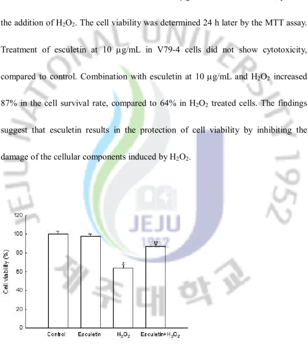
4.4 Protective effect of esculetin on cell damage induced by H2O2
The protective effect of esculetin on the cell survival in H2O2-treated V79-4 cells
was also assessed. The cells were treated with 10 mg/mL of esculetin for 1 h, prior to the addition of H2O2. The cell viability was determined 24 h later by the MTT assay.
Treatment of esculetin at 10 mg/mL in V79-4 cells did not show cytotoxicity, compared to control. Combination with esculetin at 10 mg/mL and H2O2 increased
87% in the cell survival rate, compared to 64% in H2O2 treated cells. The findings
suggest that esculetin results in the protection of cell viability by inhibiting the damage of the cellular components induced by H2O2.
Figure 7. The effect of esculetin on H2O2-induced cell death. The cells were treated
After an incubation of 24 h, cell viability was determined by the MTT assay. *Significantly different from control cells (p<0.05). **Significantly different from H2O2-treated cells (p<0.05).
5. Discussion
Coumarins are lactones of cis-o-hydroxycinnamic acid derivatives, which belong to the phenol compounds with a basic skeleton of C6 + C3[19]. Also, coumarins are a
diverse group of natural compounds essentially found in green plants. As substitutions can occur at any of the six available sites of their basic molecular moiety (1, 2-benzopyrone), these compounds are variable in structure. This structural diversity results in the multiple biological properties of coumarin that help reducing the risk of human disease. In a recent paper[20], coumarin itself and 7-hydroxycoumarin showed evidence of in vitro antiproliferative and in vivo antitumor activities. Moreover, coumarins possess anti-oxidant activities, probably due to their structural analogy with flavonoids and benzophenones[21]. For example esculetin isolated from the cortex of Fraxius chinensis showed anti-photoaging activity through antioxidant activities[22]. Coumarins in plants are present in the free form or glycosides, and are emerging as potent therapeutic drugs for free radical mediated diseases. Protection of the cellular environment from oxidative stress is dependent on the chemical structure of antioxidants, suggesting that the radical-scavenging effects of coumarins are correlated with the number and position of the hydroxyl groups. A similar correlation was also reported for flavonoids[23] and cinnamic acid[24]. In
structure and the substitution pattern of the hydroxyl groups, on the availability of phenolic hydrogens and on the possibility of stabilization of the resulting phenoxyl radicals via hydrogen bonding or by expanded electron delocalization[25]. Previous
report has also found that the phenolic hydroxyl groups, which have o-dihydroxy group (catechol structure), enhance and play an important role in antiradical efficacy of simple coumarins[26]. These results are consistent with a previous report that the
radical scavenging activity was higher for caffeic acid bearing o-dihydroxy group than methoxy-substituted derivatives[27]. Thus it can be inferred that the resonance
structures of the radical derived from esculetin is especially stable as a result of the
o-quinone form of the resonance structure. And recent paper showed consistent
results that coumarin derivatives with dihydroxy group were more potent DPPH radical scavengers than monohydroxy, free hydroxyl, methoxyl, dihydro, and coumarin derivatives[6]. Therefore, our study demonstrated that esculetin possesses antioxidant properties and provides cytoprotection against oxidative stress. Esculetin showed evidence of direct radical quenching as shown by the DPPH radical and hydroxyl radical and it might be related with catechol structure of esculetin. In addition, esculetin showed intracellular ROS scavenging activities, suggesting that it might be involved in activation of antioxidant enzymes. Oxidative stress occurs
when there is an imbalance between pro-oxidant processes and the antioxidant defense system in favor of the former, and will ultimately lead to a free radical attack of the lipid, protein, and DNA. H2O2 is a major component of the ROS produced
intracellularly and a cause of oxidative damage[28]. Lipid peroxidation is one of the
major mechanisms of cellular injury cause by H2O2[29]. In our study, cells exposed to
H2O2 showed an increase in lipid peroxidation. However, esculetin was found to
decrease the H2O2-induced lipid peroxidation, indicating the cytoprotective
properties of esculetin against H2O2 induced lipid damage. Moreover, the oxidative
damage of the amino acid residues of the proteins resulted in the formation of carbonyl derivatives and can serious compromise cellular integrity[30]. The protein carbonyl content in cells increased significantly after H2O2 treatment, whereas
esculetin treatment was found to prevent the H2O2-induced protein carbonyl
formation. In addition, H2O2 is a genotoxic agent and is known to induce oxidative
DNA damage[31]. In this study, H2O2-induced DNA damage was assessed using the
expression of the phospho histone H2A.X to detect DNA strand breakage. The exposure of cells to H2O2 increased phosphorylation of the nuclear histone H2A.X.
However, esculetin treatment in H2O2-treated cells decreased the expression of the
H2O2-induced DNA damage. Therefore, esculetin provides cytoprotection against
H2O2-induced lipid peroxidation, protein carbonyl, and DNA damage by scavenging
intracellular H2O2.
In summary, many biochemical and clinical studies suggest that natural and synthetic antioxidant compounds help treat diseases mediated by oxidative stress[32].
The results presented in this report indicate that esculetin efficiently attenuated oxidative stress-induced cell damage via its antioxidant properties. Thus, esculetin might be useful in the development of functional food and raw materials of medicine.
6. Reference
1 Halliwell B. Antioxidants in human health and disease. Annu Rev Nutr 1996; 16: 33-50.
2 Willcox JK, Ash SL, Catignani GL. Antioxidants and prevention of chronic disease. Crit Rev Food Sci Nutr 2004; 44: 275-95.
3 Berg D, Youdim MB, Riederer P. Redox imbalance. Cell Tissue Res 2004; 318: 201-13.
4 Fylaktakidou KC, Hadjipavlou-Litina DJ, Litinas KE, Nicolaides DN. Natural and synthetic coumarin derivatives with anti-inflammatory/ antioxidant activities. Curr Pharm Des 2004; 10: 3813-33.
5 Voora D, McLeod HL, Eby C, Gage BF. The pharmacogenetics of coumarin therapy. Pharmacogenomics 2005; 6: 503-13.
6 Lin HC, Tsai SH, Chen CS, Chang YC, Lee CM, Lai ZY, et al. Structure-activity relationship of coumarin derivatives on xanthine oxidase-inhibiting and free radical-scavenging activities. Biochem Pharmacol 2008; 75: 1416-25.
of esculetin on oxidative damage induced by t-butyl hydroperoxide in rat liver. Arch Toxicol 2000; 74: 467-72.
8 Joyeux M, Lobstein A, Anton R, Mortier F. Comparative antilipoperoxidant, antinecrotic and scavenging properties of terpenes and biflavones from Ginkgo and some flavonoids. Planta Med 1995; 61: 126-9.
9 Rice-Evans CA, Miller NJ, Paganga G. Structure-antioxidant activity relationships of flavonoids and phenolic acids. Free Radic Biol Med 1996; 20: 933-56.
10 Lo SF, Nalawade SM, Mulabagal V, Matthew S, Chen CL, Kuo CL, et al. In vitro propagation by asymbiotic seed germination and 1,1-Diphenyl-2-picrylhydrazyl (DPPH) radical scavenging activity studies of tissue culture raised plants of three medicinally important species of Dendrobium. Biol Pharm Bull 2004; 27: 731-5.
11 Yoshimura Y, Inomata T, Nakazawa H, Kubo H, Yamaguchi F, Ariga T. Evaluation of free radical scavenging activities of antioxidants with an H2O2/NaOH/DMSO system by electron spin resonance. J Agric Food Chem
12 Rimbach G, Weinberg PD, de Pascual-Teresa S, Alonso MG, Ewins BA, Turner R, et al. Sulfation of genistein alters its antioxidant properties and its effect on platelet aggregation and monocyte and endothelial function. Biochim Biophys Acta 2004; 1670: 229-37.
13 Rosenkranz AR, Schmaldienst S, Stuhlmeier KM, Chen W, Knapp W, Zlabinger GJ. A microplate assay for the detection of oxidative products using 2',7'-dichlorofluorescin-diacetate. J Immunol Meth 1992; 156: 39-45.
14 Ohkawa H, Ohishi N, Yagi K. Assay for lipid peroxides in animal tissues by thiobarbituric acid reaction. Anal Biochem 1979; 95: 351-8.
15 Carmichael J, DeGraff WG, Gazdar AF. Evaluation of a tetrazolium-based semiautomated colorimetric assay: assessment of chemosensitivity testing. Cancer Res 1987; 47: 936-41.
16 Szweda LI, Stadtman ER. Oxidative modification of glucose-6-phosphate dehydrogenase from Leuconostoc mesenteroides by an iron(II)-citrate complex. Arch Biochem Biophys 1993; 301: 391-5.
18 Rogakou EP, Pilch DR, Orr AH, Ivanova VS, Bonner WM. DNA double-stranded breaks induce histone H2AX phosphorylation on serine 139. J Biol Chem 1998; 273: 5858-68.
19 Cai YZ, Mei S, Jie X, Luo Q, Corke H. Structure-radical scavenging activity relationships of phenolic compounds from traditional Chinese medicinal plants. Life Sci 2006; 78: 2872-88.
20 Kostova I. Synthetic and natural coumarins as cytotoxic agents. Curr Med Chem Anticancer Agents 2005; 5: 29-46.
21 Farombi EO, Nwaokeafor IA. Anti-oxidant mechanisms of kolaviron: studies on serum lipoprotein oxidation, metal chelation and oxidative membrane damage in rats. Clin Exp Pharmacol Physiol 2005; 32: 667-74.
22 Lee BC, Lee SY, Lee HJ, Sim GS, Kim JH, Kim JH, et al. Anti-oxidative and photo-protective effects of coumarins isolated from Fraxinus chinensis. Arch Pharm Res 2007; 30: 1293-301.
23 Heim KE, Tagliaferro AR, Bobilya DJ. Flavonoid antioxidants: chemistry, metabolism and structure-activity relationships. J Nutr Biochem 2002; 13:
24 Siquet C, Paiva-Martins F, Lima JL, Reis S, Borges F. Antioxidant profile of dihydroxy- and trihydroxyphenolic acids-a structure-activity relationship study. Free Radic Res 2006; 40: 433-42.
25 Amic D, Davidovic-Amic D, Beslo D, Trinajstic N. Structure-radical scavenging activity relationships of flavonoids. Croat Chem Acta 2003; 76: 55-61.
26 Wu CR, Huang MY, Lin YT, Ju HY, Ching H. Antioxidant properties of
Cortex Fraxini and its simple coumarins. Food Chem 2007; 104: 1464-71.
27 Chang YC, Lee FW, Chen CS, Huang ST, Tsai SH, Huang SH, et al. Structure-activity relationship of C6-C3 phenylpropanoids on xanthine oxidase-inhibiting and free radical-scavenging activities. Free Radic Biol Med 2007; 43: 1541-51.
28 Krohn K, Maier J, Paschke R. Mechanisms of disease: hydrogen peroxide, DNA damage and mutagenesis in the development of thyroid tumors. Nat Clin Pract Endocrinol Metab 2007; 3: 713-20.
Essent Fatty Acids 2007; 76: 73-85.
30 Hawkins CL, Davies MJ. Generation and propagation of radical reactions on proteins. Biochim Biophys Acta 2001; 1504: 196-219.
31 Halliwell B, Aruoma OI. DNA damage by oxygen-derived species. Its mechanism and measurement in mammalian systems. FEBS Lett 1991; 281: 9-19.
32 Lee JC, Lim KT, Jang YS. Identification of Rhus verniciflua Stokes compounds that exhibit free radical scavenging and anti-apoptotic properties. Biochim Biophys Acta 2002; 1570: 181-91.
7. Abstract in Korean
Esculetin(6, 7-dihydroxycoumarin)은 phenol 성의 coumarin 유도체로서 Fraxius chinensis (물푸레나무)의 피층에 주로 함유되어 있다. 본 논문에서는 esculetin 의 항산화 작용을 조사하여 H2O2 에 의한
중국 햄스터 폐 섬유아 세포(V79-4) 손상에 대해 세포 보호 효과가 나타나는지를 연구 하였다.
1, 1-diphenyl-2-picrylhydrazyl (DPPH) radical, hydroxyl radical, 그리고 세포 내 활성 산소종에 의한 라디칼 소거율을 측정하였더니 esculetin 에 의해 DPPH radical 제거, hydroxyl radical 제거와 세포 내 활성 산소종 제거 능력을 보였다. 지방 과산화의 척도인 thiobarbituric acid 와 반응하는 물질을 측정한 결과 esculetin 이 지방 과산화를 방지함을 알 수 있었다. Protein carbonyl ELISA kit 를 이용하여 단백질 과산화의 척도인 carbonyl formation 을 측정한 결과 esculetin 에 의해 단백질에서 carbonyl formation 도 방지됨을 확인하였다. 또한, western blot 과 면역형광상을 통해 세포 DNA 손상을 측정한 결과 esculetin 을 처리하였을 때 DNA 손상을 억제함을 알 수 있었다. 3-(4, 5-dimethylthiazole-2-yl)-2, 5-diphenyltetrazolium bromide 법으로 세포 생존능을 측정하였고 역시 esculetin 이 H2O2 에 노출된 세포의
생존능을 회복시켰다.
결론적으로 esculetin 이 그 항산화 작용을 통해 산화 스트레스를 효과적으로 줄일 수 있으므로 기능성 식품이나 화장품의 원료 개발에 이용될 수 있을 것으로 생각한다.
주요어: Fraxius chinensis, Esculetin(6, 7-dihydroxycoumarin), 활성 산소종, DNA손상, 항산화 작용