Korean Circulation Journal
Introduction
Oxidative stress is caused by an imbalance between the genera- tion of oxidants and antioxidants in the body. Oxidative stress in- creases the formation of reactive oxygen species (ROS) or reactive ni-
http://dx.doi.org/10.4070/kcj.2012.42.1.23 Print ISSN 1738-5520 • On-line ISSN 1738-5555
Protective Effects of Peroxiredoxin on Hydrogen Peroxide Induced Oxidative Stress and Apoptosis in Cardiomyocytes
Keon-Jae Park, MS, Yeon-Jeong Kim, MS, Jeongeun Kim, MD, Sang Min Kim, MD, Sang Yeub Lee, MD, Jang-Whan Bae, MD, Kyung-Kuk Hwang, MD, Dong-Woon Kim, MD, and Myeong-Chan Cho, MD
Regional Cardiovascular Disease Center, Department of Internal Medicine, Chungbuk National University School of Medicine, Cheongju, Korea
Background and Objectives: The redox system is an important anti-oxidative system composed of thioredoxin, thioredoxin reductase, and peroxiredoxin (PRx). The fine details of PRx expression and its protective effects in various cells in cardiovascular tissue under oxidative stress created by hydrogen peroxide have not been fully elucidated.
Subjects and Methods: Oxidative stress was induced by adding hydrogen peroxide at 0.25 mM for 2 hours to rat neonatal cardiomyo- cytes (rCMCs), rat vascular smooth muscle cells (rVSMCs), and human umbilical vein endothelial cells (HUVECs). Apoptosis was quantified by flow cytometry and the expression patterns of the six PRx isoforms were evaluated by western blotting in the three cell lines after hydro- gen peroxide stimulation. Apoptosis and the cell survival signal pathway were evaluated by PRx1 gene delivery using lentiviral vector in hy- drogen peroxide stimulated rCMCs versus green fluorescence protein gene delivery.
Results: Hydrogen peroxide induced 25% apoptosis in rCMCs. Furthermore, the PRx1 and 5 isoforms were found to be overexpressed in hy- drogen peroxide treated rCMCs, and PRx1 overexpression by gene delivery was found to reduce hydrogen peroxide induced rCMCs apopto- sis significantly. In addition, this effect was found to originate from cell survival pathway modification.
Conclusion: Hydrogen peroxide induced significant oxidative stress in rCMCs, rVSMCs, and HUVECs, and PRx1 overexpression using a len- tiviral vector system significantly reduced hydrogen peroxide induced rCMCs apoptosis by upregulation of cell survival signals and down- regulation of apoptotic signals. These findings suggest that PRx1 could be used as a treatment strategy for myocardial salvage in conditions of oxidative stress. (Korean Circ J 2012;42:23-32)
KEY WORDS: Peroxiredoxins; Myocytes, cardiac; Oxidative stress; Apoptosis.
Received: July 5, 2011 Accepted: September 5, 2011
Correspondence: Jang-Whan Bae, MD, Regional Cardiovascular Disease Center, Department of Internal Medicine, Chungbuk National University Sch- ool of Medicine, 62 Gaesin-dong, Heungdeok-gu, Cheongju 361-763, Korea Tel: 82-43-269-6011, Fax: 82-43-273-3252
E-mail: [email protected]
• The authors have no financial conflicts of interest.
This is an Open Access article distributed under the terms of the Creative Commons Attribution Non-Commercial License (http://creativecommons.
org/licenses/by-nc/3.0) which permits unrestricted non-commercial use, distribution, and reproduction in any medium, provided the original work is properly cited.
trogen species.1) ROS contains one or more unpaired electrons in their outer orbits, causing them to be highly reactive.2) These species are generated constantly in vivo, and can cause oxidative damage to nucleic acids, lipids, and proteins, and affect cell membrane prop- erties. Furthermore, their accumulation may lead to the oxidative destruction of cells.3)
Reactive oxygen species also play central roles in cardiac physi- ology or pathophysiology, and have been shown to injure both en- dothelial cells and cardiomyocytes (CMCs) via various molecular pathways.4)5) In addition, ROS may be an important cause of athero- sclerosis, ventricular hypertrophy and its related cardiomyopathy.5) Coronary atherosclerosis is a direct cause of ischemic heart disease and oxidative stress has been suggested as a cause of atheroscle- rotic plaque instability and rupture. Thus, they are believed to play an important role in the pathophysiology of acute myocardial infarc- tion (AMI) and its related ventricular remodeling.6) The excessive generation of ROS endogenously has been directly related with met-
abolic stress, apoptosis, and necrosis in mammalian CMCs.7)8) Isch- emia and reperfusion are major causes of oxidative stress in CMCs, showing that oxidative stress provokes the damage of secondary CMCs during reperfusion therapy in AMI cases.9)
The redox system is mainly composed of thioredoxins (TRxs), TRx reductase, thioredoxin interacting protein (TRxNip), and peroxire- doxins (PRxs).10) Previously, we described the temporal expression patterns of the TRx system and their relations to cellular apoptosis in endothelial cells, in the hope that this would provide optimal con- ditions or time points for TRx system gene or protein delivery in cells and animal models to minimize TRx exhaustion under hypoxia.11) PRx family members are thiol-specific antioxidant proteins, and are also referred to as TRx peroxidases and alkyl-hydroperoxide-reduc- tase-C22 proteins.12) These enzymes are truly ubiquitous and have been identified in yeast, plant and animal cells. PRxs exert their pro- tective antioxidant role in cells through their peroxidase activities (ROOH+2e-→ ROH+H2O), which are responsible for the reductions and detoxification of hydrogen peroxide, peroxynitrite, and a wide range of organic hydroperoxides (ROOH).12)13)
Six PRx isoforms have been identified in mammals. However, the temporal expression patterns and functional significances of these isoforms in cell lines found in cardiovascular tissue, especially under conditions of hydrogen peroxide induced oxidative stress, have not yet been elucidated. Therefore, we aimed to determine the tempo- ral expression patterns of the 6 PRxs isoforms in neonatal rat cardio- myocytes (rCMCs), rat vascular smooth muscle cells (rVSMCs), and human umbilical vein endothelial cells (HUVECs) exposed to hydro- gen peroxide induced oxidative stress. In addition, we also exam- ined the functional significance of PRx1 overexpression using the lentivirus vector in rCMCs exposed to hydrogen peroxide induced oxidative stress. Changes in molecular pathways associated with cell survival and apoptosis in rCMCs exposed to the same condi- tions were also examined to further explore the causative relation between apoptosis reduction and PRx1 overexpression.
Materials and Methods
Preparation and culture of neonatal rat cardiomyocytes Isolation and primary cultures of rCMCs were performed using a modified version of a previously reported protocol.14)15) The hearts of 2 to 3 day-old rats (Sprague-Dawley, Orient Bio Inc., Seongnam, Ko- rea) were removed and the left ventricles were collected, washed 3 times with cold ADS buffer (in 116 mM NaCl, 20 mM HEPES, 0.8 mM NaH2PO4, 5.6 mM glucose, 5.4 mM KCl, 0.8 mM MgSO4, pH 7.4), ch- opped finely with surgical scissors, and digested 3 times for 20 mi- nutes with collagenase/pancreatin (0.56 mg/0.3 mg/mL). The ob- tained cells were collected selectively and enriched by differential
centrifugation through a discontinuous Percoll (Amersham Pharma- cia Biotech, Piscataway, NJ, USA) gradient with densities of 1.050, 1.062 and 1.082 g/mL.16) The band at the 1.062/1.082 density interface was collected and used as the rCMCs source. Cells were washed and suspended in Dulbecco’s modified Eagle’s medium (DMEM) (Gib- co BRL, Rockville, MD, USA) supplemented with medium 199 (M199) (Sigma-Aldrich Co., St. Louis, MO, USA), 10% horse serum, 5% fetal bovine serum (FBS), 5000 U/L of streptomycin and 5000 U/L of peni- cillin. rCMCs were then seeded at 5×10⁵ per well on 2% gelatin- coated (Sigma-Aldrich Co.) 60 mm Primarian plastic culture six-well plate dishes (BD Sciences, San Jose, CA, USA). Media changes were performed initially once daily with serum free media containing DM- EM/M199 (4 : 1). rCMCs were cultured at 37°C in 5% CO2 for 2 to 3 days before being exposed to experimental conditions. With this method, we were able to specifically select rCMCs with a negligible amount of red blood cells or fibroblasts. Purity of rCMCs was more than 98% in morphometric analysis, and immunohistochemical st- ating with MHC (Santa Cruz Biotechnology, Santa Cruz, CA, USA). All experiments were repeated 3 to 5 times and a total of 90 neonatal rats were used.
Preparation and culture of rat vascular smooth muscle cells Rat vascular smooth muscle cells from the intima-media complex of thoracic aortas of 8-week-old male Sprague-Dawley rats (Orient Bio Inc.) were prepared as previously described.16) We used 5 Sp- rague-Dawley rats for obtaining rVSMC and repeated this proce- dure at least 3 to 5 times for robustness. The cells were expanded in 60 mm Primarian plastic culture six-well plates in DMEM supple- mented with 10% of FBS at 37°C in 5% CO2.
Preparation and culture of human umbilical vein endothelial cells
We used commercialized HUVECs (ATCC, Chicago, IL, USA) ac- cording to the manufacturer’s protocol, and cells were obtained and processed as described previously.17) Briefly, umbilical cords were immersed in a culture dish, both umbilical veins were cannulated, and cord ends were bound. Veins were then washed using sterile ph- osphate buffered saline (PBS) with 0.02% collagenase, and placed in a CO2 incubator for 10 minutes at 37°C. HUVECs were isolated in centrifugation tubes by washing vessels twice with 10 mL M199.
The cell suspensions were centrifuged at 1200 rpm for 10 minutes and the supernatants were discarded. The cells were then resus- pended using the EGM-2 BulletKit system (Lonza Ltd., Basel, Swit- zerland) in 2% gelatin-coated six-well plates. Cells were passaged until confluent, dissociated with 0.05% trypsin, and grown for 3 days in fresh medium containing M199 supplemented with 10% FBS and antibiotics using the EGM-2 BulletKit system at 37°C under 5% CO2.
Quantitation of apoptosis by fluorescence-activated cell sorting
Cultured rCMCs, rVSMCs, and HUVECs were processed using the Annexin V: Phycoerythrin (PE) Apoptosis Detection kit I (BD Scienc- es) to assess cell membrane integrity. 7-Aminoacetinomycin D (7- AAD) (BD Sciences) was used as a marker for dying or dead cells. This technique used was based on assessments of cell membrane per- meability and on the intercalation of double-stranded DNA. In brief, collected rCMCs, rVSMCs, and HUVECs were washed twice with cold PBS and then resuspended in 1X binding buffer (10 mM HEPES, 140 mM NaCl, 2.5 mM CaCl2, pH 7.4) at a concentration 5×10⁶ cell/
mL. These mixtures were then transferred to a 5 mL culture tube and 5 μL of Annexin-V and 5 μL of 7-AAD were added. After gentle vor- texing, the 3 cell lines were incubated for 10 minutes at room tem- perature in the dark and analyzed using a flow cytometry analysis (FACS Calibur-S, BD Sciences) equipped with an argon ion laser sys- tem using excitation and emission wavelengths of 488 and 578 nm, respectively.
Western blot analysis
To examine the expression patterns of PRx isoforms in rCMCs, rVSMCs, and HUVECs stressed by hydrogen peroxide, western blott- ing was performed at baseline, and at 30 and 120 minutes after ad- ding 0.25 mM hydrogen peroxide. Harvested rCMCs, rVSMCs, and HUVECs were immersed in PRO-PREP Protein Extraction Solution (iNtRON Biotechnology, Seongnam, Korea) and incubated at 4°C for 2 hours. Extracts were then centrifuged at 4°C for 15 minutes at 12000 rpm to remove insoluble material. Bradford assays were then used to quantify protein expression.18) Protein concentrations were determined using a dye-binding assay (Bio-Rad protein assay kit, Bio- Rad Laboratories, Hercules, CA, USA). Equal amounts of total protein (20 μg) were diluted with sample buffer containing 100 mM dithi- othreitol and heated at 98°C for 5 minutes, then subjected to 10%
sodium dodecyl sulfate-polyacrylamide gel electrophoresis (SDS- PAGE) in a gel apparatus (Hoefer Scientific, San Francisco, CA, USA).
Samples were subsequently transferred to polyvinylidene fluoride (PVDF) membranes, and the blots were blocked with 5% skim milk in tris-buffered saline Tween-20 (TBST) for 2 hours. Membranes were then incubated with antibodies against PRx isoforms 1-6 (Ab- FRONTIER, Seoul, Korea), Bcl-2, Bax, caspase 3, and survivin (Santa Cruz Biotechnology, Santa Cruz, CA, USA) at a dilution of 1 : 2000 in TBST under shaking at room temperature for 1 hour. Membranes were then washed with TBST, incubated with horseradish peroxi- dase (HRP)-conjugated secondary antibody (1 : 2000-1 : 5000) for 1 hour, and treated with West-Zol kits (iNtRON Biotechnology) for 1 minute. Chemiluminescence was detected using a LAS-3000 (Fu- jifilm, Minato, Tokyo) for 10-300 seconds. Optical densities (OD)
were calculated using the MultiGauge Ver3.1 (Fujifilm) program.
Experiments were repeated 5 times and the results are expressed as mean±SD OD as percentages of baseline values. Percentage ch- anges in OD with time versus baseline were also calculated.
Lentiviral vector construction and production
HIV-based lentivirus stocks were generated by transient plasmid transfection into 293T cells as described previously.19) PRx1 cDNA was amplified using 5’-CGCGAATTCATGTCTTCAGGAAATGCA-3’ and 5’-CGCCTCGAGTCACTTCTGCTTAGAGAA-3’ using Taq polymerase.
The commentary DNA obtained was subcloned with the 5’ end into the EcoRI and the 3’ end into the XhoI restriction site at pRRL-cPPT- CMV-X-PRE-SIN containing lentivirus vector (LeV), and packaging was performed by transient transfection. The day prior to transfec- tion, confluent 100 mm plates of 293T cells were split. pRRL-based LeVs were generated by calcium phosphate co-transfection of the transfer vector, the HIV Gap/Pol packaging construct, a Rev expres- sion plasmid, and the VSV-G expression plasmid into 293T cells. To each 15 cm diameter dish, 23 μg transfer vector, 15 μg pMDL-g/pRRE packaging plasmid, 11.5 μg pRSV-REV, and 8 μg pCMV VSV-G enve- lope were added. DNA was resuspended in 450 μL 0.1×TE (1×TE=10 mM Tris pH 8.0, 1 mM EDTA), 50 μg of 2.5 M CaCl2 were added, and mixtures were incubated at room temperature for 10 minutes. The DNA/CaCl2 solution was then added dropwise to 500 μL (2X) HEPES- buffered saline under vigorous bubbling.
Once it was slightly turbid, the solution was immediately added to the cells. All transfection procedures were conducted over 16 hours, and this was followed by media replacement and virus collec- tion for 48-72 hours. Viral supernatant from 80 plates was filtered through 0.22 μm pore filters and stored at 4°C. The virus was con- centrated by ultracentrifugation for 2 hours at 25000 rpm. Virus pel- lets were resuspended in 1 mL phosphate-buffered saline (PBS) and stored at -80°C.
Peroxiredoxin1 gene delivery and the induction of oxidative stress with hydrogen peroxide
Rat cardiomyocytes were transfected with lentiviral vector system containing PRx1 gene (LeV-PRx1) or lentiviral vector system contain- ing the green fluorescence protein gene (LeV-GFP) at 1.5×10⁷ IU in the presence of 10 μg/mL DEAE (diethylaminoethyl cellulose) dex- tran. Media were changed 16 hours after infection and cells were cultured for another 48 hours. To generate oxidative stress, hydro- gen peroxide (0.1 or 0.25 mM) was added to cells for 2 hours. rCMCs (3×10⁵ cells) responses were compared under 3 conditions, that is, LeV-PRx1 or LeV-GFP after exposure to hydrogen peroxide and LeV- GFP not exposed to hydrogen peroxide injury.
Immunohistochemistry and the transfection efficiency of lentiviral vector system containing peroxiredoxin1 gene
To examine the transfection efficiency of LeV-PRx1 in vitro, cul- tured rCMCs were transfected with LeV-PRx1 and stained for PRx1.
In brief, cells were cultured in 6 well chamber slides, transfected with LeV-PRx1, and compared with untransfected control cells. The me- dia was changed 16 hours after transfection and cells were cultured for another 48 hours. Cells were then treated with 0.1 or 0.25 mM hydrogen peroxide for 2 hours, washed with PBS to remove hydro- gen peroxide and the media was changed. After 48 hours, these rC- MCs were fixed with 4% paraformaldehyde, washed with PBS twice for 5 minutes each and blocked with 1% horse serum for 1 hour.
Cells were washed again three times with PBS, and incubated with primary antibody for anti-PRx1 (1 : 1000 in PBS, AbFRONTIER) over- night at 4°C. Cells were then rewashed twice with PBS for 5 minutes, incubated with secondary anti-rabbit Texas Red 596 nm (1 : 200 in PBS) for 1 hour, rewashed, and stained with TO-PRO-3 iodide (a nu- clear stain; Invitrogen, Carlsbad, CA, USA). Cells were observed and images were acquired using Confocal Laser Scanning Microscope (Olympus BX51, Shinjuku, Tokyo, Japan).
Statistical analysis
Results are expressed as means±SDs. Statistical analysis was performed using the two-tailed Student’s t-test or paired t-test us-
ing Statistical Package for the Social Sciences version 16.0 (SPSS Inc., Chicago, IL, USA). Differences were considered statistically signifi- cant when p were <0.05.
Results
Flow cytometry analysis and the detection of neonatal cardiomyocyte, rat vascular smooth muscle cell and human umbilical vein endothelial cell apoptosis under hydrogen peroxide induced oxidative stress
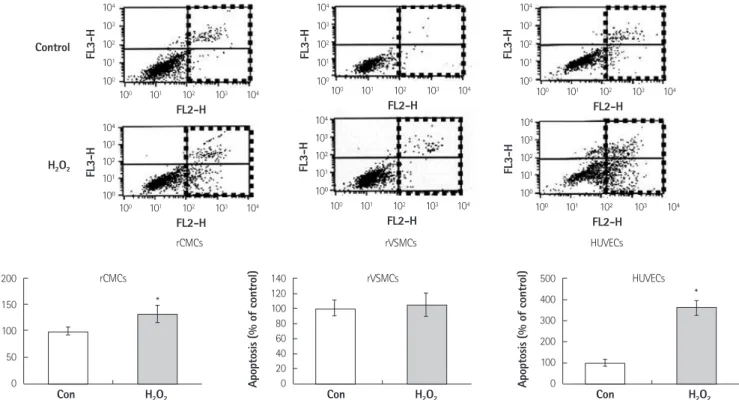
Prior to the main experiment, we checked apoptotic fractions in rCMCs, rVSMCs, and HUVECs after treatment with 0.1 mM hydro- gen peroxide for 2 hours. Apoptotic fractions were found to be sig- nificantly increased in rCMCs and HUVECs following treatment. Ap- optotic fractions were expressed as percentages of fractions at base- line, which were set at 100%. The apoptotic fraction of rCMCs increas- ed from 100±7.07% at baseline to 132±16.49% after hydrogen pe- roxide treatment (p<0.05). rVSMCs were highly resistant to hydrogen peroxide injury, and apoptotic fractions were not significantly ch- anged compared to the baseline (100±10.49% to 105±14.6%). On the other hand, HUVECs were highly susceptible to hydrogen peroxi- de, and the apoptotic fraction increased to 360.72±35.35% (p<
0.05) (Fig. 1).
Fig. 1. FACS and the detection of rCMC, rVSMC, and HUVEC apoptosis under hydrogen peroxide-induced oxidative stress. Flow cytometric analysis showed significant increases in apoptosis after treatment with 0.1 mM hydrogen peroxide for 2 hours versus non-treated control rCMCs, rVSMCs, or HUVECs.
*Compared to control, p<0.05. FACS: flow cytometry analysis, rCMCs: neonatal rat cardiomyocytes, rVSMCs: rat vascular smooth muscle cells, HUVECs: hu- man umbilical vein endothelial cells, Con: control, H2O2: hydrogen peroxide.
200 150 100 50 Apoptosis (% of control) 0
Con
rCMCs 104 103 102 101 100
104 103 102 101 100
104 103 102 101 100
104 103 102 101 100 100 101 102 103 104
100 101 102 103 104 100 101 102 103 104 100 101 102 103 104 100 101 102 103 104 100 101 102 103 104 FL2-H
FL2-H FL2-H FL2-H
FL2-H FL2-H
104 103 102 101 100
104 103 102 101 100 Control
H2O2
FL3-HFL3-H FL3-H FL3-H
FL3-H FL3-H
rVSMCs HUVECs
rCMCs
*
H2O2
140 120 100 80 60 40 20 Apoptosis (% of control) 0
Con
rVSMCs
H2O2
500 400 300 200 100 Apoptosis (% of control) 0
Con
HUVECs
*
H2O2
Expression patterns of the 6 isoforms of peroxiredoxin in neonatal cardiomyocytes, rat vascular smooth muscle cells and human umbilical vein endothelial cells under hydrogen peroxide induced oxidative stress
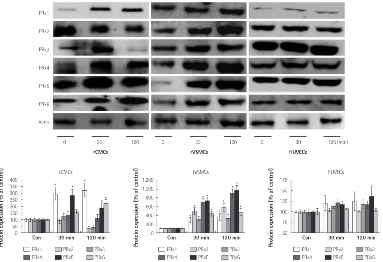
The expression patterns of the 6 PRx isoforms in rCMCs, rVSMCs, and HUVECs were examined 30 and 120 minutes after adding 0.1 mM hydrogen peroxide to cell media. PRx2 and 3 expressions in rCMCs were downregulated at 120 minutes (PRx2: 32±5.71%, PRx3:
42±7.23%), but PRx 1 and 6 expressions in rCMCs were signifi- cantly upregulated at 120 minutes versus baseline (PRx1: 325.7±
54.82% vs. 100±4.12%, PRx6: 225.82±27.34% vs. 100±2.8%, p<
0.05). PRx5 expression was upregulated at 30 minutes (285.87±
45.12%), but slightly downregulated at 120 minutes (190.42±
39.8%). In rVSMCs, the expressions of all PRx isoforms were signif- icantly upregulated (PRx1: 354.12±75.72%, PRx2: 572.3±87.45%, PRx3: 325.6±34.36%, PRx4: 892.89±97.24%, PRx5: 964.48±
112.61%, PRx6: 468.78±79.68%) at both time points. In HUVECs, PRx1, 5 were upregulated (PRx1: 125.25±22.65%, PRx5: 135.87±
34.51%) but PRx 2, 3, 4, and 6 were not changed significantly until 120 minutes after the addition of hydrogen peroxide (Fig. 2).
Conformation of gene expression after lentiviral vector system containing the green fluorescence protein gene transfection into neonatal cardiomyocytes
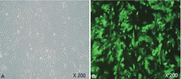
Rat cardiomyocytes (3×10⁵) were transfected with LeV-GFP for 16 hours at a multiplicity of infection of 1.5×10⁷ IU. To determine the transfection efficiency achieved using LeV-GFP, we confirmed GFP expression in rCMCs after transfection by fluorescence microscopy.
More than 80% of cells were shown to express GFP (Fig. 3).
Apoptosis fraction measurements after transfecting neonatal cardiomyocytes with lentiviral vector system containing peroxiredoxin1 gene or lentiviral vector system containing the green fluorescence protein gene and/or treating cells with hydrogen peroxide
Apoptotic fractions were significantly increased by treating cells
rCMCs rVSMCs HUVECs
PRx1
PRx2
PRx3
PRx4
PRx5
PRx6
Actin
0 30 120 0 30 120 0 30 120 (min)
Fig. 2. Temporal expression patterns of the 6 PRx isoforms in rCMCs, rVSMCs, and HUVECs exposed to hydrogen peroxide-induced oxidative stress. PRx1 and 5 were overexpressed at 30 and 120 minutes, whereas PRx6 was overexpressed at 120 minutes in rCMCs after hydrogen peroxide stimulation. All iso- forms were overexpressed in rVSMCs at both time points. No significant change in the expression of PRx isoforms was observed in HUVECs. *Compared to control, p<0.05. PRx: peroxiredoxin, rCMCs: neonatal rat cardiomyocytes, rVSMCs: rat vascular smooth muscle cells, HUVECs: human umbilical vein endo- thelial cells, con: control, min: minutes.
400 350 300 250 200 150 100 50 0
Protein expression (% of control)
Con 30 min 120 min rCMCs
PRx1 PRx2 PRx3 PRx4 PRx5 PRx6
* * *
**
1,200 1,000 800 600 400 200 0
Protein expression (% of control)
Con 30 min 120 min rVSMCs
PRx1 PRx2 PRx3 PRx4 PRx5 PRx6
*
* *
*
* *
**
*
*
* *
175 150 125 100 75 50
Protein expression (% of control)
Con 30 min 120 min HUVECs
PRx1 PRx2 PRx3 PRx4 PRx5 PRx6
with hydrogen peroxide for 2 hours (148.32±13.34% vs. 100±
13.34%, p<0.05). rCMCs/LeV-GFP (rCMCs transfected with LeV- GFP) exposed to hydrogen peroxide had a higher apoptotic frac- tion than unexposed cells (187.5±25.49% vs. 111.72±11.81%, p<
0.05). LeV-PRx1 treatment effectively reduced the apoptotic frac- tion as compared to LeV-GFP treatment in rCMCs exposed to hy- drogen peroxide (72.55±10.19% vs. 187.5±25.49%, p<0.05) (Fig. 4).
These results suggest that PRx1 overexpression protected rCMCs
from hydrogen peroxide-induced apoptosis.
The expression pattern of peroxiredoxin1 in neonatal cardiomyocytes transfected with lentiviral vector system containing peroxiredoxin1 gene in hydrogen peroxide induced oxidative stress
Peroxiredoxin 1 expression was significantly higher in rCMCs/LeV- PRx1 (rCMCs transfected with LeV-PRx1) not treated with hydro- Fig. 3. Confirmation of gene expression after the transfection of LeV-GFP into rCMCs. GFP expression in rCMCs/LeV-GFP. rCMCs (3×10⁵ cell) were trans- fected with LeV-GFP for 16 hours at a multiplicity of infection of 1.5×10⁷ IU. A: rCMCs appeared healthy under optical microscopy (×200). B: GFP expres- sion in rCMCs transfected with LeV-GFP was observed in over 80% of cells by fluorescence microscopy (×200). LeV-GFP: GFP gene containing lentivirus vector, rCMCs: neonatal rat cardiomyocytes, GFP: green fluorescence protein.
A B
Con H2O2 LeV-GFP LeV-GFP+H2O2 LeV-PRx1 LeV-PRx1+H2O2
Fig. 4. FACS and the quantitation of apoptosis in rCMCs transfected with LeV-PRx1 or LeV-GFP and exposed to hydrogen peroxide. The transfection of LeV- PRx1 significantly reduced apoptosis induced by 0.25 mM hydrogen peroxide for 2 hours as compared to LeV-GFP transfection (72.55±10.19% vs. 187.5±
25.49%, p<0.05). *Compared to control, p<0.05, †Compared to LeV-GFP+H2O2, p<0.05. FACS: flow cytometric analysis, rCMCs: neonatal rat cardiomyocytes, LeV- PRx1: PRx1 gene containing lentivirus vector, LeV-GFP: green fluorescence protein gene containing lentivirus vector, Con: control, H2O2: hydrogen peroxide, rVSMCs: rat vascular smooth muscle cells.
Con LeV-GFP LeV-PRx1 LeV-PRx1+
H2O2
LeV-GFP+
H2O2
H2O2
rVSMCs 250
200 150 100 50 0
Apoptosis (% of control)
* †
*
*
*
gen peroxide (193.02±28.44%) than in the control, and significant- ly higher in rCMCs/LeV-PRx1 treated with hydrogen peroxide than in rCMCs/LeV-GFP treated with hydrogen peroxide (221.02±27.38
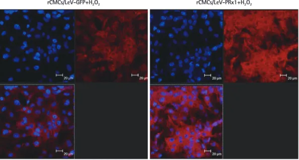
vs. 161.01±19.3%, p<0.05) (Fig. 5). Furthermore, rCMCs/LeV-PRx1 expressed significantly more PRx1 than rCMCs/LeV-GFP after treat- ment with hydrogen peroxide (Fig. 6).
The upregulation of anti-apoptotic survival signals in neonatal cardiomyocytes transfected with lentiviral vector system containing peroxiredoxin1 gene
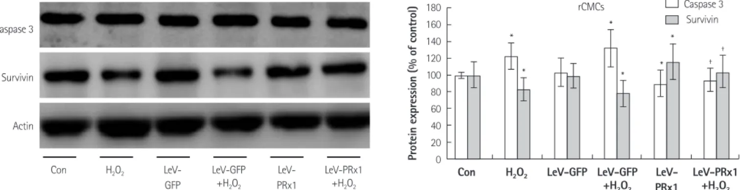
The expression of caspase 3 was lower in rCMCs/LeV-PRx1 treated with hydrogen peroxide than in rCMCs/LeV-GFP treated with hydro- gen peroxide (94.3±13.72% vs. 132.3±22.36%, p<0.05), but the expression of survivin was higher in rCMCs/LeV-PRx1 treated with hydrogen peroxide than in rCMCs/LeV-GFP treated with hydrogen peroxide (116.15±21.58% vs. 78.08±15.81%, p<0.05) (Fig. 7). Bax/
Bcl-2 ratio (a marker of apoptosis/survival) concurred with the ab- ove results, and was lower in rCMCs/LeV-PRx1 treated with hydro- gen peroxide than in rCMCs/LeV-GFP treated with hydrogen per- oxide (68.1±6.67% vs. 172.6±60%, p<0.05) (Fig. 8). These results show that PRx1 overexpression has a protective effect against hy- drogen peroxide related apoptosis in rCMCs.
Discussion
Peroxiredoxin is a member of the peroxidase family, and has been shown to be related to cell proliferation, differentiation, and apopto- sis in mammalian cells.24) Mammalian PRx consists of 6 different isoforms that are classified as typical 2-Cys PRx, the other five 2-Cys PRx isoforms have the TRx-dependent peroxidase activity utilizing TRx, thioredoxin reductase, and NADPH as a reducing system. Mam-
PRx1
Actin
Fig. 5. Expression patterns of PRx1 in rCMCs transfected with LeV-PRx1 in hydrogen peroxide induced oxidative stress. PRx1 expression patterns under various conditions of hydrogen peroxide induced oxidative stress and PRx1 gene delivery. Cells were exposed to 0.25 mM hydrogen peroxide for 2 hours.
LeV-PRx1 transfection significantly upregulated PRx1 expression versus LeV-GFP transfection in hydrogen peroxide treated rCMCs. *Compared to control, p<
0.05, †Compared to LeV-GFP+H2O2, p<0.05. rCMCs: rat cardiomyocytes, LeV- PRx1: PRx1 gene containing lentivirus vector, con: control, H2O2: hydrogen per- oxide, LeV-GFP: green fluorescence protein gene containing lentivirus vector.
Con LeV-GFP LeV-
PRx1
LeV-PRx1 +H2O2 LeV-GFP
+H2O2 H2O2
300 250 200 150 100 50
Protein expression (% of control) 0 PRx1
Con LeV-GFP LeV-
PRx1
LeV-PRx1 +H2O2 LeV-GFP
+H2O2 H2O2
* †
*
*
*
rCMCs/LeV-PRx1+H2O2
rCMCs: blue (Dapi), PRx1: red (Texas red) rCMCs/LeV-GFP+H2O2
Fig. 6. Immunohistochemistry in rCMCs transfected with LeV-PRx1. PRx1 expression was upregulated in rCMCs/LeV-PRx1 as compared to rCMCs/LeV-GFP treated with 0.25 mM hydrogen peroxide for 2 hours. Cytolsolic isoforms, PRx1 was found to be highly expressed in the rCMCs/LeV-PRx1 (stained red). rC- MCs: neonatal rat cardiomyocytes, LeV-PRx1: PRx1 gene containing lentivirus vector, LeV-GFP: green fluorescence protein gene containing lentivirus vector.
malian PRxs are 20-30 kd in size and vary in terms of their subcel- lular localizations. PRx 1, 2, 4, and 6 are found in the cytosol, PRx3 in mitochondria, whereas PRx5 has a complicated distribution, and is found in peroxisomes, mitochondria, and the cytosol.25) In particu- lar, PRxs are involved in the enzymatic degradation of hydrogen per- oxide, a wide range of organic hydroperoxides and peroxynitrite, us- ing reducing equivalents provided by thiol-containing proteins like TRxs and PRxs.26)27) Growing evidence indicates that ROS play a critical role in many disorders of the cardiovascular system, such as, ische- mia-reperfusion injury, myocardial stunning, apoptosis, and arterio-
sclerosis.20)21) Furthermore, the accumulation of ROS in mitochondria can lead to apoptotic cell death and ROS may also have direct effects on cellular structure and function, including myocardial remodeling and failure.22)23) Under physiological conditions, the toxic effects of ROS can be prevented by scavenging enzymes such as superoxide dismutase (SOD), glutathione peroxidase (GHPx), and catalase, and by non-enzymatic antioxidants. However, when the production of ROS is high, oxidative stress can have harmful effects on the func- tional and structural integrity of the heart.23)
In our experiments, the expression patterns of the 6 PRx isoforms
Caspase 3
Survivin
Actin
Fig. 7. Protein expression of caspase 3 and survivin in rCMCs transfected with LeV-Prx1. LeV-PRx transfection effectively reduced caspase 3 expression and enhanced survivin expression in rCMCs treated with 0.25 mM hydrogen peroxide for 2 hours. *Compared with control, p<0.05, †Compared with LeV-GFP+
H2O2, p<0.05. rCMCs: rat cardiomyocytes, LeV-PRx1: PRx1 gene containing lentivirus vector, Con: control, H2O2: hydrogen peroxide, LeV-GFP: GFP gene con- taining lentivirus vector.
Con LeV-
GFP
LeV- PRx1
LeV-PRx1 +H2O2 LeV-GFP
+H2O2 H2O2
Caspase 3 Survivin rCMCs
Con LeV-GFP LeV-
PRx1
LeV-PRx1 +H2O2 LeV-GFP
+H2O2 H2O2
180 160 140 120 100 80 60 40 20 Protein expression (% of control) 0
*
* *
*
*
*
†
†
Bax/Bcl-2 rCMCs
Fig. 8. Bax/Bcl-2 ratios in LeV-PRx1 versus LeV-GFP transfected rCMCs treated with hydrogen peroxide. Bax/Bcl-2 ratio was significantly lower in LeV-PRx1 than in LeV-GFP transfected rCMCs treated with 0.25 mM hydrogen peroxide for 2 hours. *Compared to control, p<0.05, †Compared to LeV-GFP+H2O2, p<0.05. LeV-PRx1: PRx1 gene containing lentivirus vector, LeV-GFP: GFP gene containing lentivirus vector, rCMCs: rat cardiomyocytes, Con: control, H2O2: hydrogen peroxide, neonatal rat cardiomyocytes.
Con LeV-GFP LeV- Con LeV-GFP
PRx1 LeV-
PRx1 LeV-PRx1
+H2O2
LeV-PRx1 +H2O2
LeV-GFP +H2O2
LeV-GFP +H2O2
H2O2 H2O2
150 125 100 75 50 25 0
250 200 150 100 50 Protein expression (% of control) Bcl-2 Bax Protein expression (% of control) 0
*
* *
*
*
*
* †
*
*
*
* † Bcl-2
Bax
Actin
Con LeV-
GFP
LeV- PRx1
LeV-PRx1 +H2O2 LeV-GFP
+H2O2
H2O2
* †
were found to be distinct in rCMCs, rVSMCs and HUVCEs. Hydrogen peroxide significantly and time dependently increased rCMC and HUVEC apoptosis in 2 hours as seen by the FACS results. HUVECs were found to be most susceptible to hydrogen peroxide induced injury, and apoptosis increased to 360.72±35.35% compared with the baseline. On the other hand, rCMCs showed a 132±16.49% in- crease in apoptosis after 2 hours of treatment, but, rVSMCs were found to be highly resistant to hydrogen peroxide. In rCMCs, the expression of PRx1 and 6 showed a continual increase at 30 and 120 minutes, whereas the expression of PRx 2 and 3 was downreg- ulated at 120 minutes. The expression of PRx 5 was elevated at 30 minutes but slightly lower than baseline at 120 minutes. In rVSMCs, the expression of all 6 isoforms of PRx was generally elevated after treatment, particularly the expression of PRx 2, 4, and 5. The ex- pression of PRx in HUVECs only slightly changed compared to that observed in rCMCs and rVSMCs. The three cell types appeared to have unique PRx expression patterns in the presence of excessive hydrogen peroxide, although apoptosis and PRx expression patterns generally appeared to be inversely related. rVSMCs were most re- sistant to hydrogen peroxide in terms of apoptosis, and this resis- tance may be attributed to elevated and constant PRx isoform ex- pression. On the other hand, HUVECs expressed PRx isoforms at low levels and were highly susceptible to hydrogen peroxide relat- ed cellular apoptosis.
Cytosolic PRx1 is a ubiquitously expressed PRx isoform that is the most abundant in mammalian cells. PRx1 has been associated with various cellular functions apparently unrelated to peroxidase activity.
For example, it has been independently identified in a Ras-trans- formed human mammary epithelial cell line after serum stimula- tion, in a human erythroleukemic cell line, in which it enhanced the activities of natural killer cells, and in stress-stimulated mouse peri- toneal macrophages.28) According to our results, PRx1 expression in- creased with time after hydrogen peroxide treatment, especially in rCMCs. Therefore, we selected PRx1 as a gene delivery candidate to rescue rCMCs from oxidative stress induced apoptosis, which was in line with previous suggestions concerning the importance of PRx1 in oxidatively stressed cells.26-28)
Peroxiredoxin 1 expression in rCMCs was significantly increased by rCMCs/LeV-PRx1 in the absence of hydrogen peroxide and PRx1 was more upregulated in rCMCs/LeV-PRx1 than in rCMCs/LeV-GFP treated with hydrogen peroxide. PRx1 overexpression protected rC- MCs against oxidative stress-induced apoptosis and cell death by hy- drogen peroxide.13) Furthermore, PRx1 transfected rCMCs treated with hydrogen peroxide were found to have a significantly lower ap- optotic cell fraction than rCMCs/LeV-GFP, and this was found to be closely related to the upregulation of the anti-apoptotic proteins Bcl-2 and survivin, as well as to the downregulation of the pro-apo-
ptotic proteins Bax and caspase 3. In addition, consistent with our findings in a previous study, the overexpression of PRx1 was found to protect thyroid cells from hydrogen peroxide-induced apoptosis, wh- ich was also found to be associated with Bax downregulation.13)
Antioxidative systems are highly complex networks, which make the direct and real time measurements of levels of antioxidative mo- lecules under oxidative stress almost impossible, because they have short biologic half-lives, are highly volatile, and are vulnerable to other cellular proteins. In the present study, our PRx1 overexpression model effectively reduced hydrogen peroxide related rCMCs apop- tosis. To determine the mechanism involved, further study is requ- ired to elucidate the interactions between PRx, TRx, TRX reductase, and TRxNip under oxidative stress. Furthermore, a PRx1 knock-down study using a suitable chemical inhibitor or siRNA is required to va- lidate and support our results. These constitute limitations of the present study, as our results do not enable us to comment on the effects of ischemia-reperfusion or oxidative stress in vivo. Also, we were not able to assess the existence of cellular necrosis in hydro- gen peroxide-induced oxidative stress. However, apoptosis and its reverse are more important than necrosis in damaged myocardial tissue. Therefore, our apoptosis focus may be justified. More detail- ed analyses focused on changes in necrosis patterns with PRx gene delivery are also required.
Up to now, there is no clinical study on PRx delivery or overex- pression. However, there are a few clinical studies ongoing on TRx inhibitor, especially in the oncology field. Compared to TRx re- search, PRx studies are not abundant and very limited. Because of the similar action and molecular relation of these two proteins, we surmise that Prx can also be an alternative candidate of human ap- plication for myocardial ischemia or oncologic disease. Protein de- livery of PRx with regards to thromolysis or emergent coronary in- tervention for AMI to relieve oxidative damage related to ischemia- reperfusion injury will be helpful.
In summary, PRx1 expression was found to be significantly in- creased by hydrogen peroxide in rCMCs, and PRx1 overexpression was found to protect rCMCs against oxidative stress-induced apop- tosis and cell death by hydrogen peroxide. We conclude that PRx provides an effective target for new drug development aimed at reducing rCMC apoptosis in ischemia-reperfusion situations.
Acknowledgments
This study was supported by a research grant from the Korean Society of Cardiology in 2008. We cordially appreciate Pf. Seung- Taik Kim in Chungbuk National University Hospital for providing us with the lentiviral vector construct.
REFERENCES
1. Tuteja N, Singh MB, Misra MK, Bhalla PL, Tuteja R. Molecular mechanisms of DNA damage and repair: progress in plants. Crit Rev Biochem Mol Biol 2001;36:337-97.
2. Tuteja N, Tuteja R. Unravelling DNA repair in human: molecular mechanisms and consequences of repair defect. Crit Rev Biochem Mol Biol 2001;36:261-90.
3. Tuteja N, Ahmad P, Panda BB, Tuteja R. Genotoxic stress in plants:
shedding light on DNA damage, repair and DNA repair helicases.
Mut Res 2009;681:134-49.
4. Kevin LG, Novalija E, Stowe DF. Reactive oxygen species as media- tors of cardiac injury and protection: the relevance to anesthesia practice. Anesth Anal 2005;101:1275-87.
5. Madamanchi NR, Runge MS. Mitochondrial dysfunction in ath- erosclerosis. Circ Res 2007;100:460-73.
6. Boersma E, Mercado N, Poldermans D, Gardien M, Vos J, Simoons ML. Acute myocardial infarction. Lancet 2003;361:847-58.
7. Byrne JA, Grieve DJ, Cave AC, Shah AM. Oxidative stress and heart failure. Arch Mal Coeur Vaiss 2003;96:214-21.
8. Ferrari R, Guardigli G, Mele D, Percoco GF, Ceconi C, Curello S. Oxi- dative stress during myocardial ischaemia and heart failure. Curr Pharm Des 2004;10:1699-711.
9. Zweier JL, Flaherty JT, Weisfeldt ML. Direct measurement of free radical generation following reperfusion of ischemic myocardi- um. Proc Natl Acad Sci U S A 1987;84:1404-7.
10. Misra MK, Sarwat M, Bhakuni P, Tuteja R, Tuteja N. Oxidative stress and ischemic myocardial syndromes. Med Sci Monit 2009;
15:RA209-19.
11. Park KJ, Kim YJ, Choi EJ, et al. Expression pattern of the thiore- doxin system in human endothelial progenitor cells and endo- thelial cells under hypoxic injury. Korean Circ J 2010;40:651-8.
12. Chae HZ, Robison K, Poole LB, Church G, Storz G, Rhee SG. Clon- ing and sequencing of thiol-specific antioxidant from mammali- an brain: alkyl hydroperoxide reductase and thiol-specific anti- oxidant define a large family of antioxidant enzymes. Proc Natl Acad Sci U S A 1994;91:7017-21.
13. Hofmann B, Hecht HJ, Flohé L. Peroxiredoxins. Biol Chem 2002;
383:347-64.
14. Poindexter BJ, Smith JR, Buja LM, Bick RJ. Calcium signaling me- chanisms in dedifferentiated cardiac myocytes: comparison with neonatal and adult cardiomyocytes. Cell Calcium 2001;30:373-82.
15. Kobayashi S, Lackey T, Huang Y, et al. Transcription factor gata4 regulates cardiac BCL2 gene expression in vitro and in vivo. FASEB J 2006;20:800-2.
16. Kau TR, Schroeder F, Ramaswamy S, et al. A chemical genetic
screen identifies inhibitors of regulated nuclear export of a fork- head transcription factor in PTEN-deficient tumor cells. Cancer Cell 2003;4:463-76.
17. Hoshi H, McKeehan WL. Brain- and liver cell-derived factors are required for growth of human endothelial cells in serum-free cul- ture. Proc Natl Acad Sci U S A 1984;81:6413-7.
18. Saraste A, Pulkki K, Kallajoki M, Henriksen K, Parvinen M, Voipio- Pulkki LM. Apoptosis in human acute myocardial infarction. Cir- culation 1997;95:320-3.
19. Zeng L, Planelles V, Sui Z, et al. HIV-1-based defective lentiviral vectors efficiently transduce human monocytes-derived macro- phages and suppress replication of wild-type HIV-1. J Gene Med 2006;8:18-28.
20. Chen HW, Chien CT, Yu SL, Lee YT, Chen WJ. Cyclosporine A regu- late oxidative stress-induced apoptosis in cardiomyocytes: me- chanisms via ROS generation, iNOS and Hsp70. Br J Pharmacol 2002;137:771-81.
21. Penna C, Mancardi D, Tullio F, Pagliaro P. Postconditioning and intermittent bradykinin induced cardioprotection require cyclo- oxygenase activation and prostacyclin release during reperfu- sion. Basic Res Cardiol 2008;103:368-77.
22. Zorov DB, Filburn CR, Klotz LO, Zweier JL, Sollott SJ. Reactive ox- ygen species (ROS)-induced ROS release: a new phenomenon accompanying induction of the mitochondrial permeability transi- tion in cardiac myocytes. J Exp Med 2000;192:1001-14.
23. Ellis HR, Poole LB. Novel application of 7-chloro-4-nitrobenzo- 2-oxa-1, 3-diazole to identify cysteine sulfenic acid in the AhpC component of alkyl hydroperoxide reductase. Biochemistry 1997;
36:15013-8.
24. Hirotsu S, Abe Y, Okada K, et al. Crystal structure of a multifunc- tional 2-Cys peroxiredoxin heme-binding protein 23 kDa/prolif- eration-associated gene product. Proc Natl Acad Sci U S A 1999;
96:12333-8.
25. Hofmann B, Hecht HJ, Flohé L. Peroxiredoxins. Biol Chem 2002;
383:347-64.
26. Wood ZA, Schröder E, Robin Harris J, Poole LB. Structure, mech- anism and regulation of peroxiredoxins. Trends Biochem Sci 2003;
28:32-40.
27. Prospéri MT, Ferbus D, Karczinski I, Goubin G. A human cDNA cor- responding to a gene overexpressed during cell proliferation en- codes a product sharing homology with amoebic and bacterial proteins. J Biol Chem 1993;268:11050-6.
28. Izumi Y, Kim-Mitsuyama S, Yoshiyama M, et al. Important role of apoptosis signal-regulating kinase 1 in ischemia-induced angio- genesis. Arterioscler Thromb Vasc Biol 2005;25:1877-83.