A Doctoral Dissertation
Cytoprotective Effect of
Triphlorethol-A from Ecklonia cava against Oxidative
Stress-Induced Cell Damage
Kyoung-Ah Kang
Department of Medicine
Graduate School
Cheju National University
산화적인 스트레스에 의해 유도된 세포손상에
대한 감태류에서 분리한 Triphlorethol-A의
세포보호 효과
지도교수
현 진 원
강
경 아
이
논문을 의학 박사학위 논문으로 제출함
2009 년 2 월
강경아의
의학 박사학위 논문을 인준함
제주대학교 대학원
2009년 2월
Cytoprotective Effect of Triphlorethol-A from
Ecklonia cava against Oxidative Stress-Induced
Cell Damage
Kyoung-Ah Kang
(Supervised by Professor Jin-Won Hyun)
A thesis submitted in partial fulfillment of the requirement for the
degree of doctor of philosophy in medicine
Department of Medicine Graduate School Cheju National University
CONTENTS CONTENTS ..............................ⅰ LIST of FIGURES ........................... ⅵ
OVERALL BACKGROUNDS
...................... 1 PART ONE .............................. ⅱ PART TWO .............................. ⅲ PART THREE ............................. ⅵ PART FOUR .............................. ⅴPART ONE
1. ABSTRACT .............................4 2. INTRODUCTION .........................5 3. MATERIALS AND METHODs......................7
3-1. Preparation of Triphlorethol-A 3-2. Reagents
3-3. Cell culture
3-4. Intracellular Reactive Oxygen Species (ROS) Measurement and Image Analysis 3-5. Lipid Peroxidation Inhibitory Activity
3-6. Cell Viability
3-7. Flow cytometry analysis
3-8. Nuclear Staining with Hoechst 33342 3-9. Superoxide Dismutase (SOD) Activity 3-10. Catalase (CAT) Activity
3-11. Glutathione Peroxidase (GPx) Activity 3-12. Statistical Analysis
4. Results ........................................................ 15 4-1. Radical Scavenging Activity of Triphlorethol-A
4- 2. Effect of Triphlorethol-A on Lipid Peroxidation
4-3. Effect of Triphlorethol-A on Cell Damage Induced by H2O2
4-4. Effect of Triphlorethol-A on ERK activation
PART TWO
1. ABSTRACT .............................28 2. INTRODUCTION .........................29 3. MATERIALS AND METHODs......................31
3-1. Cell culture 3-2. Irradiation
3-3. Intracellular reactive oxygen species measurement 3-4. Cell viability
3-5. Lipid Peroxidation Inhibitory Activity 3-6. Flow cytometry analysis
3-7. Nuclear Staining with Hoechst 33342 3-8. Single cell gel electrophoresis (Comet assay) 3-9. Statistical analysis
4. Results ........................................................ 36 4-1. Radical Scavenging Activity of Triphlorethol-A on the ROS Generated by g-ray Radiation
4- 2. Effect of Triphlorethol-A on Lipid Peroxidation Generated by g-ray Radiation 4-3. Effect of Triphlorethol-A on Cell Damage Induced by g-ray Radiation
4-4. Cytoprotective Effect of Triphlorethol-A on Radiation-Induced Apoptosis
PART THREE
1. ABSTRACT .............................47 2. INTRODUCTION .........................48 3. MATERIALS AND METHODs......................50
3-1. Reagents 3-2. Cell culture 3-3. Cell Viability 3-4. HO-1 assay 3-5. Western blot
3-6. Reverse transcriptase polymerase chain reaction
3-7. Nuclear extract preparation and electrophoretic mobility shift assay 3-8. Transient transfection and luciferase assay
3-9. Immunocytochemistry 3-10. Statistical analysis
4. Results ........................................................ 56 4-1. Effect of triphlorethol-A on HO-1 expression and activity
4-2. Triphlorethol-A increased the nuclear translocation, ARE-binding, and transcriptional activity of Nrf2
4-3. Triphlorethol-A activates Nrf2 via phosphorylation of ERK
4-4. Effect of triphlorethol-A on cell damage induced by oxidative stress
PART FOUR
1. ABSTRACT .............................71 2. INTRODUCTION .........................72 3. MATERIALS AND METHODs......................74
3-1. Reagents 3-2. Cell culture
3-3. Intracellular reactive oxygen species (ROS) measurement 3-4. Western blot
3-5. Immunocytochemistry 3-6. Comet assay
3-7. Cell viability
3-8. Reverse transcriptase polymerase chain reaction 3-9. Determination of MMP-1 activity
3-10. Preparation of nuclear extract and electrophoretic mobility shift assay 3-11. Statistical analysis
4. Results ........................................................ 81 4-1. Effect of triphlorethol-A on radical scavenging activity and catalase expression
4- 2. Effect of triphlorethol-A on cellular DNA damage and cell death induced by H2O2
4-3. Effects of triphlorethol-A on H2O2 induced MMP-1 expression and activity
4-4. Effects of triphlorethol-A on H2O2 induced ERK-AP-1 pathway
5. Discussion ..............................89 6. Part conclusion............................91 7. Reference ...........................92 8. Abstract in Korean ..........................117
LIST OF FIGURES
Figure 1. The Structures of triphlorethol-A ...... .. . .. . . . 7 Figure 2. Effect of triphlorethol-A on scavenging intracellular ROS. ........16 Figure 3. Effect of triphlorethol-A on inhibition of lipid peroxidation. ......17 Figure 4. Protective effect of triphlorethol-A on H2O2 induced oxidative damage of V79-4 cells . .. . . .. . . . . .. . . . . .. . . . . . .19 Figure 5. Effect of triphlorethol-A on ERK activation...............21 Figure 6. Effect of triphlorethol-A on the activities of antioxidative enzymes.....23 Figure 7 Effect of triphlorethol-A on scavenging intracellular ROS generated by g-ray radiation...........................36 Figure 8. Protective effect of triphlorethol-A upon g-ray radiation-induced lipid membrane da ma g e of V7 9 - 4 c el l s . . .. .. ... .. .. .. .. 3 7 Figure 9. Protective effect of triphlorethol-A upon g-ray radiation-induced cellular DNA
d a m a g e o f V 7 9 - 4 c e l l s . . . . . . . . . . . . . . 3 9 Figure 10. Protective effect of triphlorethol-A upon g-ray radiation-induced cell damage of V 7 9 - 4 c e l l s . . . . . . . . . . . . . . . . . . . 4 3 Figure 11. Effect of triphlorethol-A on heme oxygenase-1 mRNA, protein expression and a c t i v i t y i n V 7 9 - 4 c e l l s . . . . . . . . . . . . . . . 5 8 Figure 12. Effect of triphlorethol-A on Nrf2 expression, translocalization into nucleus, and
i t s t r a ns cr i p t i ona l a c t i vi t y i n V7 9 - 4 c el l s . .. .. .. .6 0 Figure 13. Induction of HO-1 and activation of Nrf2 by triphlorethol-A via phosphorylation of ERK ........................... 62 Figure 14. Protective effect of triphlorethol-A against oxidative stress induced cell damage ......................... 66
Figure 15. A proposed pathway for triphlorethol-A induced HO-1 via upregulation of ERK and Nrf2, which explains cytoprotection against oxidative stress in cells .. 69 Figure 16. Effect of triphlorethol-A on radical scavenging activity and catalase i ndu c t i o n. . .. .. .. ... .. .. .. .. .. .. . 8 2 Figure 17. The effect of triphlorethol-A on cellular DNA damage and cell death induced by H2O2 . . . . . . . . . . . . . . . . . . . . . . . 8 4
Figure 18. The effects of triphlorethol-A on H2O2 induced MMP-1 mRNA, protein expression and activity ..................... 85 Figure 19. The effect of triphlorethol-A on H2O2 induced AP-1 and ERK activation ............................... 88
OVERALL BACKGROUNDS
Reactive Oxygen species(ROS) are associated with tissue damage and are the contributing factors for inflammation, aging, cancer, arteriosclerosis, hypertension and diabetes[1-7]. Antioxidants have been shown to have some preventive or therapeutic effects against the symptoms of these diseases. Hydrogen peroxide (H2O2), which is one of the main ROS, may be involved in the formation of hydroxyl radicals[8-10]. Hydroxyl radicals are highly reactive and destructive substances which cause DNA damage in cells, and in turn result in cell death[11-12]. Some antioxidants have been shown to protect cells against DNA damage by ROS[13-15]. Therefore, substantial efforts have been made to identify both natural and synthetic antioxidants.
Ecklonia cava is a brown alga (Laminariaceae) that is abundant in the subtidal regions of
Jeju Island in Korea. Recently, it has been reported that Ecklonia species exhibits radical scavenging activity,[16,17] anti-plasmin inhibiting activity,[18-20] antimutagenic activity,[21-22] bactericidal activity, HIV-1 reverse transcriptase and protease inhibiting activity,[23] and tyrosine inhibitory activity.[24,25] Phlorotannins such as eckol (a phloroglucinol trimer, a closed-chain trimer), 6,6'-bieckol (a hexamer), dieckol (a hexamer), phlorofucofuroeckol (a pentamer) were identified to be responsible for the biological activities in Ecklonia. During the investigation of antioxidative components in E. cava, we
observed that triphloethol-A has very strong activities. Triphlorethol-A, an open-chain trimer of phloroglucinol, is one of phlorotannin components isolated from E. cava in this study, and has been previously isolated from E. Kurome.[18]
In the present study, Triphlorethol-A, phlorotannin found in Ecklonia cava, exerted cytoprotective effects against various oxidative stress and the underlying molecular mechanism was investigated. We studied the cytoprotective effect of triphlorethol-A against hydrogen peroxide (H2O2) and g-ray radiation-induced oxidative stress. We also were
investigated the capability of triphlorethol-A to induce the antioxidant enzyme HO-1. Finally, oxidative stress has been demonstrated to cause the production of reactive oxygen species (ROS) in cells, which in turn induces the synthesis of matrix metalloproteinases (MMPs) and aging. So, we investigated the protective effects of triphlorethol-A, derived from Ecklonia cava, against hydrogen peroxide (H2O2) using human skin keratinocytes.
Triphlorethol-A was observed to protect the Chinese hamster lung fibroblast (V79-4)cells against various oxidativedamage through scavenging ROS and augments cellular antioxidant defense capacity through activation of antioxidant enzyme and induction of HO-1 via ERK-Nrf2-ARE signaling pathway. Also The results suggest that the antioxidative properties of triphlorethol-A involves the inhibition of MMP-1 via ERK and AP-1 inhibition, thereby protecting human keratinocyte, HaCaT cells from oxidative stress.
PART Ⅰ
Triphlorethol-A from Ecklonia cava Protects V79-4 Lung
Fibroblast against Hydrogen Peroxide and Radiation
1. Abstract
In the present study, triphlorethol-A, a phlorotannin, was isolated from Ecklonia
cava and its antioxidant properties were investigated. Triphlorethol-A was found to
scavenge intracellular reactive oxygen species (ROS), and thus prevented lipid
peroxidation. The radical scavenging activity of triphlorethol-A protected the
Chinese hamster lung fibroblast (V79-4) cells exposed to hydrogen peroxide (H
2O
2)
against cell death, via the activation of ERK
protein. Furthermore, triphlorethol-A
reduced the apoptotic cells formation induced by H
2O
2. Triphlorethol-A increased the
activities of cellular antioxidant enzymes like, superoxide dismutase (SOD), catalase
(CAT), and glutathione peroxidase (GPx). Hence, from the present study, it is
suggestive that triphlorethol-A protects V79-4 cells against H
2O
2damage by
enhancing the cellular antioxidative activity.
2. INTRODUCTION
Reactive oxygen species (ROS) are known to cause oxidative modification of DNA, proteins, lipids and small cellular molecules. ROS are associated with tissue damage and are the contributing factors for inflammation, aging, cancer, arteriosclerosis, hypertension and diabetes.[1-7] For cytoprotection against ROS, cells have developed a variety of antioxidant defense mechanisms. Enzymatic defense mechanisms involve superoxide dismutase (SOD), which catalyzes dismutation of superoxide anion to hydrogen peroxide, catalase (CAT), which converts hydrogen peroxide into molecular oxygen and water, and glutathionine peroxidase (GPx), which destroys toxic peroxides.
Ecklonia cava is a brown alga (Laminariaceae) that is abundant in the subtidal regions of
Jeju Island in Korea. Recently, it has been reported that Ecklonia species exhibits radical scavenging activity,[16,17] anti-plasmin inhibiting activity,[18-20] antimutagenic activity,[21-23] bactericidal activity,24) HIV-1 reverse transcriptase and protease inhibiting activity,25) and tyrosine inhibitory activity[26,27] Phlorotannins such as eckol (a
phloroglucinol trimer, a closed-chain trimer), 6,6'-bieckol (a hexamer), dieckol (a hexamer), phlorofucofuroeckol (a pentamer) were identified to be responsible for the biological activities in Ecklonia. During the investigation of antioxidative components in E. cava, we observed that triphloethol-A has very strong activities. Triphlorethol-A, an open-chain trimer
of phloroglucinol, is one of phlorotannin components isolated from E. cava in this study, and has been previously isolated from E. Kurome.[18]
In the present study, we have investigated the protective effect of triphlorethol-A on cell damage induced by hydrogen peroxide and the possible mechanism of cytoprotection.
3. Material and Methods
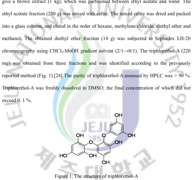
3-1. Preparation of Triphlorethol-A
The dried Ecklonia cava (4 kg), collected from Jeju Island in Korea, was immersed in 80 % methanol at room temperature for 2 days. The aqueous methanol was removed in vaccuo to give a brown extract (1 kg), which was partitioned between ethyl acetate and water. The ethyl acetate fraction (230 g) was mixed with celite. The mixed celite was dried and packed into a glass column, and eluted in the order of hexane, methylene chloride, diethyl ether and methanol. The obtained diethyl ether fraction (14 g) was subjected to Sephadex LH-20 chromatography using CHCl3-MeOH gradient solvent (2/1→0/1). The triphlorethol-A (220
mg) was obtained from these fractions and was identified according to the previously reported method (Fig. 1).[24] The purity of triphlorethol-A assessed by HPLC was > 90 %. Triphlorethol-A was freshly dissolved in DMSO; the final concentration of which did not exceed 0. 1 %.
Figure 1. The structure of triphlorethol-A
3-2. Reagents
The 1,1-diphenyl-2-picrylhydrazyl (DPPH) radical, 2',7'-dichlorodihydrofluorescein diacetate (DCF-DA), and Hoechst 33342 were purchased from Sigma Chemical Company,
St. Louis, MO, USA. The other chemicals and reagents were of analytical grade. Primary rabbit polyclonal anti-ERK 2 (42kDa ERK) and -phospho-ERK1/2 (phosphorylated 44kDa/42kDa ERK) (Thr 202/Tyr 204) antibodies were purchased from Santa Cruz Biotechnology, Santa Cruz, CA, USA.
3-3. Cell Culture
It is reported that lung is an organ sensitive to oxidative stress.[25,26] To study the effect of triphlorethol-A on oxidative stress induced by hydrogen peroxide, we used Chinese hamster lung fibroblasts (V79-4 cells). The V79-4 cells from the American Type Culture Collection, were maintained at 37 °C in an incubator with a humidified atmosphere of 5 % CO2 and
cultured in Dulbecco’s modified Eagle’s medium containing 10 % heat-inactivated fetal calf serum, streptomycin (100 mg/ml) and penicillin (100 units/ml).
3-4. Intracellular Reactive Oxygen Species (ROS) Measurement and Image Analysis The DCF-DA method was used to detect the intracellular ROS level.[27] DCF-DA diffuses into cells, where it is hydrolyzed by intracellular esterase to polar 2',7'-dichlorodihydrofluorescein. This non-fluorescent fluorescein analog gets trapped inside the cells and is oxidized by intracellular oxidants to a highly fluorescent, 2',7'-dichlorofluorescein. The V79-4 cells were seeded in a 96 well plate at 1 ´ 105 cells/ml.
triphlorethol-A and 30 minutes later, 1 mM H2O2 was added to the plate. The cells were
incubated for an additional 30 minutes at 37 °C. After addition of 25 mM of DCF-DA solution, the fluorescence of 2',7'-dichlorofluorescein was detected at 485 nm excitation and at 535 nm emission using a PerkinElmer LS-5B spectrofluorometer.
For image analysis for production of intracellular ROS, the V79-4 cells were seeded in coverslip loaded 6 well plate at 1 ´ 105 cells/ml. Sixteen hours after plating, the cells were
treated with triphlorethol-A and 30 minutes later, 1 mM H2O2 was added to the plate. After
changing media, 100 mM of DCF-DA was added in the well and was incubated for an additional 30 minutes at 37 °C. After washing with PBS, stained cells were mounted onto microscope slide in the mounting medium (DAKO, Carpinteria, CA, USA). Images were collected using the LSM 510 program on a Zeiss confocal microscope.
3-5.
Lipid Peroxidation Inhibitory ActivityLipid peroxidation was assayed by thiobarbituric acid reaction.[29] The V79-4 cells were seeded in a culture dish at 1 ´ 105 cells/ml. Sixteen hours after plating, the cells were treated
with various concentrations of triphlorethol-A. One hour later, 1 mM H2O2 was added to the
plate, and was incubated for further 1 hour. The cells were then washed with cold PBS, scraped and homogenized in ice-cold 1.15 % KCl. One hundred ml of the cell lysates was mixed with 0.2 ml of 8.1 % SDS, 1.5 ml of 20 % acetic acid (adjusted to pH 3.5) and 1.5 ml
of 0.8 % thiobarbituric acid (TBA). The mixture was made up to a final volume of 4 ml with distilled water and heated to 95 °C for 2 hours. After cooling to room temperature, 5 ml of n-butanol and pyridine mixture (15:1, v/v) was added to each sample, and the mixture was shaken well. After centrifugation at 1000 ´ g for 10 minutes, the supernatant fraction was isolated, and the absorbance was measured spectrophotometrically at 532 nm.
3-6. Cell Viability
The effect of triphlorethol-A on the viability of the V79-4 cells was determined using the [3-(4,5-dimethylthiazol-2-yl)-2,5-diphenyltetrazolium bromide (MTT) assay, which is based on the reduction of a tetrazolium salt by mitochondrial dehydrogenase in the viable cells.[30]
The V79-4 cells were seeded in a 96 well plate at 1 ´ 105 cells/ml. Sixteen hours after plating,
the cells were treated with various concentrations of triphlorethol-A. One hour later, 1 mM H2O2 was added to the plate and incubated at 37 °C for an additional 24 hours. Fifty ml of the
MTT stock solution (2 mg/ml) was then added to each well to attain a total reaction volume of 200 ml. After incubating for 4 hours, the plate was centrifuged at 800 ´ g for 5 minutes and the supernatants were aspirated. The formazan crystals in each well were dissolved in 150 ml dimethylsulfoxide and the A540 was read on a scanning multi-well spectrophotometer.
3-7. Flow cytometry analysis
The V79-4 cells were placed in a 6 well plate at 1 ´ 105 cells/ml. Sixteen hours after plating,
the cells were treated with 30 mM of triphlorethol-A. After a further incubation of 1 hour, 1 mM H2O2 was added to the culture. After 24 hours, the cells were harvested, and fixed in 1
ml of 70 % ethanol for 30 minutes at 4 °C. The cells were washed twice with PBS, and then incubated for 30 minutes in the dark at 37 °C in 1 ml of PBS containing 100 mg propidium iodide and 100 mg RNase A. Flow cytometric analysis was performed using a FACSCalibur flow cytometer (Becton Dickinson, Mountain View, CA, USA). The proportion of sub G1
hypo-diploid cells was assessed by the histograms generated using the computer program, Cell Quest and Mod-Fit.
3-8.Nuclear Staining with Hoechst 33342
The V79-4 cells were placed in a 24 well plate at 1 ´ 105 cells/ml. Sixteen hours after plating,
the cells were treated with 30 mM of triphlorethol-A and after further incubation for 1 hour, 1 mM H2O2 was added to the culture. After 24 hours, 1.5 ml of Hoechst 33342 (stock 10
mg/ml), a DNA specific fluorescent dye, was added to each well (1.5 ml) and incubated for 10 minutes at 37 °C. The stained cells were then observed under a fluorescent microscope, which was equipped with a CoolSNAP-Pro color digital camera to examine the degree of nuclear condensation.
The V79-4 cells were seeded at 1 ´ 105 cells/ml, and sixteen hours after plating, the cells
were treated with various concentrations of triphlorethol-A for 1 hour. The harvested cells were suspended in 10 mM phosphate buffer (pH 7.5) and then lysed on ice by sonication twice for 15 seconds. Triton X-100 (1 %) was then added to the lysates and was incubated for 10 minutes on ice. The lysates were centrifugated at 5000 ´ g for 30 minutes at 4 °C to remove the cellular debris. The protein content of the supernatant was determined by Bradford method,[32] with bovine serum albumin as the standard. The SOD activity was used to detect the inhibited level of auto-oxidation of epinephrine.[33] Fifty mg of the protein was added to 50 mM phosphate buffer (pH 10.2) containing 0.1 mM EDTA and 0.4 mM epinephrine. Epinephrine rapidly undergoes auto-oxidation at pH 10 to produce adrenochrome, a pink colored product, which can be measured at 480 nm using a UV/VIS spectrophotometer in kinetic mode. SOD inhibits the auto-oxidation of epinephrine. The rate of inhibition was monitored at 480 nm. The SOD activity was expressed as units/mg protein and one unit of enzyme activity was defined as the amount of enzyme required for 50 % inhibition of auto-oxidation of epinephrine.
3-10. Catalase (CAT) Activity
Fifty mg of protein was added to 50 mM phosphate buffer (pH 7) containing 100 mM (v/v) H2O2. The reaction mixture was incubated for 2 minutes at 37 °C and the absorbance was
monitored at 240 nm for 5 minutes. The change in absorbance with time was proportional to the breakdown of H2O2.[34] The CAT activity was expressed as units/mg protein and one unit of enzyme activity was defined as the amount of enzyme required to breakdown of 1 mM H2O2.
3-11. Glutathione Peroxidase (GPx) Activity
Fifty mg of the protein was added to 25 mM phosphate buffer (pH 7.5) containing 1 mM EDTA, 1 mM NaN3, 1 mM glutathione (GSH), 0.25 unit of glutathione reductase, and 0.1
mM NADPH. After incubation for 10 minutes at 37 °C, H2O2 was added to the reaction
mixture at a final concentration of 1 mM. The absorbance was monitored at 340 nm for 5 minutes. The GPx activity was measured as the rate of NADPH oxidation by change in absorbance at 340 nm.[35] The GPx activity was expressed as units/mg protein and one unit of enzyme activity was defined as the amount of enzyme required to oxidize 1 mM NADPH. 3-12. Western Blot
The V79-4 cells were placed in a plate at 1 ´ 105 cells/ml. Sixteen hours after plating, the
cells were treated with 30 mM of triphlorethol-A. The cells were harvested at the indicated times, and washed twice with PBS. The harvested cells were then lysed on ice for 30 minutes in 100 ml of lysis buffer [120 mM NaCl, 40 mM Tris (pH 8), 0.1 % NP 40] and centrifuged at 13,000 ´ g for 15 minutes. Supernatants were collected from the lysates and protein
concentrations were determined. Aliquots of the lysates (40 μg of protein) were boiled for 5 minutes and electrophoresed in 10 % SDS-polyacrylamide gel. Blots in the gels were transferred onto nitrocellulose membranes (Bio-Rad, Hercules, CA, USA), which were then incubated with primary rabbit monoclonal -ERK1/2 and -phospho ERK1/2. The membranes were further incubated with goat anti-rabbit immunoglobulin G-horseradish peroxidase conjugates (Pierce, Rockland, IL, USA), and then exposed to X-ray film. Protein bands were detected using an enhanced chemiluminescence Western blotting detection kit (Amersham, Little Chalfont, Buckinghamshire, UK).
3-13. Statistical Analysis
All the measurements were made in triplicate. The results were subjected to an analysis of the variance (ANOVA) using the Tukey test to analyze the difference. p<0.05 were considered significantly.
4. Results
4.1 Radical Scavenging Activity of Triphlorethol-A
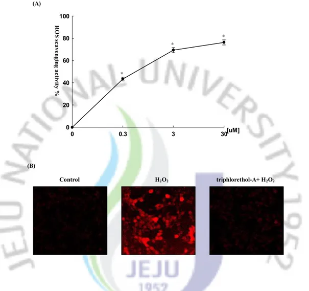
The radical scavenging effect of triphlorethol-A on the intracellular ROS and DPPH free radical scavenging activities was measured. The intracellular ROS scavenging activity of triphlorethol-A was 43, 69 and 76 % at concentrations of 0.3, 3 and 30 mM, respectively (Fig. 2A). In the presence of 2 mM of N-acetylcysteine (positive control), there was 85 % of ROS inhibition (data not shown). As shown in Fig. 2B, the fluorescent intensity of DCF-DA staining was enhanced in H2O2 treated V79-4 cells. However, triphlorethol-A reduced the red
Fig. 2. Effect of triphlorethol-A on scavenging intracellular ROS. The intracellular ROS generated was detected by DCF-DA method (A) and by confocal microscopy (B). Representative confocal images illustrate the increase in red fluorescence intensity of DCF produced by ROS in H2O2 treated V79-4 cells as compared to control and the lowered fluorescence intensity in H2O2 treated V79-4 cells in the presence of triphlorethol-A (original magnification ´ 400). *significantly different from control (p<0.05).
(B) 0 0.3 3 30 0 20 40 60 80 100 [uM] R O S s ca ve ng in g a ct iv ity % * * * Control H2O2 triphlorethol-A+ H2O2 (A)
4.2 Effect of Triphlorethol-A on Lipid Peroxidation
The ability of triphlorethol-A to inhibit lipid peroxidation in H2O2 treated V79-4 cells was
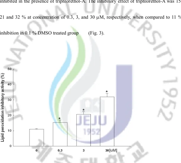
also investigated. The generation of thiobarbituric acid reactive substance (TBARS) was inhibited in the presence of triphlorethol-A. The inhibitory effect of triphlorethol-A was 15, 21 and 32 % at concentration of 0.3, 3, and 30 mM, respectively, when compared to 11 % inhibition in 0.1 % DMSO treated group (Fig. 3).
Fig. 3. Effect of triphlorethol-A on inhibition of lipid peroxidation. Lipid peroxidation was assayed by measuring the amount of TBARS. *significantly different from control (p<0.05).
4.3 Effect of Triphlorethol-A on Cell Damage Induced by H2O2
The protective effect of triphlorethol-A on cell survival in H2O2 treated V79-4 cells was also
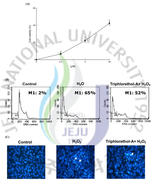
measured. Cells were treated with triphlorethol-A at various concentrations for 1 hour, prior to the addition to H2O2. The cell viability was determined 24 hours later by MTT assay. As
shown in Fig. 4A, treatment with triphlorethol-A induced a dose dependent increase in the cell survival rate; 7 % at 0.3 mM, 22 % at 3 mM, and 42 % at 30 mM. In order to study the cytoprotective effect of triphlorethol-A on apoptosis induced by H2O2, nuclei of V79-4 cells
were stained with Hoechst 33342 for microscopy and with propidium iodide for flow cytometric analysis. The microscopic pictures in Fig. 4B showed that the control cells had intact nuclei, and the H2O2 treated cells showed significant nuclear fragmentation,
characteristic of apoptosis. However, when the cells were treated with triphlorethol-A for 1 hour prior to H2O2 treatment, a dramatic decrease in nuclear fragmentation was observed. In
addition to the morphological evaluation, the protective effect of triphlorethol-A against apoptosis was confirmed by flow cytometry. As shown in Fig. 4C, an analysis of the DNA content in the H2O2 treated cells revealed an increase of 65 % of apoptotic sub-G1 DNA
content, as compared to 2 % of apoptotic sub-G1 DNA content in untreated cells. Treatment
with 30 mM of triphlorethol-A decreased the apoptotic sub-G1 DNA content to 52 %. These
apoptosis. (C) Control H2O2 Triphlorethol-A+ H2O2 [mM] 0 0.3 3 30 C el l v ia bi lit y (% ) 0 20 40 60 * * * * * * (A)
Fig. 4. Protective effect of triphlorethol-A on H2O2 induced oxidative damage of V79-4
cells. (A) The viability of V79-4 cells was determined by MTT assay. (B) Apoptotic body formation was observed under a fluorescent microscope after Hoechst 33342 staining and apoptotic bodies are indicated by arrows. (C) Apoptotic sub-G1 DNA
content was detected by flow cytometry after propidium iodide staining. M1 Control M1 M1 H2O Triphlorethol-A+ H2O2 (B) M1: 2% M1: 65% M1: 52%
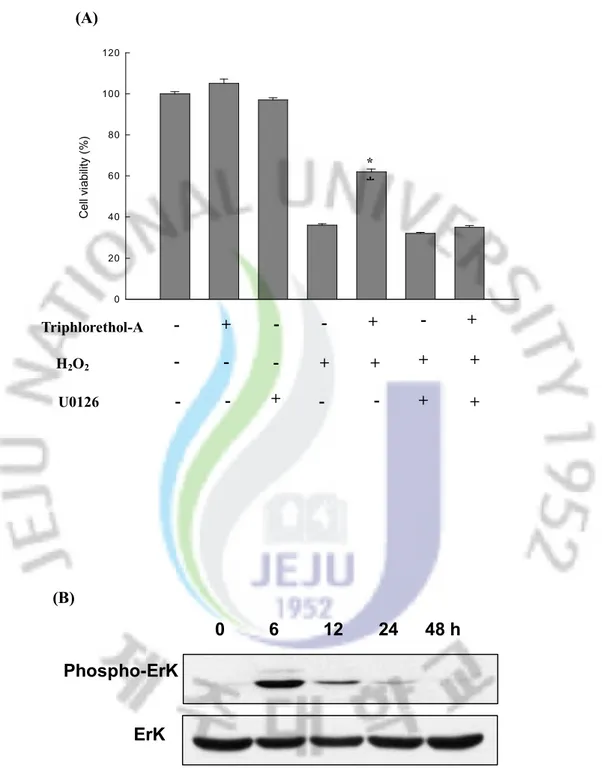
4.4 Effect of Triphlorethol-A on ERK activation
The activation of extracellular signal regulated kinase (ERK) is known to induce cell proliferation.[28] To better understand the protective mechanism of triphlorethol-A on V79-4
cells, we examined the activation of the ERK protein by western blot analysis with the phospho-ERK specific antibody. As shown in Fig. 5A, within 6 hours triphlorethol-A activated phosphorylated ERK dramatically. However, there was no change in the total ERK protein level. To determine the effect of ERK inhibitor on protection of triphlorethol-A from H2O2 induced damage, V79-4 cells were pre-treated for 30 minutes with U0126 (10 nM),
specific inhibitor of ERK kinase, followed for 30 minutes with triphlorethol-A and exposed to 1 mM H2O2 for 24 hours. As shown in Fig. 5B, U0126 treatment abolished the protection
C el l v ia bi lit y (% ) 0 20 40 60 80 100 120
*
*Fig. 5. Effect of triphlorethol-A on ERK activation. (A) Cell lysates were electrophoresed and proteins of ERK1/2 and phospho-ERK1/2 were detected by their respective antibodies. (B) After treatment of U0126, triphlorethol-A or/and H2O2, the viability of V79-4 cells was determined by MTT assay. *significantly different from H2O2 treated cells (p<0.05).
0 6 12 24 48 h
Phospho-ErK
ErK
(A) (B) H2O2 Triphlorethol-A U0126 + + + - + + - - - - - + + - - + - - + + -4.5 Effect of Triphlorethol-A on the Intracellular Antioxidant Systems
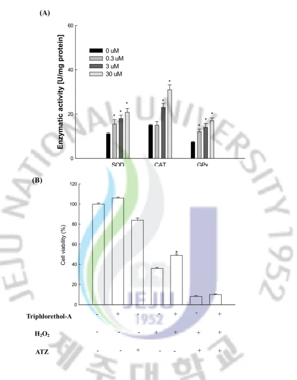
In order to investigate whether the radical scavenging activity of triphlorethol-A was mediated by the activities of antioxidant enzymes, the SOD, CAT and GPx activities in triphlorethol-A treated V79-4 cells were measured. Triphlorethol-A increased the activities of these three enzymes (Fig. 6A); in the case of SOD activity, at concentration of 0.3, 3, and 30 mM of triphlorethol-A, the activity was 16, 18, and 21, as compared to 11 U/mg protein of the control; in the case of CAT activity, it was 15, 23, and 31 U/mg protein at concentration of 0.3, 3, and 30 mM, as compared to 15 U/mg protein of the control; in the case of GPx activity, it was 12, 14, and 17 U/mg protein at concentration of 0.3, 3, and 30 mM, as compared to 7 U/mg protein of the control. The 3-amino-1,2,4 triazol (ATZ) is known as a specific inhibitor of catalase.[36] To determine the effect of catalse inhibitor on protection of
triphlorethol-A from H2O2 induced damage, V79-4 cells were pre-treated with 20 mM of
ATZ for 1 hour, followed for 30 minutes with triphlorethol-A and exposed to 1 mM H2O2 for
24 hours. As shown in Fig. 6B, ATZ treatment abolished the protection activity of triphlorethol-A in H2O2 damaged cells. It is reported that most polyphenolic compounds
interact with commonly used cell culture media to generate H2O2.[37] This generated low
(A) SOD CAT GPx E n zy m a ti c a ct iv it y [U /m g p ro te in ] 0 20 40 60 0 uM 0.3 uM 3 uM 30 uM * * * * * * * * (B) C e ll vi a b ili ty ( % ) 0 20 40 60 80 100 120 * + - - - + + + - + + - - H2O2 Triphlorethol-A ATZ + + - - - - + + -
Fig. 6. Effect of triphlorethol-A on the activities of antioxidative enzymes. (A) The data represents three experiments and are expressed as average enzyme unit per mg protein ± S.E. *significantly different from control (p<0.05). (B) After treatment of ATZ, triphlorethol-A or/and H2O2, the viability of V79-4 cells was
determined by MTT assay. *significantly different from H2O2 treated cells
4. Discussion
Phlorotannins, which are marine algal polyphenols and are also known as brown
algae, are polymers of phloroglucinol.
[38]Although some reports suggest that
phlorotannins from algae exhibit the antioxidant effect on free radicals,
[13,39,40]there
are no reports on the antioxdant activity of triphlorethol-A, isolated from Ecklonia
cava. In our present study, it was observed that upon exposure to H
2O
2,
triphlorethol-A decreased intracellular ROS generation and DPPH radical level.
Triphloroethol-A
has a polyphenol structure. Polyphenols are electron-rich compounds and prone to
enter into efficient electron-donation reactions with oxidizing agents to produce
phenoxyl radical (PhO∙) species as intermediates. Phenoxyl radicals are stabilized by
resonance delocaliztion of the unpaired electron to the ortho and para positions of
the ring. In addition to the resonance stability, phenoxyl radicals can also be
stabilized by hydrogen bonding with an adjacent hydroxyl group. Phenoxyl radicals
also undergo dimerization (“phenol coupling”) to produce new C-C or C-O
linkage.[
41]These intrinsic stability of phenolic structures might be related to
antioxidative activity of
triphloroethol A.
The cells exposed to H
2O
2exhibited
distinct morphological features of apoptosis, such as nuclear fragmentation and an
increase in sub G
1DNA content. However, cells that were pretreated with
triphlorethol-A had significantly reduced percentage of apoptotic cells, as shown by
morphological changes and reduction in sub G
1DNA content. Our results are also
consistent with the antioxidant activity of N-acetylcysteine, which also prevents
H
2O
2induced apoptosis, indicating that the inhibition of ROS formation may be
important for cytoprotection against oxidative damage. In many cell types,
extracellular signal regulated kinase (ERK) pathway is induced by a variety of
extracellular stimuli.
[42]The phosphorylation of ERK can phosphorylate cytoplasmic
and nuclear targets and participates in a wide range of cellular programs including
proliferation, differentiation, and movement.
[43-46]The level of phosphorylated ERK
in triphlorethol-A treated V79-4 cells was induced, and treatment of U0126, specific
inhibitor of ERK kinase, suppressed the protection activity of triphlorethol-A in H
2O
2damaged cells,
suggesting that the protective effect of triphlorethol-A on cells may
also be involved in activating ERK pathway. Antioxidant enzymes, like SOD, CAT,
and GPx, play significant roles in effective augmentation of antioxidant defense
mechanisms in cells. SOD converts superoxide radicals to hydrogen peroxide and
subsequently to water by the activity of CAT and GPx. Triphlorethol-A increased
SOD, CAT, and GPx activities, suggesting that the scavenging of ROS may be
related to the increased antioxidant activity. Therefore, the effects of triphlorethol-A
on cell viability might involve dual actions: direct action on oxygen radical
scavenging, as shown by intracellular ROS scavenging, and indirect action through
induction of anti-oxidative enzymes.
In conclusion, triphlorethol-A, a phlorotannin, exerted intracellular ROS
scavenging activity, promoted cell viability via activation of ERK protein, inhibited
H
2O
2induced apoptosis, and enhanced the effects of antioxidant enzymes.
PART Ⅱ
Protective Effect of Triphlorethol-A from Ecklonia cava
against Ionizing Radiation in vitro
3. Abstract
We studied the cytoprotective effect of triphlorethol-A against
g-ray radiation-
induced oxidative stress. Triphlorethol-A was observed to scavenge intracellular
reactive oxygen species (ROS) that were generated by
g-ray radiation. This
compound protected membrane lipid peroxidation and cellular DNA damage which
are the main targets of radiation-induced damage. In addition, triphlorethol-A
reduced the apoptotic cells formation induced by radiation. Taken together, the
results suggest that triphlorethol-A protects cells against oxidative
damage induced
by radiation through scavenging ROS.
2. INTRODUCTION
Most of Ionizing radiation's toxic effects are attributed to the free radicals (HO
.,
H
., H
2
O
2etc) generated by the radiolysis of water. Interaction of free radicals with
DNA can induce DNA damage lead to mutagenesis and carcinogenesis. [47] The
damaging effects of free radicals lead to cell death are associated with an increased
risk for numerous genetically determined diseased by ionizing radiation. [48] The
potential application of radioprotective chemicals in the event of planned exposure or
radiation accidents has been investigated from the beginning of the nuclear era [49].
Historically, sulfhydryl compounds have been among the first radioprotective agents.
Amifostine as a thiol compound is a powerful radioprotective agent, but its
application is clinically limited due to side effects and toxicity.[50,51] In recent years,
The screening of the compounds having the ability to scavenge these ROS could lead
to promising radioprotectors. Most of the antioxidants identified till date are derived
from the natural sources and exploration of the phytochemicals, as radioprotectors is
one among the competent strategies in lead identification. [51] Most of these plant
products have many compounds with medicinal value that protect biological systems
from ionizing radiation with minimum side effects. Recently, we reported that
triphlorethol-A, derived from Ecklonia cava, decreased H
2O
2-induced cell damage
through activation of an antioxidant system.[52]
In this study, we investigated the protective effects of triphlorethol-A on cell
damage that is induced by
g-ray radiation and the possible mechanism of the
cytoprotection.
3. Material and Methods
3-1. Cell culture
Chinese hamster lung fibroblasts (V79-4) cells from the American Type Culture Collection were maintained at 37 °C in an incubator with a humidified atmosphere of 5 % CO2 and cultured in Dulbecco’s modified Eagle’s medium containing 10 % heat-inactivated
fetal calf serum, streptomycin (100 mg/ml) and penicillin (100 units/ml).
3-2. Irradiation
Cells were exposed to g-ray from a 60Co g-ray source (MDS Nordion C-188 standard
source, located in Cheju National University, Jeju, Korea).
3-3. Intracellular reactive oxygen species measurement
The DCF-DA method was used to detect the intracellular ROS level.[8] The V79-4 cells were treated with triphlorethol-A at 30 mM and 1 h later, g-ray radiation at 10 Gy was applied to the cells. The cells were incubated for an additional 24 h at 37 °C. After the addition of 25 mM of DCF-DA solution, the fluorescence of 2',7'-dichlorofluorescein was detected at 485 nm excitation and at 535 nm emission using a PerkinElmer LS-5B spectrofluorometer.
3-5.
Lipid Peroxidation Inhibitory ActivityLipid peroxidation was assayed by a thiobarbituric acid reaction.[29] The V79-4 cells were treated with triphlorethol-A at 30 mM and 1 h later, g-ray radiation at 10 Gy was applied to the cells. The cells were incubated for 24 h at 37 °C. The cells were then washed with cold PBS, scraped and homogenized in ice-cold 1.15 % KCl. One hundred ml of the cell lysates was mixed with 0.2 ml of 8.1 % SDS, 1.5 ml of 20 % acetic acid (adjusted to pH 3.5) and 1.5 ml of 0.8 % thiobarbituric acid (TBA). Distilled water was added to the mixture to a final volume of 4 ml and heated to 95 °C for 2 h. After cooling to room temperature, 5 ml of an n-3-4.Cell viability
The effect of triphlorethol-A on the viability of the V79-4 cells was determined using the [3-(4,5-dimethylthiazol-2-yl)-2,5-diphenyltetrazolium] bromide (MTT) assay, which is based
on the reduction of a tetrazolium salt by mitochondrial dehydrogenase in viable cells.[30] The V79-4 cells were treated with triphlorethol-A at 30 mM and 1 h later, g-ray radiation at 10 Gy was applied to the cells. The cells were incubated for an additional 48 h at 37 °C. Fifty
ml of the MTT stock solution (2 mg/ml) was then added to each well to attain a total reaction volume of 200 ml. After incubating for 4 h, the plate was centrifuged at 800 ´ g for 5 min and the supernatants were aspirated. The formazan crystals in each well were dissolved in 150 ml
butanol and pyridine mixture (15:1, v/v) was added to each sample, and the mixture was shaken. After centrifugation at 1000 ´ g for 10 min, the supernatant fraction was isolated, and the absorbance was measured spectrophotometrically at 532 nm.
3-6. Flow cytometry analysis
Flow cytometry was performed in order to determine the apoptotic sub-G1 hypodiploid
cells.[31]The V79-4 cells were treated with triphlorethol-A at 30 mM and 1 h later, g-ray radiation at 10 Gy was applied to the cells. The cells were then incubated for an additional 48 h at 37 °C, subsequently harvested, and fixed in 1 ml of 70 % ethanol for 30 min at 4 °C. The cells were washed twice with PBS, and then incubated for 30 min in the dark at 37 °C in 1 ml of PBS containing 100 mg propidium iodide and 100 mg RNase A. Flow cytometric analysis was performed using a FACS Calibur flow cytometer (Becton Dickinson, Mountain View, CA, USA). The proportion of sub-G1 hypodiploid cells was assessed based on the
histograms generated using the computer programs, Cell Quest and Mod-Fit.
3-7. Nuclear Staining with Hoechst 33342
The V79-4 cells were treated with triphlorethol-A at 30 mM and 1 h later, g-ray radiation at 10 Gy was applied to the cells. The cells were then incubated for 48 h at 37 °C. After 24 h, 1.5 ml of Hoechst 33342 (stock 10 mg/ml), a DNA-specific fluorescent dye, was added to
each well (1.5 ml) and incubated for 10 min at 37 °C. The stained cells were observed under a fluorescent microscope, which was equipped with a CoolSNAP-Pro color digital camera to order to examine the degree of nuclear condensation.
3-8. Single cell gel electrophoresis (Comet assay)
Comet assay was performed to determine the oxidative DNA damage.[54,55] The cell suspension was mixed with 75 ml of 0.5 % low melting agarose (LMA) at 39 °C and spread on a fully frosted microscopic slide that was pre-coated with 200 ml of 1 % normal melting agarose (NMA). After solidification of the agarose, the slide was covered with another 75 ml of 0.5 % LMA and then immersed in a lysis solution (2.5 M NaCl, 100 mM Na-EDTA, 10 mM Tris, 1 % Trion X-100, and 10 % DMSO, pH 10) for 1 h at 4 °C. The slides were then placed in a gel-electrophoresis apparatus containing 300 mM NaOH and 10 mM Na-EDTA (pH 13) for 40 min to allow DNA unwinding and the expression of the alkali labile damage. An electrical field was then applied (300 mA, 25 V) for 20 min at 4 °C to draw negatively charged DNA toward an anode. After electrophoresis, the slides were washed three times for 5 min at 4 °C in a neutralizing buffer (0.4 M Tris, pH 7.5) and then stained with 75 ml of propidium iodide (20 mg/ml). The slides were observed with a fluorescence microscope and image analysis (Kinetic Imaging, Komet 5.5, UK). The percentage of total fluorescence in
the tail and the tail length of the 50 cells per slide were recorded. 3-9. Statistical analysis
All measurements were made in triplicate and all values were represented as means ± S.E. The results were subjected to an analysis of the variance (ANOVA) using the Tukey test to analyze the difference. p<0.05 were considered significantly.
4. Results
4.1 Radical Scavenging Activity of Triphlorethol-A on the ROS Generated by g-ray
Radiation
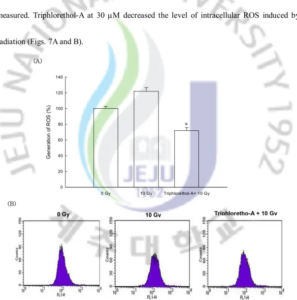
The radical scavenging effect of triphlorethol-A on the ROS generated by g-ray radiation was measured. Triphlorethol-A at 30 mM decreased the level of intracellular ROS induced by radiation (Figs. 7A and B).
Fig. 7. Effect of triphlorethol-A on scavenging intracellular ROS generated by
g-ray
radiation. The intracellular ROS was detected using fluorescence spectrophotometer
(A) and flow cytometry (B) after DCF-DA staining. The measurements were made in
triplicate and values are expressed as means ± S.E. FI means fluorescence intensity.
*Significantly different from radiation (p<0.05).
0 Gy 10 Gy Triphlorethol-A+ 10 Gy G en er at io n o f R O S ( % ) 0 20 40 60 80 100 120 140 (A) 0 Gy 10 Gy Triphloretho-A + 10 Gy (B) *
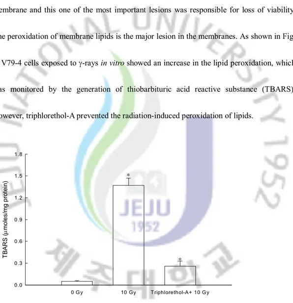
4.2 Effect of Triphlorethol-A on Lipid Peroxidation
The ability of triphlorethol-A to inhibit membrane lipid peroxidation and cellular DNA damage in radiated cells was also investigated. Ionizing radiation induced damage to cell membrane and this one of the most important lesions was responsible for loss of viability. The peroxidation of membrane lipids is the major lesion in the membranes. As shown in Fig. 8, V79-4 cells exposed to g-rays in vitro showed an increase in the lipid peroxidation, which was monitored by the generation of thiobarbituric acid reactive substance (TBARS). However, triphlorethol-A prevented the radiation-induced peroxidation of lipids.
Fig. 8. Protective effect of triphlorethol-A upon g-ray radiation-induced lipid membrane damage of V79-4 cells. Lipid peroxidation was assayed by measuring the amount of TBARS. The measurement was made in triplicate and values are expressed as means ± S.E. *Significantly different from control (p<0.05).
0 G y 10 Gy T riphlorethol-A+ 10 G y T B A R S ( mm o le s/ m g p ro te in ) 0.0 0.3 0.6 0.9 1.2 1.5 1.8 * *
4.3 Effect of Triphlorethol-A on Cell Damage Induced by Radiation
Damage to cellular DNA induced by g-radiation exposure was detected using an alkaline comet assay. The exposure of cells to radiation increased comet parameters like tail length and percentage of DNA in the tails of cells, which suggested radiation-induced damage from g-radiation. Treatment with triphlorethol-A before irradiation resulted in a decrease of tail length (Fig. 9A). When the cells were exposed to g-radiation at 10 Gy, the percent of DNA in the tail was increased 50.1 ± 3.4 as shown in Fig. 9B. Treatment with triphlorethol-A resulted in a decrease to 29.5 ± 2.3, which indicated a protective effect of triphloroethol-A on radiation-induced DNA damage in vitro.
(A)
(B)
Fig. 9. Protective effect of triphlorethol-A upon
g-ray radiation-induced cellular
DNA damage of V79-4 cells. The cellular DNA damage was detected by an alkaline
comet assay. The measurement was made in triplicate and values are expressed as
means ± S.E. *Significantly different from control (p<0.05).
0 Gy 10 Gy Triphorethol-A + 10 Gy 0 Gy 10 Gy Triphlorethol-A+10Gy % F lu o re sc en ce in ta il 0 10 20 30 40 50 60 * *
4.4 Cytoprotective Effect of Triphlorethol-A on Radiation-Induced Apoptosis
The protective effect of triphlorethol-A on cell survival in V79-4 cells exposed to radiation was also measured. As demonstrated in Fig. 10A, treatment with triphlorethol-A at 30 mM increased 74 % of the cell survival compared with 54 % of the cell survival in irradiated cells. In order to study the cytoprotective effect of triphlorethol-A on radiation-induced apoptosis, nuclei of V79-4 cells were stained with Hoechst 33342 for microscopy and with propidium iodide for flow cytometric analysis. The microscopic pictures in Fig. 10B demonstrated that the control cells had intact nuclei, and the radiation-exposed cells demonstrated significant nuclear fragmentation, which is characteristic of apoptosis. However, when the cells were treated with triphlorethol-A for 1 h prior to radiation, dramatic decrease in nuclear fragmentation was observed. In addition to the morphological evaluation, the protective effect of triphlorethol-A against apoptosis was confirmed by flow cytometry. As demonstrated in Fig. 10C, an analysis of the DNA content in the radiation-exposed cells revealed an increase of 35 % in apoptotic sub-G1 DNA content, compared with 2 % of
apoptotic sub-G1 DNA content in untreated cells. Treatment with 30 mM of triphlorethol-A
decreased the apoptotic sub-G1 DNA content to 18 %. These results suggested that
triphlorethol-A protects cell viability by inhibiting radiation-induced apoptosis. To understand how triphlorethol-A protects radiation-induced apoptosis, we examined changes
of Bcl-2 families and caspase activities by western blot.
As shown in Fig. 10. D the Bcl-2, an anti-apoptotic protein, recovered in treatment with triphlorethol-A and radiation compared to irradiated cells and the Bax, a pro-apoptotic protein, decreased in treatment with triphlorethol-A and radiation compared to irradiated cells. t was also observed the decreased activation of caspase 3(17kDa), the major effector caspase of the apoptotic process, and PARP cleavage(89kDa), one of substrates of activated caspase 3, in combination with triphlorethol-A and radiation compared to irradiated cells.
0 Gy 10 Gy Triphlorethol-A + 10 Gy C el l v ia bi lit y (% ) 0 20 40 60 80 100 120 * * 0 Gy 10 Gy Triphlorethol-A + 10 Gy DNA content (A) (B) (C) Triphlorethol-A + 10 Gy 10 Gy 0 Gy
Fig. 10. Protective effect of triphlorethol-A upon g-ray radiation-induced cell damage of V79-4 cells. (A) The viability of V79-4 cells upon radiation was determined by MTT assay. The measurements were made in triplicate and values are expressed as means ± S.E. *Significantly different from control (p<0.05). (B) Apoptotic body formation was observed under a fluorescence microscope after Hoechst 33342 staining and apoptotic bodies are indicated by arrows. (C) Apoptotic sub-G1 DNA content was detected by flow cytometry
after propidium iodide staining. (D) Western blot analysis was performed using antibcl2, -bax,-caspase 3 and -PARP antibodies.
4. Discussion
Exposure to ionizing radiation causes cell damage through the production of ROS
which induces oxidative stress.[55] The role of ROS in radiation injury and the
potential of antioxidants to reduce these deleterious effects have been studied for
more than 50 years.[56] Radioprotective agents minimize this damage by reducing
the generation of free radicals.[57] Increased understandings of the interrelationship
between the effects of oxygen and radiation exposure lead to a rational application of
naturally occurring antioxidants.[58] Our study demonstrated that in addition to an
antioxidant effect,[59]triphlorethol-A has protective activity against
g-radiation in
vitro. Triphlorethol-A scavenged the ROS generated by radiation. Radiation-induced
cellular lethality occurs through the formation of aqueous free radicals, which attack
vital cellular sites, such as DNA and cell membranes. The
cellular membrane and
DNA are the two main targets of radiation-induced lethality. The formation of lipid
peroxides in cells exposed to
g-radiation is one of the markers of the membrane
damage. Triphloroethol-A protected the cell membrane lipids from the peroxidation
damage induced by radiation. In addition, triphlorethol-A inhibited the increase of
radiation-induced comet parameters, which indicated the protection of cellular DNA.
These inhibition effects of triphlorethol-A against lipid and DNA damage resuled in
protective effects against radiation- induced cell death. Triphlorethol-A is polymer of
phloroglucinol and has a polyphenol structure. The presence of a phenolic group with
aromatic conjugation, which exists in the structure of triphlorethol-A, contributes to
the scavenging ROS generated by irradiation. Our study demonstrated that
triphlorethol-A has a radioprotective effect through scavenging ROS generated by
radiation.
PART Ⅲ
Triphlorethol-A induces heme oxygenase-1 via activation of
ERK and NF-E2 related factor 2 transcription factor
1. Abstract
Triphlorethol-A, phlorotannin found in Ecklonia cava, exerted cytoprotective effects against oxidative stress and the underlying molecular mechanism was investigated. Triphlorethol-A induced heme oxygenase-1 (HO-1) expression at mRNA and protein levels, leading to increased HO-1 activity. NF-E2 related factor 2 (Nrf2), transcription factor involved in the cellular protection against oxidative stress, regulates antioxidant response element (ARE)-directed induction of several phase 2 detoxifying and antioxidant enzymes. Triphlorethol-A increased nuclear translocation, ARE binding, and transcriptional activity of Nrf2. Moreover, triphlorethol-A exhibited activation of extracellular signal regulated protein kinase (ERK). U0126, inhibitor of ERK kinase, suppressed triphlorethol-A induced activation of Nrf2 and finally decreased HO-1 protein level. Pharmacological inhibitors of HO-1 and ERK attenuated the cytoprotective effect of triphlorethol-A on H2O2 or radiation
induced oxidative stress. Taken together, these data suggest that triphlorethol-A augments cellular antioxidant defense capacity through induction of HO-1 via ERK-Nrf2-ARE signaling pathway, thereby protecting cells from oxidative stress.
Keywords: triphlorethol-A; heme oxygenase-1; NF-E2 related factor 2; antioxidant response element
2. INTRODUCTION
Heme oxygenase-1 (HO-1) catalyzes the rate-limiting step in heme catabolism,
leading to the formation of biliveridin, free iron and carbon monoxide. In the
presence of biliveridin reductase, biliveridin is further converted to bilirubin, which
is a potent antioxidant [60-62]. HO-1 expression is induced in response to various
chemically or physiologically produced oxidative stresses in cells and tissues [63-66].
The HO-1 pathway represents a primer cellular defense system against oxidative
stress [63, 67-71], Recently, a new class of AP-1 related sequences has been shown
to mediate the oxidative stress responsiveness of the HO-1 gene. These regions,
termed stress responses elements (StRE) or antioxidant responses elements (ARE),
are tightly regulated by the redox-sensitive transcription factor, NF-E2 regulated
factor 2 (Nrf2) [72]. ARE is a cis-acting enhancer sequence that mediates
transcriptional activation of Nrf2 in response to oxidative stress [73]. ARE is found
in the promoters of genes that encode HO-1 [74] and other cellular antioxidant
enzymes, such as g-glutamylcysteine synthetase [75]. Therefore, genes regulated by
the ARE encode proteins that help control of the cellular redox status and protect the
cells from oxidative damage [76]. Mitogen activated protein kinase (MAPK) family
are important signaling intermediates involved transducing a variety of signals from
the cell surface to the nucleus. MAPKs pathway associates with the modulation of
ARE driven gene expression via Nrf2 activation. ERK and p38 MAPK pathways
involved the nucleus binding of Nrf2 to the ARE [77]. Inhibition of ERK and p38
abrogated binding of Nrf2 and induction of ARE-driven
g-glutamylcysteine
synthetase [78].
In view of the increasing evidence explaining the importance of carbon
monoxide and bilirubin in counteracting cellular dysfunction [79-82], attention has
recently been focused on up-regulation of HO-1 by natural compounds [83-87].
Recently, we reported that triphlorethol-A protected cells from H
2O
2induced
oxidative stress via radical quenching and catalase activation
42and from ionizing
radiation induced cellular damage [94].
In the present study, we investigated the capability of triphlorethol-A to induce
the antioxidant enzyme HO-1 via activation of the Nrf2 transcription factor.
3. Material and Methods
3-1. Reagents
[3-(4,5-dimethylthiazol-2-yl)-2,5-diphenyltetrazolium] bromide (MTT) and zinc protoporphyrin (ZnPP) were purchased from Sigma Chemical Company, St. Louis, MO, USA. Primary rabbit polyclonal anti-ERK, phospho-ERK, Nrf2, and actin antibodies were purchased from Santa Cruz Biotechnology, Santa Cruz, CA, USA. U0126 was provided from Calbiochem, San Diego, CA, USA. The ARE-luciferase reporter gene was kindly provided byDr. Young-Joon Surh (Seoul National University, Seoul, Korea).The other chemicals and reagents were of analytical grade.
3-2. Cell culture
The lung is an organ sensitive to oxidative stress [110-111]. To study the effect of triphlorethol-A on oxidative stress, we used Chinese hamster lung fibroblasts (V79-4 cells). The V79-4 cells (American type culture collection) were maintained at 37°C in an incubator with a humidified atmosphere of 5% CO2 and cultured in Dulbecco’s modified Eagle’s
medium containing 10% heat-inactivated fetal calf serum, streptomycin (100 mg/ml) and penicillin (100 units/ml).
3-3. Cell Viability
The effect of triphlorethol-A on the viability of the V79-4 cells was determined by the MTT assay [63]. The cells were treated with 30 mM triphlorethol-A, followd 1 h later by H2O2 or
γ-irradiation at 5 Gy. The cells were seeded in a 96 well plate at a density of 1 ´ 105 cells/ml
and incubated for 48 h at 37°C. Fifty ml of the MTT stock solution (2 mg/ml) was then added to each well to attain a total reaction volume of 200 ml. After incubation for 4 h, the plate was centrifuged at 800 ´ g for 5 min and the supernatants were aspirated. The formazan crystals in each well were dissolved in 150 ml DMSO, and the A540 was read on a scanning
multi-well spectrophotometer. 3-4.HO-1 assay
HO-1 enzyme activity was measured as described previously [114]. Cells were homogenized in 0.5 ml ice-cold 0.25 M sucrose solution containing 50 mM potassium phosphate buffer (pH 7.4). Homogenates were centrifuged at 200 ´ g for 10 min. The supernatants were centrifuged at 9000 ´ g for 20 min, and further centrifuged at 105,000 ´ g for 60 min. The pellet was then resuspended in 50 mM potassium phosphate buffer (pH 7.4) and the amount of protein was determined. The reaction mixture (200 ml) containing 0.2 mM of the substrate hemin, 500 mg/ml of cell lysate, 0.5 mg/ml rat liver cytosol as a source of biliveridin reductase, 0.2 mM MgCl2, 2 mM glucose-6-phosphate, 1U/ml glucose-6-phosphate
dehydrogenase, 1 mM NADPH and 50 mM potassium phosphate buffer (pH 7.4) was incubated at 37°C for 2 h. The reaction was stopped with 0.6 ml of chloroform and after extraction, the chloroform layer was measured spectrophotometrically. Bilirubin formation
was calculated from the difference in absorption between 464 and 530 nm. 3-5.Western blot
The V79-4 cells were plated at 1 ´ 105 cells/ml, and sixteen hours after plating, the cells were
treated with 30 mM triphlorethol-A. The cells were harvested at the indicated times, and washed twice with PBS. The harvested cells were then lysed on ice for 30 min in 100 ml of lysis buffer [120 mM NaCl, 40 mM Tris (pH 8), 0.1% NP 40] and centrifuged at 13,000 ´ g for 15 min. Supernatants were collected from the lysates and protein concentrations were determined. Aliquots of the lysates (40 μg of protein) were boiled for 5 min and electrophoresed on a 10% SDS-polyacrylamide gel. Blots in the gels were transferred onto nitrocellulose membranes (Bio-Rad, Hercules, CA, USA), which were then incubated with primary antibodies. The membranes were further incubated with secondary antibody-horseradish peroxidase conjugates (Pierce, Rockland, IL, USA), and exposed to X-ray film. Protein bands were detected using an enhanced chemiluminescence Western blotting detection kit (Amersham, Little Chalfont, Buckinghamshire, UK).
3-6.Reverse transcriptase polymerase chain reaction
Total RNA was isolated from V79-4 cells using Trizol (GibcoBRL, Grand Island, NY, USA). Reverse transcriptase polymerase chain reaction (RT-PCR) was performed as described previously.[115] PCR conditions for HO-1 and for the housekeeping gene, b-actin, were: 35
cycles of 94°C for 45 sec; 53°C for 45 sec; and 72°C for 60 sec. The primer pairs (Bionics, Seoul, Korea) were follows (forward and reverse, respectively): HO-1, 5'-GAG AAT GCT GAG TTC ATG -3' and 5'-ATG TTG AGC AGG AAG GC-3'; and b-actin, 5'-GTG GGC CGC CCT AGG CAC CAG G-3'; and 5'-GGA GGA AGA GGA TGC GGC AGT G-3'. Amplified products were resolved by 1% agarose gel electrophoresis, stained with ethidium bromide, and photographed under ultraviolet light.
3-7. Nuclear extract preparation and electrophoretic mobility shift assay
The cells were harvested at the indicated times, and were then lysed on ice with 1 ml of lysis buffer (10 mM Tris-HCl, pH 7.9, 10 mM NaCl, 3 mM MgCl2, and 1% NP-40) for 4 min.
After 10 min of centrifugation at 3,000 ´ g, the pellets were re-suspended in 50 ml of extraction buffer (20 mM HEPES, pH 7.9, 20% glycerol, 1.5 mM MgCl2, 0.2 mM EDTA, 1
mM DTT, and 1 mM PMSF), incubated on ice for 30 min, and centrifuged at 13,000 ´ g for 5 min. The supernatant was then harvested as nuclear protein extracts and stored at -70ºC after determination of protein concentration. Synthetic double strand oligonucleotide containing the Nrf2-binfing domain (ARE) was labeled with [g-32P] ATP using T4
polynucleotide kinase, and used as probes. The probes (50,000 cpm) were incubated with 6 mg of the nuclear extracts at 4oC for 30 min in a final volume of 20 ml containing 12.5%