• 검색 결과가 없습니다.

Antioxidant Effect of Edaravone on the Development of Preimplantation Porcine Embryos against Hydrogen Peroxide-Induced Oxidative Stress

N/A
N/A
Protected

Academic year: 2021

Share "Antioxidant Effect of Edaravone on the Development of Preimplantation Porcine Embryos against Hydrogen Peroxide-Induced Oxidative Stress"

Copied!
10
0
0

로드 중.... (전체 텍스트 보기)

전체 글

(1)

Antioxidant Effect of Edaravone on the Development of Preimplantation Porcine Embryos against Hydrogen Peroxide-Induced Oxidative Stress

Geon-Yeop Do, Jin-Woo Kim, Sung-Kyu Chae, Jae-Hyun Ahn, Hyo-Jin Park, Jae-Young Park, Seul-Gi Yang and Deog-Bon Koo

Dept. of Biotechnology, College of Engineering, Daegu University, Gyeongsan 38453, Korea

ABSTRACT

Edaravone (Eda) is a potent scavenger of inhibiting free radicals including hydroxyl radicals (H

2

O

2

). Reactive oxygen species (ROS) such as H

2

O

2

can alter most kinds of cellular molecules such as lipids, proteins and nucleic acids, cellular apoptosis. In addition, oxidative stress from over-production of ROS is involved in the defective embryo development of porcine. Previous study reported that Eda has protective effects against oxidative stress-like cellular damage. However, the effect of Eda on the preimplantation porcine embryos development under oxidative stress is unclear. Therefore, in this study, the effects of Eda on blastocyst development, expression levels of ROS, and apoptotic index were first investigated in preimplantation porcine embryos. After in vitro fertilization, porcine embryos were cultured for 6 days in PZM medium with Eda (10 μM), H

2

O

2

(200 μM), and Eda+H

2

O

2

treated group, respectively.

Rate of blastocyst development was significantly increased (P<0.05) in the Eda treated group compared with only H

2

O

2

treated group. And, we measured intracellular levels of ROS by DCF-DA staining methods and investigated numbers of apoptotic nuclei by TUNEL assay analysis is in porcine blastocyst, respectively. Both intracellular ROS levels and the numbers of apoptotic nucleic were significantly decreased (P<0.05) in porcine blastocysts cultured with Eda (10 μM). More over, the total cell number of blastocysts were significantly increased (P<0.05) in the Eda-treated group compared with untreated group and the only H

2

O

2

treated group. Based on the results, Eda was related to regulate as antioxidant-like function according to the reducing ROS levels during preimplantation periods. Also, Eda is beneficial for developmental competence and preimplantation quality of porcine embryos. Therefore, we concluded that Eda has protective effect to ROS derived apoptotic stress in preimplantation porcine embryos.

(Key words: edaravone (Eda), antioxidants, porcine embryo, reactive oxygen species (ROS), apoptosis)

This work was supported by grants from the Next-Generation BioGreen 21 Program (PJ01117604) and the Bio-Industry Technology Development Program (112130031HD030) through the Rural Development Administration and the Ministry of Agriculture, Food and Rural Affairs, Republic of Korea.

Correspondence : [email protected]

INTRODUCTION

In vitro production (IVP) of porcine embryos has been extensively studied for improving embryonic development and reproductive technologies (Choi et al., 2013). IVP embryos are very susceptible to oxidative damage because their defense mechanisms are insufficient to protect their delicate cellular structure (Rocha-Frigoni et al., 2015). Therefore, many resear- chers are investigating ways to optimize the condition of in vitro maturation (IVM) of oocytes or in vitro culture (IVC) of embryos, including temperature, gas tension, composition of media, etc. (Booth et al., 2005; Jin et al., 2007). It is well known that one of the problems that impair IVP of porcine embryos is the oxidative stress that is mainly caused by reac-

tive oxygen species (ROS) production from highly reactive molecules such as hydrogen peroxide (H

2

O

2

), hydroxyl radicals ( ․OH), superoxide anions (O

2

) and nitric oxide (NO). In particular, oocytes and early stage embryos are more vulner- able to oxidative stress, and the developmental competence of embryos is impaired by the resulting damage (Harvey et al., 2002).

Edaravone (Eda) is a potent free radical scavenger which

has been shown to provide neuroprotection against cerebral

ischemia-reperfusion injuries in experimental animal models

(Yan et al., 2012). Eda is a potent and novel synthetic sca-

venger of free radicals inhibiting not only hydroxyl radicals

but also iron-induced peroxidative injuries. In addition, Eda

has been prescribed clinically in Japan, since 2001 to treat

(2)

patients with cerebral ischemia (Tsuji et al., 2005). Eda has been used as a free novel radical scavenger to inhibit oxidative stress and apoptosis. Also, Eda has antioxidant effects and decreases hydroxyl radicals and superoxide radical production (Cheng et al., 2014). Eda has been reported to exert antioxidant effects because it can quench hydroxyl radicals and hydroxyl radical-dependent lipid peroxidation (Kikuchi et al., 2013).

Most of all, the generation and production of intracellular level ROS are involved in several signaling mechanisms. ROS can alter most kinds of cellular molecules such as lipids, pro- teins and nucleic acids, which results in mitochondrial damage, embryo cell block, ATP depletion, and apoptosis (Guerin et al., 2001; Gualtieri et al., 2014). The level of ROS production in mammalian embryos is particularly important during various stages of preimplantation development in vitro. Furthermore, during in vitro culture periods, embryos are exposed to relatively high oxidative stress compared to the environment of in vivo, thus the production of ROS within embryos is increased (Choi et al., 2008). Therefore, low rate of preimplantation embryo development is one of the main problems due to elevation of ROS during in vitro procedures (Dehghani-Mohammadabadi et al., 2014). Oxidative stress, depending on its severity, can lead to either cell necrosis or apoptosis. Also, apoptosis is charac- terized by cell shrinkage and chromatin condensation (Liu et al., 2000). Then, apoptosis, in response to inappropriate cultural conditions and stress, is a common physiological process in in vitro embryo development (Choi et al., 2008). And, cellular apoptosis has been assayed in mammalian blastocysts stage using TUNEL staining, which is influenced by suboptimal developmental conditions and stress, but mainly in in vitro fer- tilized (IVF) and parthenogenetic embryos (Isom et al., 2007;

Neuber et al., 2002).

Although an antioxidant effect of Eda has been reported in some mammalian cell models, there is currently no research regarding the effect of Eda on porcine embryo development.

Therefore, in this study, we evaluated the effects of Eda on the developmental competence of preimplantation porcine embryos cultured under oxidative-stress conditions. In addition, the ex- pression levels of ROS and the apoptotic index in blastocyst stage embryos derived from Eda treatment were measured.

MATERIALS AND METHODS

1. Chemicals

All chemical used in this study were purchased from Sigma Chemical Co. (St. Louis, Mo, USA) unless otherwise indicated.

2. In Vitro Maturation

Porcine ovaries were collected from a local slaughterhouse and transported to the laboratory at 30 ∼35℃ in 0.9% saline supplemented with 75 μg/ml potassium penicillin G. Cumulus oocyte complexes (COCs) were aspirated through an 18 gauge needle into a disposable 10 ml syringe from follicles of 3 to 6 mm in diameter (Funahashi et al., 1994). After washing three times with Tyrode’s lactate (TL)-N-2-hydroxyenthylpipera- zine-N'-2-ethanesulfoic acid (HEPES) medium, approximately immature 50 COCs were matured in 500 μl of IVM medium in a four-well multidish (Nunc, Roskilde, Denmark) at 38.5 ℃, 5% CO

2

in air. The medium used for oocyte maturation was North Carolina State University (NCSU) 23 medium (Petters and Wells, 1993) supplemented with 10% follicular fluid, 0.57 mM cysteine,10 ng/ml β-mercaptoethanol, 10 ng/ml epidermal growth factor, 10 IU/ml pregnant mare's serum gonadotropin (PMSG) and 10 IU/ml human chorionic gonadotropin (hCG).

After 22 h of culture, oocytes were washed three times and then further cultured in maturation medium without hormones supplement (PMSG and/or hCG) for 22 h.

3. In Vitro Fertilization

After IVM stage, the oocytes were subjected to IVF as des- cribed by Abeydeera and Day (1997). This medium was desig- nated as modified Tris-buffered medium (mTBM). Fresh semen was kindly supplied twice a week by artificial insemination company (Darby Pig AI Center, Anseong, Korea) and kept at 17 ℃ for 4 days. Semen was washed three times by centrifugation with Dulbecco's phosphate buffered saline (DPBS; Gibco-BRL, Grand Island, NY, USA) supplemented with 1 mg/ml bovine serum albumin (BSA; Fraction V), 100 mg/ml penicillin G and 75 mg/ml streptomycin sulfate. After washing, the spermatozoa were suspended in mTBM at pH 7.8. Oocytes were washed three times in mTBM with 2.5 mM caffeine sodium benzoate and 1 mg/ml BSA (fatty acid free) and placed into 48 μl of mTBM under paraffin oil. Diluted spermatozoa (2 μl) were added to 48 μl drop of the medium containing 20 oocytes to give a final concentration of 1.5×10

5

sperms/ml. The oocytes were co- incubated with the spermatozoa for 6 h at 38.5 ℃, 5% CO

2

in air.

4. In Vitro Culture and Chemical Treatment

(3)

For all experiments the embryos were cultured in 50 μl drops of porcine zygote medium 3 (PZM-3) medium with 3 mg/ml BSA at 38.5 ℃ under 5% CO

2

in air. After 48 h of culture, cleaved embryos were further cultured in a 50 μl drop of PZM-3 medium supplemented with 3 mg/ml BSA at 38.5 ℃ under 5% CO

2

in air for 4 days. To investigate oxidative stress, fertilized embryos were treated with oxidative inducer H

2

O

2

(200 μM) or antioxidant Eda (1, 10 or 20 μM) by direct addition to the culture medium. In addition, porcine embryos were cultured for 6 days in IVC medium with or without Eda (10 μM) under oxidative stress condition though H

2

O

2

200 μM treatment. Blastocyst formation was evaluated by stereomicro- scopic observation at Day 6 after insemination.

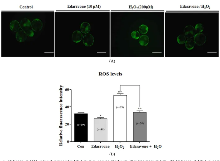
5. Measurement of Reactive Oxygen Species Levels

The level of H

2

O

2

in each embryo was measured using the dichlorofluorescin diacetate method (DCF-DA; Molecular Pro- bes, Eugene, OR, USA) described previously (Choi et al., 2008). At Day 6, in vitro produced blastocysts were recovered and used for the experiment. After three time washes in IVC medium, blastocysts were transferred into IVC medium con- taining 5 μM DCF-DA for 20 min at 38.5 ℃, 5% CO

2

in air.

A stock solution of DCF-DA dissolved in dimethylsulfoxide (DMSO) was then diluted in IVC medium, after which the permeabilised blastocysts in DCF-DA were washed three times with 0.1% polyvinylalcohol PVA in DPBS and placed in to a 50 μl drop covered with mineral oil. The fluorescent emissions from the embryos were recorded with a fluorescent microscope (Olympus) equipped with a cooled charge coupled device (CCD) camera where filters at 488 and 520 nm were used for excitation and emission, respectively. The recorded fluorescent images were analyzed by subtracting background and measuring inte- grated density with Image J software Version 1.38 (National Institutes of Health, Bethesda, MD, USA).

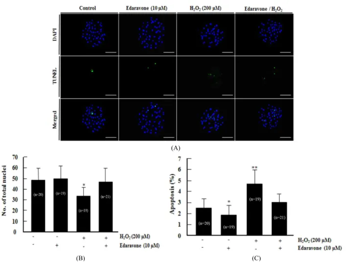
6. TUNEL Assay Analysis

Apoptotic cells in blastocysts were detected using the in situ Cell Death Detection Kit (Roche diagnostics GmbH, Mannheim, Germany). The blastocysts were recovered from fixed in 4%

(v/v) formaldehyde diluted in DPBS for 1 h at 4 ℃. For mem- brane permeabilization, the fixed embryos were incubated in DPBS containing 0.1% (v/v) Triton X-100 for 30 min at 4 ℃.

The fixed embryos were incubated in terminal deoxynucleotidyl transferase-mediated dUTP nick-end labeling (TUNEL) reac-

tion medium for 1 h at 38.5 ℃ in the dark and then washed and transferred into 2 mg/ml of DAPI and mounted on slides.

After TUNEL and DAPI staining, whole-mount embryos were examined under an epifluorescence microscope (Olympus) to determine the number of apoptotic nuclei and total number of nuclei.

7. Statistical Analysis

All experiments were repeated more than three times. All percentage data and data sets obtained in the present study are presented as the mean±standard deviation (SD) and mean±

standard error of the mean (SEM). The results were analyzed using a one-way ANOVA followed by Dunnett’s multiple Com- parison Test and the Student’s t-test. All calculations were per- formed using the GraphPad Prism 5.0 software package (San Diego, CA). Differences were considered significant at

*

P<

0.05,

**

P<0.01, and

***

P<0.001.

RESULTS

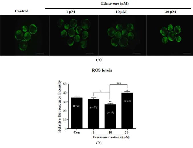
1. Effects of Eda on Developmental Competence and Fertilization Rate of Porcine Embryos

To investigate the effect of Eda on embryonic development,

we confirmed the blastocyst development and fertilization rate

in embryo of porcine after Eda treatment. First, the effect of Eda

on embryonic development was investigated. We investigated

the optimal Eda concentration of culture medium in development

competence of porcine embryos. After IVM and IVF, porcine

embryos were cultured in IVC medium supplemented with va-

rious concentrations (1, 10 or 20 μM) during full-time culture

periods at 38.5 ℃ and under 5% CO

2

. Table 1 showed that the

proportion of blastocysts development was significantly increased

(P<0.05) in culture media supplemented with Eda at a con-

centration of 10 μM. Based on the results, we proceeded in a

following set of experiments the concentration of 10 μM. Next,

we investigated the effects of Eda on development of porcine

embryos against oxidative stress induced by H

2

O

2

. Presumptive

zygotes were cultured in the presence or absence of Eda and

H

2

O

2

for 6 days at 38.5 ℃ and under 5% CO

2

in air. These re-

sults showed that the rates of cleavage and blastocyst formation

under H

2

O

2

-induced oxidative stress were significantly improved

in the presence of Eda group when compared with absence of

the only Eda treatement group (Table 2), suggesting that de-

velopment competence improved due to Eda treatment.

(4)

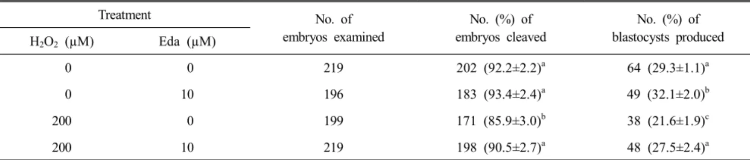
Table 2. Effect of Eda treatment on the development of porcine embryos against oxidative stress induced by H

2

O

2

Treatment No. of

embryos examined

No. (%) of embryos cleaved

No. (%) of blastocysts produced H

2

O

2

(µM) Eda (µM)

0 0 219 202 (92.2±2.2)

a

64 (29.3±1.1)

a

0 10 196 183 (93.4±2.4)

a

49 (32.1±2.0)

b

200 0 199 171 (85.9±3.0)

b

38 (21.6±1.9)

c

200 10 219 198 (90.5±2.7)

a

48 (27.5±2.4)

a

Data are the mean±SD.

a∼c

Values from ten replicates with different superscripts denote a significant difference relative to other groups (P<0.05).

Table 1. Effect of Eda treatment on the developmental competence in porcine embryos

Groups (µM)

No. of embryos examined

No.(%) of embryos cleaved

No.(%) of blastocysts produced 0 285 257 (90.1±3.8) 79 (27.6±2.1)

a

1 265 239 (90.2±2.8) 79 (29.8±1.1)

a

10 265 241 (91.9±2.9) 84 (31.7±2.7)

b

20 265 234 (88.2±2.7) 65 (24.3±2.6)

c

Data are the mean±SD.

a∼c

Values from ten replicates with different superscripts denote a significant difference relative to other groups (P<0.05).

2. Effects of Eda on Intracellular ROS Level, Cell Number and Frequency of Apoptotic Nuclei in Porcine Blastocysts

This study was carried out to assess the effect of ROS by measuring the effects of H

2

O

2

on blastocyst by Eda treatments.

After determining the optimal concentration of Eda groups, an experiment was designed to analyze the effect of Eda on the ROS level of porcine blastocysts (Fig. 1A). As shown in Fig.

1B, the intracellular ROS level in blastocysts did not differ significantly between the control and groups treated with 1 μM Eda. However, the quantification analysis showed that Eda 10 μM exposure significantly decreased (P<0.05) the level of in- tracellular ROS in blastocysts. Next, we used merge data stained by blue and green find out for apoptosis in blastocysts.

In order to determine the frequency of apoptosis in porcine blastocysts, TUNEL assay was used (Fig. 2A). As shown in Fig. 2B, the total cell number in blastocysts did not differ sig- nificantly between the control and groups treated with 1 and 10 μM Eda. Furthermore, the number of TUNEL positive cells decreased (P<0.05) significantly in blastocysts derived from

concentration of 10 μM Eda treated group (Fig. 2C). These results the number of TUNEL positive cells decreased signifi- cantly in blastocysts derived from Eda treated groups as well, suggesting a protective effect of Eda against apoptosis in blastocysts.

3. Effects of Eda Treatment on Intracellular ROS Level, Cell Number and Frequency of Apoptotic Nuclei in Porcine Blastocysts against Oxidative Stress induced by H

2

O

2

We next investigated the intracellular levels of ROS in blastocyst-stage embryos treated with or without Eda under oxi- dative stress induced by H

2

O

2

(Fig. 3A). As shown in Fig. 3B, the intracellular ROS level in blastocysts formation under H

2

O

2

- induced oxidative stress did not differ significantly between control and Eda treated group. These results in the quantification analysis is showing that Eda exposure significantly decreased the level of intracellular ROS in blastocysts, suggesting that Eda can prevent an increase in ROS induced by H

2

O

2

. We also investigated the apoptotic index in blastocyststage embryos treated with or without Eda under oxidative stress induced by H

2

O

2

(Fig. 4A). As shown in Fig. 4B, the total cell number in blastocyst formation under H

2

O

2

-induced oxidative stress did not differ significantly between the control and groups treated Eda group. However, the proportion of TUNEL positive cells decreased significantly in blastocysts derived from Eda treated group under H

2

O

2

-induced oxidative stress (Fig. 4C). These result indicated that Eda treatment effect on blastocyst quality and development proportion.

DISCUSSION

The aim of this study was to investigate the protective role

(5)

(A)

(B)

Fig. 1. Detection of intracellular ROS level in porcine blastocyst. (A) To determine the production of ROS in porcine blastocyst, we per- formed the DCF-DA staining. (B) Eda of relative fluorescence intensity of ROS per blastocysts. Scale bars = 100 µm. The quan- tification analysis of fluorescence intensity in DCF-DA stained porcine blastocyst obtained by the image J program. Data in the bar graph represents the means±SEM of three independent experiments. Statistically differences are indicated by asterisks (

*

P<

0.05;

**

P<0.01;

***

P<0.001, compared to control group).

of the Eda from oxidative stress through the preimplantation embryo development. Oxidative stress is directly related to the quality and developmental competence of blastocyst stage embryos. Previous report documented that Eda, a potent anti- oxidant, reduced oxidative stress in cells (Zong et al., 2014).

However, the anti-oxidative role of Eda in development process of porcine preimplantation embryos and the effect of Eda on apoptosis mechanisms of early embryogenesis are not fully understood. Therefore, our study investigated the Eda known to have antioxidant affects in early embryo development. As expected, cell death is decreased by oxidative stress of the preimplantation embryo through the treatment of Eda. Through this reduction in apoptosis in the early embryo development, the efficiency of fertilization and the embryo development is increased.

In a healthy body, ROS and antioxidants remain in balance

(Agarwal et al., 2005). ROS is known as regulation of embryo

metabolism, these metabolism generates ROS via several enzy-

matic mechanisms (Guerin et al., 2001). Oxidative stress caused

by excessive ROS production and/or breakdown mechanisms

of the antioxidant defense has been shown to be detrimental

to fertilized eggs, resulting in the impairment of early embryonic

development (Ruder et al., 2008). In particular, many studies

reported a sustaining increase in oxidative stress by loss of

balance between ROS and antioxidants in embryo of in vitro

production (Combelles et al., 2009). The development until

blastocyst stage is an important period during preimplantation

embryogenesis, and quality of blastocyst portends their ability

to implant (Fabian et al., 2005). Our result showed that de-

velopmental competence of blastocyst formation increased

(6)

(A)

(B)

(C)

Fig. 2. Evaluation of cellular apoptosis by using TUNEL assay in porcine blastocyst. (A) Detection of apoptosis by TUNEL assay using fluorescent images in porcine blastocyst-stage embryos cultured with or without Eda undergoing apoptosis in vitro. Hoechst 33342 staining identifying nuclei, TUNEL staining identifying apoptotic cells and merged photograph of Hoechst 33342 and TUNEL-stained porcine blastocyst (scale bar = 100 µm). (B) Eda of the total cell numbers per blastocyst. (C) Percentage of TUNEL positive cell per blastocyst. The quantification analysis of fluorescence intensity in TUNEL stained porcine blastocyst obtained by the image J program. Data in the bar graph represents the means±SEM of three independent experiments. Statistically differences are indicated by asterisks (P<0.05).

significantly in 10 μM Eda treated groups (Table 1). We also found the antioxidant and anti-apoptotic effects of Eda con- firmed in porcine early embryo development process for the first time in this field of research.

As shown in the Fig. 1, we confirmed the level of intra- cellular ROS in porcine embryo of preimplantation development by DCF-DA staining method via culture medium within Eda.

Eda significantly reduced ROS levels in dose-dependent manner at the blastocyst stage of porcine. A similar effect of antioxidant addition such as Eda, vitamin E on development has previously been reported in porcine embryo studies (Kitagawa et al., 2004).

Based on the results, Eda acts as an oxygen free radical sca-

venger to protect porcine embryos from oxidative stress and

improve blastocyst development. Moreover, ROS detected the by

DCF-DA staining analysis in blastocyst of control group during

in vitro production. ROS production by embryo has been

reported on many occasions and culture environment (Guerin

et al., 2001). This being so, the IVP embryos by in vitro fer-

tilization is known that ROS are generated in culture (Agarwal

et al., 2012). To investigate the detailed functional role of Eda

as antioxidant, we performed dividing into four treatment groups

(control, Eda, H

2

O

2

and Eda+H

2

O

2

). H

2

O

2

treatment group had

(7)

(A)

(B)

Fig. 3. Detection of H

2

O

2

-induced intracellular ROS level in porcine blastocyst after treatment of Eda. (A) Detection of ROS in porcine blastocysts derived from Eda treated and H

2

O

2

treated groups. (B) Eda of relative fluorescence in tensity of ROS per blastocysts.

Scale bars = 100 µm. The quantification analysis of fluorescence intensity in DCF-DA stained porcine blastocyst obtained by the image J program. Data in the bar graph represents the means±SEM of three independent experiments. Statistically differences are indicated by asterisks (

*

P<0.05;

**

P<0.001, compared to control group).

decrease in blastocysts developmental rate. However, the group treated with H

2

O

2

and Eda recovered as the control group.

Under oxidative stress condition, the Eda may also play a sig- nificant role as antioxidant in regulating oxidative stress during porcine preimplantation embryo development.

Also, we found significant differences in the development competence of blastocyst formation embryos cultured with and without Eda under oxidative stress (Table 2). Thus, our data showed that Eda treatment greatly improved the quality of H

2

O

2

-induced oxidant stress by increased blastocyst formation, reducing apoptosis and fewer incidences of fragmentation and developmental retardation. We believe that the Eda may also play a significant role as an antioxidant in regulating apoptosis and quality of embryo in preimplantation development until blastocyst.

The regulation of ROS production and oxidative stress in the

early embryonic development of the porcine affects apoptosis

(Choi et al., 2008). Moreover, the regulation of apoptosis in

vitro fertilization (IVF) since the early embryo development

process is very important for embryo development efficiency

and determining of quality of blastocyst (Lichnovsky et al.,

2000). In addition, apoptosis during early embryonic develop-

ment supports embryo survival under healthy conditions by

selectively eliminating abnormal cells with nuclear and chro-

mosomal abnormalities and by preventing massive cell death,

which may lead to the loss of the whole embryo (Han et al.,

2008). Previously studies observed elevated increasing of the

apoptotic factors of the mRNA expression levels and apoptotic

cell in embryo development via ROS increasing condition (Cui

and Kim, 2005). These studies are supported that regulating of

(8)

(A)

(B)

(C)

Fig. 4. Evaluation of H

2

O

2

-induced apoptosis by using TUNEL assay in porcine blastocysts cultured with Eda. (A) Apoptosis scanning images and anti-apoptotic characteristics in porcine blastocysts derived from H

2

O

2

and/or Eda treatment groups. The chromatin content is stained by DAPI (blue), fragmented DNA is labeled by the TUNEL reaction (green), and colocalization with DAP Iappears sky-blue. (B) Eda of the total cell numbers per blastocyst. (C) Percentage of TUNEL positive cell per blastocyst. The quantification analysis of fluorescence intensity in TUNEL stained porcine blastocyst obtained by the image J program. Data in the bar graph represents the means±SEM of three independent experiments. Statistically differences are indicated by asterisks (P<0.05).

apoptosis and apoptotic gene expression is critical role in preim- plantation embryo development. So, we performed the analysis of TUNEL assay that revealed the number of apoptotic nuclei in blastocysts derived from embryos cultured with Eda was lower than that in those cultured without Eda following the H

2

O

2

induction of oxidative stress. The total number of cells in blastocysts derived from embryos cultured with Eda was significantly higher than that of those cultured with out Eda (Fig. 4). Overall, these finding suggest that reduction of oxi- dative stress by Eda treatment may improve porcine blastocyst quality.

In conclusions, the results of the present study suggest that H

2

O

2

-induced oxidative stress results in decreased developmental

competence as shown by an increased number of apoptotic

nuclei and decreased cell numbers in blastocysts. Moreover, the

addition of Eda under H

2

O

2

-induced oxidative stress improved

developmental competence to the blastocyst stage, reducing the

number of apoptotic nuclei and increasing the cell number in

porcine IVP embryos by preventing oxidative stress. Therefore,

ours study has provided the first evidence of Eda functional

role associated with antioxidant and/or blocking of apoptosis

in early embryo development stage until blastocyst of porcine

in vitro production. Also, our results suggest that Eda may

improve the development and quality of porcine embryos by

preventing oxidative stress. Furthermore, these data will be

beneficial for better understanding the antioxidant mechanisms

(9)

of in vitro culture condition in early embryo development stage of porcine in vitro production.

REFERENCES

Abeydeera LR and Day BN. 1997. Fertilization and subsequent development in vitro of pig oocytes inseminated in a mo- dified tris-buffered medium with frozen-thawed ejaculated spermatozoa. Biology of Reproduction 57: 729-734.

Agarwal A, Aponte-Mellado A, Premkumar BJ, Shaman A and Gupta S. 2012. The effects of oxidative stress on female reproduction: A review. Reproductive Biology and Endocri- nology 10: 49.

Agarwal A, Gupta S and Sharma RK. 2005. Role of oxidative stress in female reproduction. Reproductive Biology and Endocrinology 3: 28.

Booth PJ, Holm P and Callesen H. 2005. The effect of oxygen tension on porcine embryonic development is dependent on embryo type. Theriogenology 63: 2040-2052.

Cheng B, Guo Y, Li C, Ji B, Pan Y, Chen J and Bai B. 2014.

Eda protected PC12 cells against MPP(+)-cytoxicity via in- hibiting oxidative stress and up-regulating heme oxygenase-1 expression. Journal of the Neurological Sciences 343: 115- 119.

Choi J, Park SM, Lee E, Kim JH, Jeong YI, Lee JY, Park SW, Kim HS, Hossein MS, Jeong YW, Kim S, Hyun SH and Hwang WS. 2008. Anti-apoptotic effect of melatonin on preimplantation development of porcine parthenogenetic embryos. Molecular Reproduction and Development 75:

1127-1135.

Choi JY, Kang JT, Park SJ, Kim SJ, Moon JH, Saadeldin IM, Jang G and Lee BC. 2013. Effect of 7,8-dihydroxyflavone as an antioxidant on in vitro maturation of oocytes and de- velopment of parthenogenetic embryos in pigs. The Journal of Reproduction and Development 59: 450-456.

Combelles CM, Gupta S and Agarwal A. 2009. Could oxidative stress influence the in-vitro maturation of oocytes? Repro- ductive Biomedicine Online 18: 864-880.

Cui XS and Kim NH. 2005. Polyamines inhibit apoptosis in porcine parthenotes developing in vitro. Molecular Repro- duction and Development 70: 471-477.

Dehghani-Mohammadabadi M, Salehi M, Farifteh F, Nematol- lahi S, Arefian E, Hajjarizadeh A, Parivar K and Nourmo- hammadi Z. 2014. Melatonin modulates the expression of

BCL-xl and improve the development of vitrified embryos obtained by IVF in mice. Journal of Assisted Reproduction and Genetics 31: 453-461.

Fabian D, Koppel J and Maddox-Hyttel P. 2005. Apoptotic processes during mammalian preimplantation development.

Theriogenology 64: 221-231.

Funahashi H, Cantley TC, Stumpf TT, Terlouw SL and Day BN. 1994. In vitro development of in vitro-matured porcine oocytes following chemical activation or in vitro fertiliza- tion. Biology of Reproduction 50: 1072-1077.

Gualtieri R, Barbato V, Fiorentino I, Braun S, Rizos D, Longo- bardi S and Talevi R. 2014. Treatment with zinc, d-aspartate, and coenzyme Q10 protects bull sperm against damage and improves their ability to support embryo development. The- riogenology 82: 592-598.

Guerin P, El Mouatassim S and Menezo Y. 2001. Oxidative stress and protection against reactive oxygen species in the pre-implantation embryo and its surroundings. Human Re- production Update 7: 175-189.

Han Z, Mtango NR, Patel BG, Sapienza C and Latham KE.

2008. Hybrid vigor and transgenerational epigenetic effects on early mouse embryo phenotype. Biology of Reproduction 79: 638-648.

Harvey AJ, Kind KL and Thompson JG. 2002. REDOX re- gulation of early embryo development. Reproduction 123:

479-486.

Isom SC, Prather RS and Rucker EB. 2007. Heat stress-induced apoptosis in porcine in vitro fertilized and parthenogenetic preimplantation-stage embryos. Molecular Reproduction and Development 74: 574-581.

Jin YX, Lee JY, Choi SH, Kim T, Cui XS and Kim NH.

2007. Heat shock induces apoptosis related gene expression and apoptosis in porcine parthenotes developing in vitro.

Animal Reproduction Science 100: 118-127.

Kikuchi K, Tancharoen S, Takeshige N, Yoshitomi M, Morioka M, Murai Y and Tanaka E. 2013. The efficacy of Eda (ra- dicut), a free radical scavenger, for cardiovascular disease.

International Journal of Molecular Sciences 14: 13909-13930.

Kitagawa Y, Suzuki K, Yoneda A and Watanabe T. 2004.

Effects of oxygen concentration and antioxidants on the in vitro developmental ability, production of reactive oxygen species (ROS), and DNA fragmentation in porcine embryos.

Theriogenology 62: 1186-1197.

Lichnovsky V, Prochazkova J, Erdosova B, Nepozitkova D,

(10)

Aernochova D and Pospisilova E. 2000. Apoptosis and expression of bcl-2 and bax during early human embryoge- nesis. Scripta Medica 73: 245 –250.

Liu L, Trimarchi JR and Keefe DL. 2000. Involvement of mi- tochondria in oxidative stress-induced cell death in mouse zygotes. Biology of Reproduction 62: 1745-1753.

Neuber E, Luetjens CM, Chan AW and Schatten GP. 2002.

Analysis of DNA fragmentation of in vitro cultured bovine blastocysts using TUNEL. Theriogenology 57: 2193-2202.

Petters RM and Wells KD. 1993. Culture of pig embryos.

Journal of Reproduction and Fertility Supplement 48: 61- 73.

Rocha-Frigoni NA, Leao BC, Nogueira E, Accorsi MF and Mingoti GZ. 2015. Effects of gaseous atmosphere and anti- oxidants on the development and cryotolerance of bovine embryos at different periods of in vitro culture. Zygote 23:

159-168.

Ruder EH, Hartman TJ, Blumberg J and Goldman MB. 2008.

Oxidative stress and antioxidants: exposure and impact on female fertility. Human Reproduction Update 14: 345-357.

Tsuji K, Kwon AH, Yoshida H, Qiu Z, Kaibori M, Okumura T and Kamiyama Y. 2005. Free radical scavenger (Eda) prevents endotoxin-induced liver injury after partial hepa- tectomy in rats. Journal of Hepatology 42: 94-101.

Yan Y, Gong K, Ma T, Zhang L, Zhao N, Zhang X, Tang P and Gong Y. 2012. Protective effect of Eda against Alzhei- mer's disease-relevant insults in neuroblastoma N2a cells.

Neuroscience Letters 531: 160-165.

Zong L, Yu QH, Du YX and Deng XM. 2014. Eda protects endotoxin-induced liver injury by inhibiting apoptosis and reducing proinflammatory cytokines. Brazilian Journal of Medical and Biological Research 47: 231-236.

Received November 25, 2015, Revised December 18, 2015,

Accepted December 21, 2015

수치

Table  1.  Effect  of  Eda  treatment  on  the  developmental  competence  in  porcine  embryos Groups (µM) No
Fig.  1.  Detection  of  intracellular  ROS  level  in  porcine  blastocyst.  (A)  To  determine  the  production  of  ROS  in  porcine  blastocyst,  we  per-  formed  the  DCF-DA  staining
Fig.  2.  Evaluation  of  cellular  apoptosis  by  using  TUNEL  assay  in  porcine  blastocyst
Fig.  3.  Detection  of  H 2 O 2 -induced  intracellular  ROS  level  in  porcine  blastocyst  after  treatment  of  Eda
+2

참조

관련 문서

Kuwait will celebrate on Sunday the fourth anniversary of the UN honoring and proclamation of His Highness the Amir, Sheikh Sabah Al-Ahmad Al-Jaber Al-Sabah as

The Joseon government designed and promulgated the Taegeukgi as a national flag for diplomatic and political purposes, but it was the independence movement that made it

• 이명의 치료에 대한 매커니즘과 디지털 음향 기술에 대한 상업적으로의 급속한 발전으로 인해 치료 옵션은 증가했 지만, 선택 가이드 라인은 거의 없음.. •

Since every classical or virtual knot is equivalent to the unknot via a sequence of the extended Reidmeister moves together with the forbidden moves, illustrated in Section 2,

웹 표준을 지원하는 플랫폼에서 큰 수정없이 실행 가능함 패키징을 통해 다양한 기기를 위한 앱을 작성할 수 있음 네이티브 앱과

_____ culture appears to be attractive (도시의) to the

【판결요지】[1] [다수의견] 동일인의 소유에 속하는 토지 및 그 지상 건물에 관하여 공동저 당권이 설정된 후 그 지상 건물이 철거되고 새로 건물이 신축된 경우에는

After first field tests, we expect electric passenger drones or eVTOL aircraft (short for electric vertical take-off and landing) to start providing commercial mobility