• 검색 결과가 없습니다.

Lysophosphatidic acid enhances breast cancer cells-mediated osteoclastogenesis

N/A
N/A
Protected

Academic year: 2022

Share "Lysophosphatidic acid enhances breast cancer cells-mediated osteoclastogenesis"

Copied!
9
0
0

로드 중.... (전체 텍스트 보기)

전체 글

(1)

Author contributions: J.S.N. and S.S.L. conceptualized and designed the studies. J.S.N., A.R.S. and L.T.N. performed the experiments and analyzed the data. S.J., Y.H.L. and G.S. contributed to the acquisition of data, analysis and/or interpretation of data. A.R.S. and L.T.N. drafted the manuscript. J.S.N.

and S.S.L. reviewed and edited the manuscript.

This is an Open Access article distributed under the terms of the Creative Commons Attribution Non-Commercial License, which permits unrestricted non-commercial use, distribution, and reproduction in any medium, provided the original work is properly cited.

Copyright © Korean J Physiol Pharmacol, pISSN 1226-4512, eISSN 2093-3827

INTRODUCTION

Breast cancer is the most common malignant tumor diagnosed among women worldwide. Despite continued improvement in treatment and earlier detection, breast cancer is the second lead- ing cause of death from cancer in women. As per survey, there are 1.7 million new cases and 521,900 deaths in 2012 [1]. Reportedly, metastasis was found more responsible for the vast majority of cancer patient deaths. Breast cancer cell metastasizes to several organs, however bone is the most preferential metastatic target of breast cancer cells.

To support cancer cell growth, progression and metastasis,

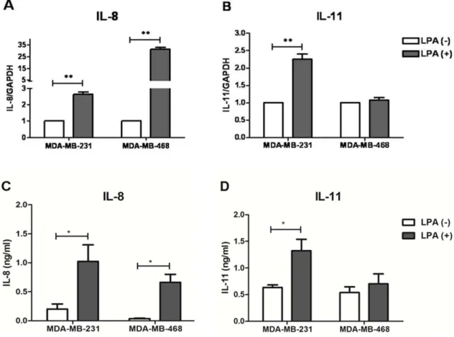
tumor cells communicate with other surrounding cells via pro- ducing cytokines. Ones of such tumor-derived cytokines influ- encing progression and metastasis process of breast cancer cells are interleukin (IL)-8 and IL-11. These cytokines are known to possess multiple effects on primary tumor and bone metastasis microenvironment. Studies has elucidated that IL-8 can promote breast tumor cells growth [2,3], migration, invasion [2,3] and an- giogenesis [4,5]. Similar to IL-8, IL-11 has also been demonstrated to support breast tumor growth via non-autonomous effects [6].

IL-8 is able to promote early micro-metastatic colonies formation in bone [7]. While, transcription level of IL-11 in patients with breast cancer was found associated with subsequent development

Original Article

Lysophosphatidic acid enhances breast cancer cells-mediated osteoclastogenesis

Ju-Suk Nam*

,#

, Ashish Ranjan Sharma

#

, Lich Thi Nguyen

#

, Supriya Jagga, Yeon-Hee Lee, Garima Sharma, and Sang-Soo Lee*

Institute for Skeletal Aging & Orthopedic Surgery, Hallym University-Chuncheon Sacred Heart Hospital, Chuncheon 24252, Korea

ARTICLE INFO

Received January 21, 2018 Revised April 22, 2018 Accepted May 18, 2018

*Correspondence Ju-Suk Nam

E-mail: [email protected] Sang-Soo Lee

E-mail: [email protected] Key Words

Breast cancer Interleukin-8 Interleukin-11 Lysophosphatidic acid Osteoclastogenesis

#

These authors contributed equally to this work.

ABSTRACT Lysophosphatidic acid (LPA) is known to play a critical role in breast can-

cer metastasis to bone. In this study, we tried to investigate any role of LPA in the

regulation of osteoclastogenic cytokines from breast cancer cells and the possibility

of these secretory factors in affecting osteoclastogenesis. Effect of secreted cytokines

on osteoclastogenesis was analyzed by treating conditioned media from LPA-stimu-

lated breast cancer cells to differentiating osteoclasts. Result demonstrated that IL-8

and IL-11 expression were upregulated in LPA-treated MDA-MB-231 cells. IL-8 was

induced in both MDA-MB-231 and MDA-MB-468, however, IL-11 was induced only in

MDA-MB-231, suggesting differential LPARs participation in the expression of these

cytokines. Expression of IL-8 but not IL-11 was suppressed by inhibitors of PI3K, NF-

kB, ROCK and PKC pathways. In the case of PKC activation, it was observed that PKCd

and PKCμ might regulate LPA-induced expression of IL-11 and IL-8, respectively, by

using specific PKC subtype inhibitors. Finally, conditioned Medium from LPA-stimu-

lated breast cancer cells induced osteoclastogenesis. In conclusion, LPA induced the

expression of osteolytic cytokines (IL-8 and IL-11) in breast cancer cells by involving

different LPA receptors. Enhanced expression of IL-8 by LPA may be via ROCK, PKCu,

PI3K, and NFkB signaling pathways, while enhanced expression of IL-11 might involve

PKCd signaling pathway. LPA has the ability to enhance breast cancer cells-mediated

osteoclastogenesis by inducing the secretion of cytokines such as IL-8 and IL-11.

(2)

of bone metastasis [8]. Moreover, both the cytokines are known to act as osteolytic factors by supporting osteoclastogenesis [9- 11]. Taken together, IL-8 and IL-11 can be considered for playing important roles in breast cancer progression and osteolytic bone metastasis.

Lysophosphatidic acid (LPA) has been known as a bioactive phospholipid mainly derived from active platelets and several other cell types. Recent study suggested that Autotaxin (ATX)- LPA axis was responsible for bone metastasis by breast cancer cells [12]. As growth factor, LPA has diverse biological activities such as regulation of cell function, proliferation, differentiation, migration and survival in many cell types [13]. The biological action of LPA is mediated by a family of G protein-coupled recep- tors (GPCRs) called endothelial cell differentiation gene (EDG) family. Breast cancer cells express almost three LPA receptors (LPARs) EDG-2 (LPAR1), EDG-4 (LPAR2) and EDG-7 (LPAR3) which have high affinity for LPA [14]. It has recently emerged that LPA is involved in cancer metastasis and osteolytic bone me- tastasis although its mechanism of action is poorly understood.

Previous studies have shown that LPA induces breast cancer cells proliferation and migration involving signaling pathways like Phosphoinositide 3-kinase (PI3K), mitogen-activated protein ki- nase (MAPK) and b-catenin [15-17]. In addition, LPA may initiate metastasis process via inductive effect on angiogenesis which is mediated by IL-8 and vascular endothelial growth factor (VEGF) [18,19]. Several evidences in animal models also suggest the role of LPA in bone metastasis of breast cancer cells [12,20].

Therefore, this study was designed to investigate whether LPA can regulate osteolytic factors IL-8 and IL-11 secretion from breast cancer cells and whether can influence breast cancer cells- mediated bone osteoclastogensis via these secretory factors.

METHODS

Reagents and antibodies

Recombinant mouse receptor activator of nuclear factor kappa- b ligand (RANKL) was purchased from BioVision Inc. (Milpitas, CA, US). Oleoyl-L-a-lysophosphatidic acid sodium salt (LPA) was from Sigma (US). LPA receptor-1/2/3inhibitor Ki16425 was bought from Santa Cruz Biotechnology (CA, US). 3-[4, 5-dimeth- ylthiazol-2-yl]-2, 5-diphenyltetrazolium bromide (MTT) were purchased from Amresco (Albany, NY, US). PD98059, SB203580, SP600125, LY294002, BAY-11-7082, Go6976 and GF109203X were purchased from Tocris Bioscience (Bristol, UK), Y27632 from Cayman Chemical (Ann Arbor, MI, US). Phospho-(pan) PKC (Ser660) antibody was from Cell Signaling Technology (Boston, US). Anti-b-actin was obtained from Santa Cruz Biotechnology.

Peroxidase-AffiniPure goat anti-rabbit IgG (H+L) and peroxi- dase-AffiniPure goat anti-mouse IgG (H+L) were supplied from Jackson ImmunoResearch (Baltimore, US).

Cell culture

Human breast cancer cell line MDA-MB-231 (American Type Culture Collection, ATCC, HTB-26) and MDA-MB-468 (ATCC, HTB-132) were cultured in Dulbecco’s Modified Eagle Medium (DMEM) supplemented with 10% fetal bovine serum (FBS) and 1% penicillin-streptomycin (all from Gibco, Invitrogen). Cells were maintained at 37°C and 5% CO

2

in a humidified atmo- sphere. Cells were starved in serum-free media for 12 h prior to LPA treatment.

Murine macrophage cell line RAW264.7 (ATCC, TIB-71) was maintained in an undifferentiated state by culturing in DMEM containing 10% FBS and 1% penicillin-streptomycin at conflu- ences of less than 80%. To induce osteoclastogenesis, RAW264.7 cells were seeded at density of 2×10

3

cells/cm

2

of culture plate for overnight. Then cells were shifted into differentiation media con- taining Minimum Essential Medium Eagle-Alpha Modification (Alpha-MEM) (Gibco) supplemented with 10% FBS, 1% penicil- lin-streptomycin and 30 ng/ml RANKL.

Collection of conditioned media (CM)

MDA-MB-231 and MDA-MB-468 cells were plated at a den- sity of 1.0×10

6

cells/60 mm dish in complete DMEM media for overnight. Thereafter, cells were starved with serum-free DMEM for 12 h. Cells were then incubated with fresh serum-free DMEM containing LPA. After 12 h of incubation, conditioned media (CM) were collected, centrifuged to remove the cell debris if any, and was used to treat RAW264.7 cells.

Treatment of conditioned media

To induce osteoclastogenesis, RAW264.7 cells were cultured in differentiation media containing 30 ng/ml RANKL for 2.5 days.

After that, cells were incubated with fresh Alpha-MEM (without RANKL) containing 50% Conditioned Medium (CM) for another 2.5 days. Serum-free DMEM (50%) was used as vehicle treatment.

At day 5, cells were collected for RNA extraction or were stained for tartrate resistant acid phosphatase (TRAP assay) to observe osteoclast formation.

Cell viability assay

RAW264.7 cells were cultured in differentiation media for 2.5 days and treated with CM for another 2.5 days. Thereafter, media were replaced with fresh media containing MTT solution at a final concentration of 0.5 mg/ml. After 2 h of incubation at 37°C, supernatant was carefully removed and the insoluble formazan crystal was dissolved in 200 ml of dimethyl sulfoxide (DMSO).

Optical density (OD) of the dissolved crystals was measured at

570 nm.

(3)

TRAP staining

At day 5, cells were fixed and stained for TRAP using the Acid Phosphatase Leukocyte kit (Sigma-Aldrich). The TRAP (+) mul- tinucleated cells that contained three or more nuclei were counted as osteoclasts using Ziess AxioCam digital microscope.

Reverse transcription-quantitative polymerase chain reaction (RT-PCR)

Total RNA (RNAs) was extracted from cultured cells using Trizol reagent (Invitrogen, Carlsbad, CA). After that, 2 mg of RNAs was reverse-transcribed into cDNA using SuperScript II Reverse Transcriptase (Invitrogen). One ml of cDNA was diluted into 20 ml of qPCR reaction mixture containing SYBR Green (2xGreen star qPCR MasterMix, Bioneer, Korea). qPCR was performed on Rotor-Gene Q (Qiagen, Hilden, Germany) with cy- cling conditions as follow: initial denaturation at 95°C for 10 min, and then 40 cycles of denaturation at 95°C for 20 s, annealing at 60°C for 20 s and extension at 72°C for 25 s. Relative mRNA expression levels of the selected genes were analyzed using DDCT method and were normalized to GAPDH. The sequences of qPCR primers are listed in Table 1.

Protein isolation and western blot analysis

Cells cultured in multiple well-plate were washed with PBS and lysed with cold RIPA buffer (20 mM Tris-HCl, pH 7.5, 200 mM NaCl, 1% Triton X-100, 1 mM dithiothreitol) containing protease inhibitor cocktail (Roche, Mannheim, Germany) and phospha- tase inhibitors. Protein concentrations were determined using micro BCA protein assay kit (Thermo Scientific, Massachusetts, USA) following the manufacture’s protocol. Total protein was separated on 9% SDS-polyacryramide gel electrophoresis and transferred to PVDF membrane (Millipore, Billerica, MA). The membrane was then blocked with 5% skimmed milk and incu- bated with primary antibodies as follow: polyclonal anti-rabbit p- PKC (1:1000 dilution in 1% BSA) and monoclonal anti-mouse b- actin (1:1000 dilution in 1% BSA) for 2 h at room temperature.

After washing, membrane was incubated with horseradish per- oxidase-conjugated anti-rabbit IgG secondary antibody (1:2000 dilution in 5% skim milk) or anti-mouse IgG secondary antibody (1:5000 dilution in 5% skim milk). Protein bands were visualized

using Luminata Forte western HRP substrate (Millipore) and sig- nals were detected and quantified using Fusion-Fxsystem (Vilber Lourmat, Eberhardzell, Germany).

ELISA

After 12 h of LPA stimulation, supernatant from both MDA- MB-231 and MDA-MB-468 cells were collected. Human IL-8 and IL-11 ELISA kit (R&D systems, Minneapolis, MN) were utilized for a quantitative measurement of cytokines according to the manufacturer’s recommendations.

Statistical analysis

All the statistical data were analyzed by Graphpad Prism 5.0 (San Diego, CA) and evaluated using two-tailed Student’s t-test.

All data was presented as mean±SEM of at least three indepen- dent experiments. Value of p<0.05 was considered to indicate a statistically significant difference.

RESULTS

LPA induced expression of IL-8 and IL-11 in breast cancer cells

IL-8 and IL-11 have important roles in breast cancer progres- sion and bone metastasis mechanism. To determine whether LPA stimulates expression of IL-8 and IL-11 in MDA-MB-231 breast cancer cells, serum-starved cells were treated with 10 mM of LPA and the expression of IL-8 and IL-11 mRNA were examined after 1 h, 3 h and 6 h of LPA treatment. A dose of 10 mM of LPA has been shown to induce no effect on cell cycle of the MDA-MB-231 cells, ruling out any possibility of proliferative or cytotoxic ef- fect [21]. The result indicates that treatment with LPA caused significant increases in IL-8 and IL-11 expression levels in MDA- MB-231 cells with maximal increments of about 5-6 folds of un- treated control (Figs. 1A and B).

LPA has been known to act on breast cancer cells via it recep- tors, LPARs. Therefore, identification of specific LPA receptors responsible for induction of each osteolytic factor was required. It has been known that MDA-MB-231 and MDA-MB-468 are both triple-negative breast cancer cells. These cell types do not express

Table 1. Primers for RT-PCR

Target Forward primer (5'->3') Reverse primer (5'->3')

Human IL-8 AGAAACCACCGGAAGGAACCATCT AGAGCTGCAGAAATCAGGAAGGCT

Human IL-11 AGATATCCTGACATTGGCCAGGCA ACTTCAGTGATCCACTCGCTTCGT

Human GAPDH TTCAGCTCAGGGATGACCTT ACCCAGAAGACTGTGGATGG

Mouse TRAP ACATGACCACAACCTGCAG CCTCAGATCCATAGTGAAACCG

Mouse GAPDH TCGTGGATCTGACGTGCCGCCTG CACCACCCTGTTGCTGTAGCCGTAT

(4)

three specific receptors: estrogen receptor (ER), human epidermal growth factor receptor 2 (Her2) and progesterone receptor (PR).

These two cell lines differ from each other in expression of specif- ic LPA receptors i.e; MDA-MB-468 cells predominantly express LPAR2 [14,22] while MDA-MB-231 mainly expresses LPAR1 and LPAR2 [23,24]. MDA-MB-231 is known as highly aggressive and invasive, poorly differentiated breast cancer cells. Compared to MDA-MB-231 cells, MDA-MB-468 cells are less aggressive and invasive (Table 2) [25-27]. Both MDA-MB-231 and MDA-MB-468 were treated with LPA for 3 h and the mRNA expression level of IL-8 and IL-11 was analyzed by RT-PCR. We observed that treat- ment of LPA significantly increased the mRNA expression level of IL-8 in both the cell lines (Fig. 2A). LPA induced significant mRNA expression of IL-11 in MDA-MB-231 cells, but not in MDA- MB-468 cells (Fig. 2B). We also quantified the amount of IL-8 and IL-11 secreted from LPA stimulated MDA-MB-231 and MDA-MB-468 after 12 h of treatment (Figs. 2C and D). Similar to mRNA expression results, a significant increase of IL-8 was observed in the media of both the human breast cancer cell lines.

However, amount of secreted IL-11 was only significant in MDA- MB-231 cells. The data shows that MDA-MB-231 cells expressing dominantly both LPAR1 and LPAR2, has induction of IL-8 and IL-11 after LPA stimulation, while, MDA-MB-468 cells express- ing dominantly LPAR2, has induction of IL-8 only.

Analysis of signaling pathways involved in regulation of expression of IL-8 and IL-11 after LPA stimulation

In addition to LPA receptors, we also hypothesized that specific signaling pathways might be involved in regulating expression of IL-8 and IL-11 in LPA-treated breast cancer cells. It has been re- ported that LPA induces IL-8 expression through signaling mech- anisms like PI3K/Akt, ROCK, ERK, p38/MAPK, NFkB and JNK- dependent pathways in different cell types [28-31]. Similarly, IL-11 expression was found to be dependent on the activation of ERK, PKC, p38/MAPK and Smad signaling pathways [32,33]. There- fore, involvement of these signaling pathways in LPA-induced IL-8 and IL-11 expression in MDA-MB-231 cells was examined.

For this, inhibitor to specific pathways were used including ERK inhibitor PD98059 (10 mM), PI3K inhibitor LY294002 (10 mM), p38/MAPK inhibitor SB203580 (10 mM), JNK inhibitor SP600125 (2 mM), NFkB inhibitor BAY-11-7082 (5 mM), ROCK inhibitor Y27632 (5 mM) and PKC inhibitor Go6976 (1 mM). Moreover, a specific inhibitor for LPA receptors (Ki16425, 5 mM) was used to confirm the involvement of LPARs in induction of cytokines.

The data revealed that expression of both the cytokines was inhibited by LPARs inhibitor, Ki16425. Results also indicated that LPA-stimulated expression of both cytokines was not inhibited by ERK, p38/MAPK or JNK inhibitor (data not shown), confirming their non-involvement in LPA stimulated expression of both the cytokines. However, induction of IL-8 expression was strongly

Fig. 1. Stimulation of IL-8 and IL-11 expression in MDA-MB-231 cells by LPA. MDA-MB-231 cells were treated with 10 mM LPA for indicated times (1h, 3 h and 6 h), quantitative expression of IL-8 (A) and IL-11 (B) mRNA were determined by RT-PCR in normalization with GAPDH. *p<0.05, **p<0.01, significant difference compared to control at 1 h.

Table 2. Characteristics of MDA-MB-231 and MDA-MB-468 cell lines

Characteristics MDA-MB-231 MDA-MB-468 References

Tumorigenic property in vitro Highly aggressive, invasive Moderately aggressive, invasive [26]

Tumorigenic property in mice Locally invasive, metastatic Non- invasive, non- metastatic [25]

LPARs expression pattern Strong expression of LPAR1, moderate expression of LPAR2, almost no expression of LPAR3

Strong expression of LPAR2, faint

expression of LPAR1 and LPAR3 [14,22]

[23,24]

(5)

inhibited by ROCK and PKC inhibitors, also partially blocked by PI3K and NFkB inhibitors. But, none of these inhibitors affected the LPA-stimulated IL-11 expression (Fig. 3 ).

LPA induced IL-8 and IL-11 expressions were regulated by specific PKC subunits

PKC is one of the main downstream signaling molecules for LPA receptors [18]. We observed the regulation of IL-8 induction by LPA via PKC pathway. Moreover, PKC has been reported in regulation of IL-11 in osteoblasts [34]. Hence, we examined acti- vation of PKC after stimulation by LPA in MDA-MB-231 breast cancer cells. Cancer cells were treated with LPA for several time points from 15 min to 4 h. Phosphorylation of PKC was detected by western blotting. Fig. 4 shows that LPA increased level of p- PKC from 15 min to 2 h with maximal increment at 15 min and thereafter returning to normal level at 4 h after treatment.

Next, we aimed to clarify the involvement of PKCs subtypes in regulation of IL-8 and IL-11 expression after LPA stimulation. For this, different PKC inhibitors were used, including Go6976 (100 nM, selective inhibitor for PKCa, b1 and m inhibitor), GF109203X (1 mM, selective inhibitor for PKCa, bI, bII, g, d and e) and rot- tlerin (10 mM, selective PKCd inhibitor) [35]. As shown in Fig. 4,

IL-8 induction was strongly inhibited by Go6976 (similar result shown in Fig. 3), but not by GF109203X or rottlerin. However, IL-11 induction was inhibited by GF109203X and rottlerin. This result suggests that different PKC subtypes regulate expression of IL-8 and IL-11 in LPA-stimulated breast cancer cells. Namely, PKCm is responsible for regulation of IL-8 because only PKCm is inhibited by Go6976 but not by GF109203X or rottlerin, and PKCd is responsible for IL-11 expression as it is inhibited by both GF109203X and rottlerin.

CM from LPA-stimulated breast cancer cells induced osteoclastogenesis

Upregulation of LPA signaling has been shown to enhance the formation of bone metastasis in mouse xenograft model [36].

Hence, we tried to inspect whether LPA-induced secretory factors from breast cancer cells can affect osteoclastogenesis. CM from breast cancer cells treated with or without LPA was collected and added to culture of RANKL-stimulated RAW264.7 cells. Results shows that LPA-treated breast cancer cells CM (MDA-MB-231 and MDA-MB-468) induced expression of differentiation marker, TRAP (about 1.5 folds), and incited formation of significant number of mature osteoclasts (about 2 folds), compared to vehicle

Fig. 2. Stimulation of IL-8 and IL-11 expression by LPA in MDA-MB-231 and MDA-MB-468 cells. MDA-MB-231 and MDA-MB-468 cells were treat-

ed with 10 mM LPA for 3 h, quantitative expression of IL-8 (A) and IL-11 (B) mRNA was determined by RT-PCR in normalization with GAPDH. After 12 h

of stimulation of LPA, the concentrations of IL-8 (C) and IL-11 (D) were measured by ELISA. *p<0.05, **p<0.01.

(6)

CM. CM did not confer any effect on cell viability of RANKL- differentiated RAW264.7 cells in comparison to vehicle CM. To order to exclude the possibility that LPA existing in CM, due to treatment, might affected osteoclastogenesis, we directly added LPA into culture of RAW264.7 cells. The result shows that LPA did not affect osteoclastogenesis, compared to vehicle control (Figs. 5 A-D).

DISCUSSION

LPA, a growth factor-like phospholipid has diversity of effects on cell physiological activities in many cell types. Role of LPA

in cancer progression has been elucidated in numerous studies.

Recently, studies on the involvement of LPA on cancer metastasis such as bone invasion has become an emerging research area [20].

Our study analyzed the possible role of LPA in breast cancer cells-mediated osteoclastogenesis via upregulation of osteoclas- togenic cytokines, IL-8 and IL-11. Treatment of LPA to MDA- MB-231 cells induced the expression of IL-8 and IL-11 (Figs. 1 and 2). LPA-induced IL-8 expression and secretion was reported in several cell types including breast cancer cells [28-30]. More- over, breast cancer cells stimulated with TGFb displays increased expression of IL-8 and IL-11 [32,37]. Our result revealed that LPA also induces IL-11 expression in MDA-MB-231 cells. Simi- lar to previous study of Mu et al, LPA stimulation increased the

Fig. 4. LPA stimulated IL-8 and IL-11 mRNA expression through regulation of different PKC subtypes. (A) MDA-MB-231 cells were treated with LPA (10 mM). Protein levels of p-PKC were detected by western blot at 0-4 h. b-actin was used as loading control, (B) MDA-MB-231 cells were pretreat- ed with specific inhibitors for 30 min, prior to LPA (10 mM) treatment for 3 h. Quantitative expression of IL-8 and IL-11 mRNA were evaluated by RT- qPCR in normalization with GAPDH. *p<0.05, **p<0.01.

Fig. 3. Effects of PI3K, NFKB, ROCK and PKC inhibitors on induction of IL-8 and IL-11 by LPA in MDA-MB-231 cells. MDA-MB-231 cells were

pretreated with specific inhibitors for 30 min, prior to LPA (10 mM) treatment for 3 h. Quantitative expression of IL-8 (A) and IL-11 (B) mRNA were evalu-

ated by RT-PCR in normalization with GAPDH. *p<0.05, **p<0.01.

(7)

mRNA expression of IL-8 (maximally, about 6 folds) after 3 h of treatment [38]. Moreover, our results also demonstrates that LPA enhanced mRNA expression of IL-11 up to 5-6 folds at the same time point. ELISA results also confirmed the induction of IL-8 and IL-11 in protein after LPA stimulation (Figs. 2C and D).

Since, LPA induced the expression level of IL-8 and IL-11 in MDA-MB-231 breast cancer cells, association of LPA-stimulated expression of these cytokines with LPARs and downstream sig- naling pathways of LPARs were investigated. In breast cancer, LPARs are expressed in many cancer cell lines, but at various levels. Till now, there are only few studies addressed the involve- ment of LPARs and cytokine expression. Our study suggests that LPAR1 might be responsible for IL-11 and LPAR2 for IL-8 induc- tion in stimulated Triple-Negative Breast Cancer (TNBC) cells (Fig. 2 ). LPAR2 is highly expressed in less aggressive breast cancer cells (MDA-MB-468) than in more aggressive cells (MDA-MB-231 cells) [14,22]. This might explain for a strong induction of IL-8 in MDA-MB-468 (about 30 folds) after LPA treatment. More- over, our result is consistent with previous study of Hartman et al, indicating that regulation of IL-8 is mediated by LPAR2 in TNBC cells [30]. The other evidence for involvement of LPARs in LPA-stimulated cytokines expression is that treatment with

LPA receptors inhibitor (Ki16425) reduced the induction of IL-8 and IL-11 in breast cancer cells (Fig. 3). Thus, it may be concluded that specific LPA receptors might be involved in regulating the induction of IL-8 and IL-11 expression in TNBC cells. Recently, a number of naturally occurring or synthetic analogue of LPA has been recognized by various studies [39,40]. These LPA analogues promises to be a selective type of LPA receptor ligand (agonist or antagonist) providing an opportunity to probe complex overlap- ping LPA signaling pathways. Further studies might utilize them for bifurcating the complex LPA receptors signaling mechanism in induction of cytokines for bone metastasis by breast cancer cells.

LPARs have been known as G protein-coupled receptors. Ac- tivation of LPARs leads to activation of three different G alpha subunits G

i/o

, G

q

, G

12/13

, in turn, it triggers activation of many cel- lular signaling such as PKC, Ras/ERK, PI3K/Akt, RhoA/ROCK and NFkB [15]. Our study indicates that IL-8 induction was mostly dependent on ROCK, PKC, and to a lesser extent on PI3K and NFkB signaling pathways. Previous study have reported that LPA-regulated IL-8 secretion in Sum159, a TNBC cell line, was completely inhibited by the treatment of NFkB inhibitor, BAY-11- 7082 (10 mM) [30]. The difference between our study and previ-

Fig. 5. LPA-stimulated breast cancer cells conditioned media induced osteoclast formation. RAW264.7 cells after stimulation with 30 ng/ml

RANKL for 2.5 days were exposed to CM from breast cancer cells (MDA-MB-231 or MDA-MB-468) treated with or without LPA for 2.5 days (see Materi-

als and Methods). (A) RT real-time PCR analysis for expression of TRAP mRNA. (B) Cell viability was assessed by MTT. (C) Osteoclast formation was as-

sessed by TRAP staining and osteoclast number was counted. (D) Representative microcopy picture of cells stained with TRAP.

(8)

ous study may be due to the difference in dose of inhibitor used for the treatment. In our case, we have used a lower dose of NFkB inhibitor (5 mM) while other study used a higher dose of 10 mM.

Moreover, the difference in the cell type utilized in the two stud- ies might be a factor. However, the signaling pathways responsible for IL-8 induction were not involved in the regulation of IL-11.

PKCs belong to a serine/threonine kinase family which in- cludes three classes of isoenzymes: conventional isoforms (a, b and g) that are calcium-dependent and require diacylglycerol (DAG) for activation; novel isoforms (d, e, h, q and m) that are cal- cium-independent but requires DAG for activation; and atypical isoforms (x, i, and l) that are independent of both Ca

2+

and DAG.

It was observed that MDA-MB-231 breast cancer cells express high levels of PKC subtypes [41]. In our study, LPA treatment in- duced rapid activation of PKC as observed by the increase in the levels of p-(pan) PKC from 15 min to 2 h of treatment (Fig. 4A).

The normalization of phosphorylation of PKC at 4 h of treatment of LPA implies it can only stimulate PKC signaling pathway at a very early time point and is for a short duration. Moreover, the result obtained shows that just PKCm and no other PKC subtypes are involved the regulation of IL-8 expression in LPA-stimulated breast cancer cells (Fig. 4B). For the first time, this study indicated a possibility that LPA-induced IL-11 expression in breast cancer cells might involve the PKCd subtype (Fig. 4B).

Breast cancer cells have been known to secret many factors including osteolytic factors. Among osteolytic factors, cytokines IL-8 and IL-11 are mostly implied in breast cancer for bone me- tastasis. Our results demonstrated that LPA-stimulated breast cancer cells CM increased the expression of osteoclast differentia- tion marker, TRAP, in macrophage cell line, as well an increase in the number of mature osteoclasts were also observed (Fig. 5).

It appears that secreted products from LPA-stimulated breast cancer cells can promote osteoclastogenesis. Since, our RT-PCR data showed an enhanced mRNA expression of IL-8 and IL-11 after stimulation of breast cancer cells, role of IL-8 and Il-11 in increased osteoclastogensis can be assumed.

Metastasis is complex process in which breast cancer cells leave the original tumor site and migrate into other part of body via circulatory systems (bloodstream or lymphatic system). In the current study, LPA increased the expression levels of IL-8 in MDA-MB-231 and MDA-MB-468 cells and conditioned media from LPA-stimulated breast cancer cells MDA-MB-231 and MDA-MB-468 increased osteoclastogenesis, therefore, IL-8 might mediate osteoclastogensis in this context. In addition, as shown in Table 2, MDA-MB-231 cells are highly aggressive, metastatic cells and LPA-stimulated MDA-MB-231 cells expressed higher levels of IL-11, suggesting possible role of IL-11 in metastasis. LPA is a component of serum, mainly generated from platelets and breast cancer cells are also known to secret LPA. Hence, it can be presumed that elevated presence of LPA in tumor micro-environ- ment can stimulate breast cancer cells via an autocrine pathway to help in metastasis process. However, more detailed studies will

be required to delineate the exact mechanism of induction of cy- tokines by LPA stimulated breast cancers and their participatory role in metastasis. Better understanding of mechanism of LPA regulation of its products may support to finding the good treat- ment for breast cancer metastasis.

ACKNOWLEDGEMENTS

This research was supported by Hallym University Re- search Fund, by Basic Science Research Program through the National Research Foundation of Korea (NRF) funded by the Ministry of Education (NRF-2016R1D1A1B03931318 and NRF- 2017R1A2B4012944).

CONFLICTS OF INTEREST

The authors declare no conflicts of interest.

REFERENCES

1. Torre LA, Bray F, Siegel RL, Ferlay J, Lortet-Tieulent J, Jemal A.

Global cancer statistics, 2012. CA Cancer J Clin. 2015;65:87-108.

2. Lin Y, Huang R, Chen L, Li S, Shi Q, Jordan C, Huang RP. Identi- fication of interleukin-8 as estrogen receptor-regulated factor in- volved in breast cancer invasion and angiogenesis by protein arrays.

Int J Cancer. 2004;109:507-515.

3. Yao C, Lin Y, Chua MS, Ye CS, Bi J, Li W, Zhu YF, Wang SM. Inter- leukin-8 modulates growth and invasiveness of estrogen receptor- negative breast cancer cells. Int J Cancer. 2007;121:1949-1957.

4. Li A, Dubey S, Varney ML, Dave BJ, Singh RK. IL-8 directly en- hanced endothelial cell survival, proliferation, and matrix metal- loproteinases production and regulated angiogenesis. J Immunol.

2003;170:3369-3376.

5. Martin D, Galisteo R, Gutkind JS. CXCL8/IL8 stimulates vascular endothelial growth factor (VEGF) expression and the autocrine activation of VEGFR2 in endothelial cells by activating NFkappaB through the CBM (Carma3/Bcl10/Malt1) complex. J Biol Chem.

2009;284:6038-6042.

6. Marusyk A, Tabassum DP, Altrock PM, Almendro V, Michor F, Polyak K. Non-cell-autonomous driving of tumour growth supports sub-clonal heterogeneity. Nature. 2014;514:54-58.

7. Benoy IH, Salgado R, Van Dam P, Geboers K, Van Marck E, Schar- pé S, Vermeulen PB, Dirix LY. Increased serum interleukin-8 in patients with early and metastatic breast cancer correlates with early dissemination and survival. Clin Cancer Res. 2004;10:7157-7162.

8. Sotiriou C, Lacroix M, Lespagnard L, Larsimont D, Paesmans M, Body JJ. Interleukins-6 and -11 expression in primary breast can- cer and subsequent development of bone metastases. Cancer Lett.

2001;169:87-95.

9. Morgan H, Tumber A, Hill PA. Breast cancer cells induce osteoclast

formation by stimulating host IL-11 production and downregulat-

ing granulocyte/macrophage colony-stimulating factor. Int J Can-

(9)

cer. 2004;109:653-660.

10. McCoy EM, Hong H, Pruitt HC, Feng X. IL-11 produced by breast cancer cells augments osteoclastogenesis by sustaining the pool of osteoclast progenitor cells. BMC Cancer. 2013;13:16.

11. Bendre MS, Montague DC, Peery T, Akel NS, Gaddy D, Suva LJ. In- terleukin-8 stimulation of osteoclastogenesis and bone resorption is a mechanism for the increased osteolysis of metastatic bone disease.

Bone. 2003;33:28-37.

12. Boucharaba A, Serre CM, Guglielmi J, Bordet JC, Clézardin P, Pey- ruchaud O. The type 1 lysophosphatidic acid receptor is a target for therapy in bone metastases. Proc Natl Acad Sci U S A. 2006;103:

9643-9648.

13. Mills GB, Moolenaar WH. The emerging role of lysophosphatidic acid in cancer. Nat Rev Cancer. 2003;3:582-591.

14. Chen M, Towers LN, O’Connor KL. LPA2 (EDG4) mediates Rho-de- pendent chemotaxis with lower efficacy than LPA1 (EDG2) in breast carcinoma cells. Am J Physiol Cell Physiol. 2007;292:C1927-1933.

15. Liu S, Umezu-Goto M, Murph M, Lu Y, Liu W, Zhang F, Yu S, Ste- phens LC, Cui X, Murrow G, Coombes K, Muller W, Hung MC, Perou CM, Lee AV, Fang X, Mills GB. Expression of autotaxin and lysophosphatidic acid receptors increases mammary tumorigenesis, invasion, and metastases. Cancer Cell. 2009;15:539-550.

16. Reya T, Clevers H. Wnt signalling in stem cells and cancer. Nature.

2005;434:843-850.

17. Li Y, Welm B, Podsypanina K, Huang S, Chamorro M, Zhang X, Rowlands T, Egeblad M, Cowin P, Werb Z, Tan LK, Rosen JM, Var- mus HE. Evidence that transgenes encoding components of the Wnt signaling pathway preferentially induce mammary cancers from progenitor cells. Proc Natl Acad Sci U S A. 2003;100:15853-15858.

18. Liu S, Murph M, Panupinthu N, Mills GB. ATX-LPA receptor axis in inflammation and cancer. Cell Cycle. 2009;8:3695-3701.

19. Panupinthu N, Lee HY, Mills GB. Lysophosphatidic acid production and action: critical new players in breast cancer initiation and pro- gression. Br J Cancer. 2010;102:941-946.

20. Boucharaba A, Serre CM, Grès S, Saulnier-Blache JS, Bordet JC, Guglielmi J, Clézardin P, Peyruchaud O. Platelet-derived lysophos- phatidic acid supports the progression of osteolytic bone metastases in breast cancer. J Clin Invest. 2004;114:1714-1725.

21. Du J, Sun C, Hu Z, Yang Y, Zhu Y, Zheng D, Gu L, Lu X. Lysophos- phatidic acid induces MDA-MB-231 breast cancer cells migration through activation of PI3K/PAK1/ERK signaling. PLoS One. 2010;5:

e15940.

22. Samadi N, Bekele RT, Goping IS, Schang LM, Brindley DN. Lyso- phosphatidate induces chemo-resistance by releasing breast cancer cells from taxol-induced mitotic arrest. PLoS One. 2011;6:e20608.

23. Hama K, Aoki J, Fukaya M, Kishi Y, Sakai T, Suzuki R, Ohta H, Yamori T, Watanabe M, Chun J, Arai H. Lysophosphatidic acid and autotaxin stimulate cell motility of neoplastic and non-neoplastic cells through LPA1. J Biol Chem. 2004;279:17634-17639.

24. Swamydas M, Nguyen D, Allen LD, Eddy J, Dréau D. Progranulin stimulated by LPA promotes the migration of aggressive breast can- cer cells. Cell Commun Adhes. 2011;18:119-130.

25. Brünner N, Boysen B, Rømer J, Spang-Thomsen M. The nude mouse as an in vivo model for human breast cancer invasion and metastasis. Breast Cancer Res Treat. 1993;24:257-264.

26. Chekhun S, Bezdenezhnykh N, Shvets J, Lukianova N. Expression of biomarkers related to cell adhesion, metastasis and invasion of

breast cancer cell lines of different molecular subtype. Exp Oncol.

2013;35:174-179.

27. Dickson RB, Bates SE, McManaway ME, Lippman ME. Charac- terization of estrogen responsive transforming activity in human breast cancer cell lines. Cancer Res. 1986;46:1707-1713.

28. Hwang YS, Lee SK, Park KK, Chung WY. Secretion of IL-6 and IL-8 from lysophosphatidic acid-stimulated oral squamous cell carcino- ma promotes osteoclastogenesis and bone resorption. Oral Oncol.

2012;48:40-48.

29. Shimada H, Rajagopalan LE. Rho-kinase mediates lysophosphatidic acid-induced IL-8 and MCP-1 production via p38 and JNK path- ways in human endothelial cells. FEBS Lett. 2010;584:2827-2832.

30. Hartman ZC, Poage GM, den Hollander P, Tsimelzon A, Hill J, Panupinthu N, Zhang Y, Mazumdar A, Hilsenbeck SG, Mills GB, Brown PH. Growth of triple-negative breast cancer cells relies upon coordinate autocrine expression of the proinflammatory cytokines IL-6 and IL-8. Cancer Res. 2013;73:3470-3480.

31. Chen RJ, Chen SU, Chou CH, Lin MC. Lysophosphatidic acid recep- tor 2/3-mediated IL-8-dependent angiogenesis in cervical cancer cells. Int J Cancer. 2012;131:789-802.

32. Gupta J, Robbins J, Jilling T, Seth P. TGFβ-dependent induction of interleukin-11 and interleukin-8 involves SMAD and p38 MAPK pathways in breast tumor models with varied bone metastases po- tential. Cancer Biol Ther. 2011;11:311-316.

33. Matsumoto T, Kuriwaka-Kido R, Kondo T, Endo I, Kido S. Regula- tion of osteoblast differentiation by interleukin-11 via AP-1 and Smad signaling. Endocr J. 2012;59:91-101.

34. Kido S, Kuriwaka-Kido R, Umino-Miyatani Y, Endo I, Inoue D, Taniguchi H, Inoue Y, Imamura T, Matsumoto T. Mechanical stress activates Smad pathway through PKCδ to enhance interleukin-11 gene transcription in osteoblasts. PLoS One. 2010;5:e13090.

35. Brenner W, Beitz S, Schneider E, Benzing F, Unger RE, Roos FC, Thüroff JW, Hampel C. Adhesion of renal carcinoma cells to endo- thelial cells depends on PKCmu. BMC Cancer. 2010;10:183.

36. David M, Wannecq E, Descotes F, Jansen S, Deux B, Ribeiro J, Serre CM, Grès S, Bendriss-Vermare N, Bollen M, Saez S, Aoki J, Saulnier- Blache JS, Clézardin P, Peyruchaud O. Cancer cell expression of autotaxin controls bone metastasis formation in mouse through ly- sophosphatidic acid-dependent activation of osteoclasts. PLoS One.

2010;5:e9741.

37. Kang Y, He W, Tulley S, Gupta GP, Serganova I, Chen CR, Manova- Todorova K, Blasberg R, Gerald WL, Massagué J. Breast cancer bone metastasis mediated by the Smad tumor suppressor pathway.

Proc Natl Acad Sci U S A. 2005;102:13909-13914.

38. Mu H, Calderone TL, Davies MA, Prieto VG, Wang H, Mills GB, Bar-Eli M, Gershenwald JE. Lysophosphatidic acid induces lym- phangiogenesis and IL-8 production in vitro in human lymphatic endothelial cells. Am J Pathol. 2012;180:2170-2181.

39. Yung YC, Stoddard NC, Chun J. LPA receptor signaling: pharma- cology, physiology, and pathophysiology. J Lipid Res. 2014;55:1192- 1214.

40. Tigyi G. Aiming drug discovery at lysophosphatidic acid targets. Br J Pharmacol. 2010;161:241-270.

41. Morse-Gaudio M, Connolly JM, Rose DP. Protein kinase C and its

isoforms in human breast cancer cells: relationship to the invasive

phenotype. Int J Oncol. 1998;12:1349-1354.

수치

Table 1. Primers for RT-PCR
Table 2. Characteristics of MDA-MB-231 and MDA-MB-468 cell lines
Fig. 2. Stimulation of IL-8 and IL-11 expression by LPA in MDA-MB-231 and MDA-MB-468 cells
Fig. 4. LPA stimulated IL-8 and IL-11 mRNA expression through regulation of different PKC subtypes
+2

참조

관련 문서

Expression of cIAP2 and EMT-associated molecules were examined in luminal-type (BT474, MCF7) and triple-negative (MDA-MB-231, Hs 578T) breast cancer cells at (A) protein and (B)

MCF-7 and MDA-MB-231 breast cancer cells were treated with different concentrations of Glycyrrhiza root extracts and the cell viability was measured using MTT assay.. glabra showed

Objectives: In this study, we investigated the anti-proliferative and apoptosis inducing effect of water extract of Chelidonium majus (CM) on human breast cancer

Synergistic effects of the combination of trabectedin and olaparib on poly(ADP-ribosyl)ation and DNA damage in four different breast cancer cell lines. MCF7, MDA-MB-231,

Effect of natural compounds on the expression of p21 WAF1 , DNMT1, HDAC1, and methyl-CpG binding proteins (MeCP2 and MBD2) in human breast cancer cell lines.. MCF 7 and MDA MB 231

To determine whether direct interaction with breast cancer cells potentiates ECFC-mediated angiogenesis, ECFCs were co-cultured directly with MDA-MB-231 or MCF7 to assess

Among the isolates, xanthoxylol (4) exhibited significant cytotoxicity against human breast cancer cells MCF-7 and MDA-MB-231 in vitro with IC 50 values of 9.15 and 13.95

Estrogen-receptor (ER) positive MCF-7 and ER-negative MDA-MB-231 human breast cancer cells were treated with different doses of each RA isomers, all-trans RA, 13-cis RA, or 9-cis