Natural Product Sciences 25(1) : 72-75 (2019)
https://doi.org/10.20307/nps.2019.25.1.72
72
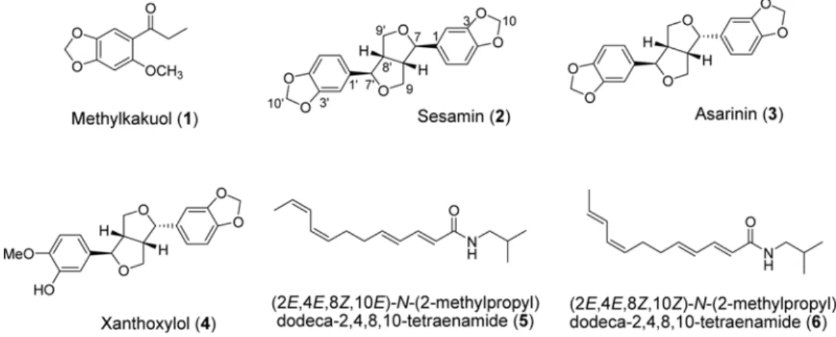
Cytotoxic Constituents from the Roots of Asarum sieboldii in Human Breast Cancer Cells
Eunae Kim
1, Hyun Jung Kim
2, Ha-Na Oh
2, Ah-Won Kwak
2, Su-Nam Kim
3, Bok Yun Kang
4, Seung-Sik Cho
2, Jung-Hyun Shim
2,*, and Goo Yoon
2,*
1
College of Pharmacy, Chosun University, Gwangju 61452, Republic of Korea
2
College of Pharmacy, Mokpo National University, Muan, Jeonnam 58554, South Korea
3
KIST Gangneung Institute, Gangneung 25451, Republic of Korea
4