INTRODUCTION
DNA hypermethylation plays an important role in silencing the tumor suppressor genes being one of the most consistent hallmarks of the human cancers, and the phenomenon is of a comparable significance to classic genetic mutations [1]. In
recent years, DNA methylation has emerged as an attractive target for the cancer therapeutics [2]. DNA methylation is cat- alyzed by a family of enzymes called DNA methyltransferases (DNMTs) [3]. The human genome contains four DNA methyl- transferase genes, DNMT1, DNMT2, DNMT3A, and DNMT3B which encode proteins with distinct functional specificities.
Among these DNMTs, DNMT1 is the most abundant DNA methyltransferase in the mammalian cells, and considered to be the key maintenance methyltransferase in the mammals. It has been established that the inhibition of DNA methyltrans- ferase activity can strongly inhibit the formation of tumors [4].
The repressive effects of DNA methylation are mediated in large part by the methyl-CpG binding proteins (MCBPs) and also associated with histone modifications. MCBPs, such as MeCP2, methyl-CpG binding domain 1 (MBD1), and MBD2,
Expression of DNA Methyltransferases in Breast Cancer Patients and to Analyze the Effect of Natural Compounds on DNA Methyltransferases and Associated Proteins
Sameer Mirza1,*, Gayatri Sharma1,2*, Rajinder Parshad3, Sidhartha Datta Gupta4, Pranav Pandya2, Ranju Ralhan1,5,6,7,8
1Department of Biochemistry, All India Institute of Medical Sciences, New Delhi; 2Dev Sanskriti Vishwa Vidhyalaya, Hardwar; Departments of 3Surgery and 4Pathology, All India Institute of Medical Sciences, New Delhi, India; 5Alex and Simona Shnaider Laboratory of Molecular Oncology, 6Department of Pathology and Laboratory Medicine, 7Sonshine Family Centre for Head & Neck Diseases, and 8Department of Otolaryngology, Mount Sinai Hospital, Toronto, Canada
ORIGINAL ARTICLE
Purpose: The DNA methylation mediated by specific DNA meth- yltransferases (DNMTs), results in the epigenetic silencing of multiple genes which are implicated in human breast cancer. We hypothesized that the natural compounds modulate the expres- sion of DNMTs and their associated proteins in the breast cancer cell lines and affect the methylation mediated gene silencing.
Methods: The DNMTs transcript expression was analyzed by reverse transcription-polymerase chain reaction (RT-PCR) in the tumors and the adjacent normal breast tissues of the patients with invasive ductal breast carcinoma. We tested the hypothesis that the natural compounds, viz., epigallocatechin gallate (EGCG), genistein, withaferin A, curcumin, resveratrol, and guggulsterone, have demethylation potential. To investigate this hypothesis, we analyzed the DNMTs expression at the transcript levels, followed by the analysis of DNMT1 and its associated proteins (HDAC1, MeCP2, and MBD2). Results: The increased DNMTs transcripts expression, viz., DNMT1, DNMT3a, and DNMT3b, in the breast
cancer tissues suggest involvement of the DNMTs in the breast carcinogenesis. Quantitative RT-PCR analysis revealed that the treatment with natural compounds, viz., EGCG, genistein, with- aferin A, curcumin, resveratrol, and guggulsterone, resulted in a significant decrease in the transcript levels of all the DNMTs investigated. Importantly, these natural compounds decreased the protein levels of DNMT1, HDAC1, and MeCP2. Conclusion:
Our results demonstrate that the natural compounds, EGCG, genistein, withaferin A, curcumin, resveratrol, and guggulsterone, have the potential to reverse the epigenetic changes. Moreover, their lack of toxicity makes these natural compounds promising candidates for the chemoprevention of the breast cancer. In-depth future mechanistic studies aimed to elucidate how these com- pounds affect the gene transcription are warranted.
Key Words: Breast neoplasms, Chemoprevention, DNA methylation, Epigenomics
Correspondence to: Ranju Ralhan
Alex and Simona Shnaider Laboratory of Molecular Oncology, Department of Pathology and Laboratory Medicine, Room 6-500, Mount Sinai Hospital, Joseph & Wolf Lebovic Health Complex, 600 University Avenue, Toronto, Ontario M5G 1X5, Canada
Tel: +1-416-586-4800 (ext. 6426), Fax: +1-416-586-8628.
E-mail: [email protected]
*These authors contributed equally to this work.
Received: December 11, 2012 Accepted: February 27, 2013
Cancer
specifically bind to CpG methylated DNA and are associated with the histone deacetylase (HDAC)-containing complexes, to “erase” the transcription-activating histone acetyl marks.
Natural products have received increasing attention in the recent years as novel anticancer agents [5-9]. Interest in the potential cancer chemopreventive and therapeutic properties of the diet-derived compounds, including those of the plant polyphenols, has increased tremendously. These compounds can be found in many fruits and vegetables including soya, turmeric, grapes, celery, apples, onions, parsley, capsicum, green tea, pepper, etc. and have been shown to possess anti- cancer activities [10]. The mechanisms by which the flavo- noids exert the anticancer effects are varied and may include action through anti-inflammation [11], free radical scaveng- ing [12], modulation of survival and proliferation pathways [13,14], and inhibition of the ubiquitin-proteasome pathway [15,16]. The potential of the nutraceutical agents in combina- tion therapies is being increasingly considered based on the the findings of the improved animal model outcome when these compounds are combined with radiotherapy and che- motherapy [17].
The primary rationale of this work was to explore the effects of the natural compounds, viz., epigallocatechin gallate (EGCG), genistein, withaferin A, curcumin, resveratrol, and guggulsterone, on the hypermethylation of specific genes and determine their inhibitory effects on the key proteins involved in the DNA hypermethylation mechanism.
METHODS
Tissue specimens
Surgically resected tissue samples were collected from the untreated primary breast carcinoma patients (n=40), along with the paired normal breast tissues (n=10) (taken 5-10 cm away from the site of the tumor). The patients were enrolled as outpatients at the Department of Surgical Disciplines, All India Institute of Medical Sciences, New Delhi, India between 2004 and 2008, following the study approval by the Institutional Human Ethics Committee. Written consent was obtained from all patients enrolled in the study. The patient age ranged 30 to 81 years in age (median, 50 years). All patients were diagnosed with invasive ductal carcinoma (IDC) of the breast. A section of each tumor and a matched normal breast tissue were sam- pled and stored in formalin for the histopathological charac- terization in the diagnosis conformation and immunohisto- chemistry. The rest of the tissues were immediately snap fro- zen and stored at -80°C for further use. The clinicopathologi- cal characteristics of the patients analyzed in this study are summarized in Table 1.
Chemicals
EGCG, genistein, curcumin, resveratrol, guggulsterone, 5- aza-2´-deoxycytidine (Decitabine) and MTT 3-(4,5-dimethyl- thiazol-2-yl)-2,5-diphenyltetrazolium bromide were purchased from Sigma Chemical Co. (Bangalore, India). Curcumin, resve- ratrol, and guggulsterone were dissolved in dimethyl sulfoxide (DMSO) and stored in dark at -20°C while genistein, EGCG and withaferin A were dissolved in water and stored at 4°C.
Decitabine was dissolved in PBS buffer and stored as a 1 mM solution at -20°C.
Cell culture and treatment
Human breast carcinoma cell lines MCF7 and MDA MB 231 were obtained from the American Type Culture Collec- tion (Manassas, USA) and cultured in Dulbecco’s Modified Eagle Medium (DMEM). The effect of the natural compounds on cellular proliferation was assessed by MTT assay, accord- ing to standard protocols. Briefly, 104 cells were seeded per well in a 96-well plate. After 24 hours of preincubation in their respective media containing 2% FBS, the tested natural com- pounds were added to the culture medium and the cells were incubated for 96 hours. Control cells were treated with DMSO only, where the concentration of DMSO did not exceed 0.2%.
Media were replenished after every 48 hours in both the treat- ed and control cells. After 96 hours, the cells were washed twice with PBS, and a fresh medium containing MTT (0.5 mg/mL) was added. After 4-hour incubation, the formazan Table 1. Patients’ characteristics
Characteristic No.
Total 40
Age (yr)
≤50 20
>50 20
Tumor size (cm)
≤3 13
>3 27
Node involvement
Negative 10
Positive 30
Tumor stage
T1+T2 16
T3+T4 24
Menopausal status
Premenopausal 11
Postmenopausal 29
Estrogen receptor α
Positive 13
Negative 27
Progesterone receptor
Positive 12
Negative 28
crystals were dissolved in acidic isopropanol and the absor- bance was measured at 540 nm. All experiments were repeat- ed three times, with at least three measurements (triplicates).
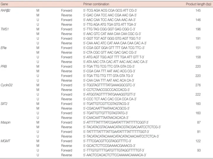
Bisulfite conversion, methylation-specific polymerase chain reaction
Isolation of genomic DNA and treatment with sodium bisulfite were done according to protocols optimized in our laboratory. The MSP of genes: ERα, PRB, RARβ2, TMS1, Cyclin D2, MGMT, SLIT2, and Maspin were carried out using the primers and conditions as described previously. The details are given in Table 2 [18-20]. All PCRs were performed with positive controls for both the unmethylated and methylated alleles, and with no DNA to check for contamination.
Real-time RT-PCR analysis
Real-time reverse transcription-polymerase chain reaction (RT-PCR) for DNMT1, DNMT3a and DNMT3b, was done
with cDNA synthesized from the total RNAs of the control and the drug-treated cells as described using Syber Green Quantitative-RTPCR kit (Stratagene, Vancouver, Canada) following the manufacturer’s instructions. The primer sequen- ces are provided in Table 3. The comparative Ct method was used to calculate the relative changes in gene expression using 7500 Fast Real Time PCR System (Applied Biosystems, Foster City, USA). The relative changes of gene expression were calcu-
Table 2. Methylation specific polymerase chain reaction primers used in the study
Gene Primer combination Product length (bp)
RARβ2 M Forward 5´-TCG AGA ACG CGA GCG ATT CG-3´ 145
M Reverse 5´-GAC CAA TCC AAC CGA AAC GA-3´
U Forward 5´-AAC CAA TCC AAC CAA AAC AA-3´ 146
U Reverse 5´-TTG AGA ATG TGA GTG ATT TGA-3´
TMS1 M Forward 5´-TTG TAG CGG GGT GAG CGG C-3´ 196
M Reverse 5´-AAC GTC CAT AAA CAA CAA CGC G-3´
U Forward 5´-GGT TGT AGT GGG GTG AGT TGG T-3´ 191
U Reverse 5´-CAA AAC ATC CAT AAA CAA CAA CAC A-3´
ERα M Forward 5´-CGA GGT GGA GTT TTT GAA TCG TTC–3´ 151
M Reverse 5´-CTA CGC GTT AAC GAC GAC CG-3´
U Forward 5´-ATG AGT TGG AGT TTT TGA ATT GTT T-3´ 158
U Reverse 5´-ATA AAC CTA CAC ATT AAC AAC AAC CA-3´
PRB M Forward 5´-TGA TTG TCG TTC GTA GTA CG-3´ 220
M Reverse 5´-CGA CAA TTT AAT AAC ACG CG-3´
U Forward 5´-TGA TTG TTG TTT GTA GTA TG-3´ 220
U Reverse 5´-CAA CAA TTT AAT AAC ACA CA-3´
CyclinD2 M Forward 5´-TGGTAGTTTTTATGAAAGGCGTC-3´ 276
M Reverse 5´-CCTCTAACCGCCCACCACG-3´
U Forward 5´-ATGGTAGTTTTTATGAAAGGTGTT-3´ 222
U Reverse 5´-CCC TCT AAC CAC CCA CCA CA-3´
SlIT2 M Forward 5´-TGATTGTCGTTCGTAGTACG-3´ 160
M Reverse 5´-CGACAATTTAATAACACGCG-3´
U Forward 5´-TGATTGTTGTTTGTAGTATG-3´ 160
U Reverse 5´-CAACAATTTAATAACACACA-3´
Maspin M Forward 5´-ATTTTTATTTTATCGAATATTTTATTTTTCGGT-3´ 87
M Reverse 5´-TACATACGTACAAACATACGTACGACAATCCTCTCG-3´
U Forward 5´-TATTTTTATTTTATTGAATATTTTATTTTTTGGT-3´ 81
U Reverse 5´-TACATACATACAAACATACATACAACAATCCTCTCA-3´
MGMT M Forward 5´-TTTCGACGTTCGTAGGTTTTC-3´ 122
M Reverse 5´-GCACTCTTCCGAAAACGAAACG-3´
U Forward 5´-TTTGTGTTTTGATGTTTGTAGGTTTTTGT-3´ 93
U Reverse 5´-AACTCCACACTCTTCCAAAAACAAAACA-3´
M=methylated; U=unmethylated.
Table 3. Real time polymerase chain reaction primers used in the study
Gene Primer combination
DNMT1 Forward 5´-TAC CTG GAC GAC CCT GAC CTC-3´
Reverse 5´-CGT TGG CAT CAA AGA TGG ACA-3´
DNMT3a Forward 5´-TAT TGA TGA GCG CAC AAG AGA GC-3´
Reverse 5´-GGG TGT TCC AGG GTA ACA TTG AG-3´
DNMT3b Forward 5´-GGC AAG TTC TCC GAG GTC TCT G-3´
Reverse 5´-TGG TAC ATG GCT TTT CGA TAG GA-3´
18S rRNA Forward 5´-GTAACCCGTTGAACCCCATT-3´
Reverse 5´-CCATCCAATCGGTAGTAGCG-3´
lated using the following formula: Fold change in gene expres- sion, 2-∆∆Ct=2-[∆Ct (natural compound treated samples)-
∆Ct (untreated control)], where ∆Ct=Ct (detected genes)-Ct (18S rRNA) and Ct represent the threshold cycle number.
Western blot
Cells were lysed on ice using lysis buffer (0.05 mol/L Tris- HCl, pH 7.4, 0.15 mol/L NaCl, 0.25% deoxycholic acid, 1%
NP-40, 1 mmol/L ethylenediaminetetraacetic acid, 0.5 mmol/L dithiothreitol, 1 mmol/L phenylmethylsulfonyl fluoride, 5 mg/
mL leupeptin, and 10 mg/mL aprotinin). The lysates were then centrifuged at 13,000 rpm at 4°C for 10 minutes. Protein extracts were solubilized in sodium dodecyl sulfate (SDS) gel loading buffer (60 mmol/L Tris base, 2% SDS, 10% glycerol, and 5% β-mercaptoethanol). Samples containing equal amounts of protein (80 µg) were separated on an 8% SDS-polyacryl- amide gel electrophoresis and electroblotted onto Immobilon-P membranes (Millipore, Bedford, USA) in a transfer buffer.
Immunoblotting was performed using antibodies against p21 (1:500; Santa Cruz Biotechnology, Santa Cruz, USA), DNMT1 (1:1,000), HDAC1 (1:2,000), MeCP2 (1:3,000), MBD2 (1:2,000) (Santa Cruz Biotechnology, Billerica, USA), and anti-β-actin antibodies (1:5,000; Santa Cruz), as internal control. The signal was developed with enhanced chemiluminescence (Pierce, Rockland, USA) after incubation with appropriate secondary antibodies.
Statistical analysis
Statistical analysis was performed using the SPSS version 16.0 statistical software (SPSS Inc., Chicago, USA). The relationships between the expression of DNMTs and the clinicopathological variables were tested using chi-square test. The strength of the association between the expression levels of each DNMT in different sample categories was calculated by the Spearman rank-correlation coefficient.
RESULTS
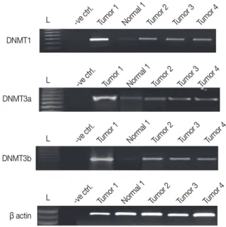
Increased mRNA expression of DNMT1, DNMT3a, and DNMT3b in breast cancer patients
All breast carcinoma patients included in this study showed amplicons of DNMT1, DNMT3a, and DNMT3b, respectively, as shown in Figure 1. The level of expression of DNMTs in the breast cancer patients was compared, after normalization with β-actin, with the adjacent normal breast tissues. The levels of DNMT1, DNMT3a, and DNMT3b mRNA were observed to be 1.2- to 4.4-folds, 1.1- to 3.77-folds, and 1.06- to 4.01-folds elevated in breast cancer tissues, respectively, as compared to the adjacent normal breast tissues.
In summary, DNMT1 and DNMT3b showed moderately higher transcript levels (mean±SE, 2.9±0.2 and 2.6±0.1, respectively) as compared to DNMT3a (mean±SE, 2.0±0.1).
To elucidate the effects of the DNMTs expression on the hor- monal receptor status, the correlation was determined between the hormone receptor status and the DNMT expression level.
A moderate level of correlation was observed between ER/PR and DNMT1 (r=0.5, p=0.01), DNMT3a (r=0.4, p=0.05), and DNMT3b (r=0.5, p=0.001). Further, the transcript levels were analyzed to determine whether these three DNMTs were expressed coordinately or independently. The mRNA levels of these three DNMTs were observed to be moderately correlated with each other as follows: DNMT1 with DNMT3a (r=+0.6, p=0.001), DNMT1 with DNMT3b (r=+0.6, p≤0.001), and DNMT3a with DNMT3b (r=+0.5, p=0.004).
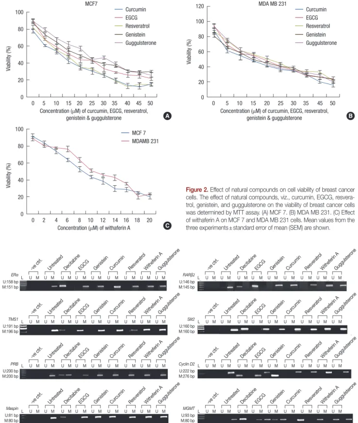
MTT assay
The effects of the various natural compounds on the cell vi- ability after 96 hours of exposure were assessed by MTT assay.
The IC50s for MCF 7 cells were 10, 15, 10, 10, 8, and 20 μM, respectively for EGCG, genistein, curcumin, resveratrol, with- aferin A, and guggulsterone (Figure 2). MDA MB 231 cells were more sensitive to genistein and guggulsterone than MCF 7. The IC50s for MDA MB 231 cells were 15, 10, 10, 15, 10, and 15 μM, respectively for EGCG, genistein, curcumin, resvera- trol, withaferin A, and guggulsterone (Figure 2).
Figure 1. Expression levels of DNMT1, DNMT 3a, and DNMT3b in breast cancer tissues. The levels of DNMT1, DNMT3a, and DNMT3b mRNA were observed to be 1.2- to 4.4-folds, 1.1- to 3.77-folds, and 1.06- to 4.01-folds elevated in most of the breast cancer tissues as compared to the adjacent normal breast tissues.
DNMT1
-ve ctrl.
-ve ctrl.
-ve ctrl.
-ve ctrl.
Tumor 1
Tumor 1
Tumor 1
Tumor 1 Normal 1
Normal 1
Normal 1
Normal 1 Tumor 2
Tumor 2
Tumor 2
Tumor 2 Tumor 3
Tumor 3
Tumor 3
Tumor 3 Tumor 4
Tumor 4
Tumor 4
Tumor 4 L
L
L
L DNMT3a
DNMT3b
β actin
Figure 2. Effect of natural compounds on cell viability of breast cancer cells. The effect of natural compounds, viz., curcumin, EGCG, resvera- trol, genistein, and guggulsterone on the viability of breast cancer cells was determined by MTT assay. (A) MCF 7. (B) MDA MB 231. (C) Effect of withaferin A on MCF 7 and MDA MB 231 cells. Mean values from the three experiments±standard error of mean (SEM) are shown.
Viability (%)
0 5 10 15 20 25 30 35 40 45 50 Concentration (μM) of curcumin, EGCG, resveratrol,
genistein & guggulsterone 100
80
60
40
20
0
Curcumin EGCG Resveratrol Genistein Guggulsterone MCF7
Viability (%)
0 5 10 15 20 25 30 35 45 50 MDA MB 231
Concentration (μM) of curcumin, EGCG, resveratrol, genistein & guggulsterone
120 100 80 60 40 20 0
Curcumin EGCG Resveratrol Genistein Guggulsterone
Viability (%)
0 2 4 6 8 10 12 14 16 18 20 Concentration (μM) of withaferin A 100
80
60
40
20
0
MCF 7 MDAMB 231
A B
C
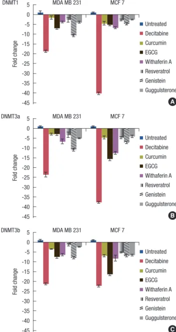
Figure 3. Effect of natural compounds on the methylation status of ERα, TMS1, PRB, Maspin, RARβ2, SLIT2, Cyclin D2, and MGMT. MCF 7 and MDA MB 231 cells were treated with EGCG, genistein, withaferin A, curcumin, resveratrol, guggulsterone (conc. of each natural compound used equals their IC50 value) and decitabine (at conc. of 10 μM for MDA MB 231 cells, and 12 μM for MCF 7 cells) for 96 hours. MSP was performed for ERα, TMS1, PRB, Maspin, Cyclin D2, and MGMT in MDA MB 231 cells and RARβ2 and SLIT2 in MCF 7 cells.
ERα RARβ2
U:158 bp
M:151 bp U:146 bp
M:145 bp
TMS1 Slit2
U:191 bp M:196 bp
U:160 bp M:160 bp
PRB Cyclin D2
U:200 bp M:200 bp
U:222 bp M:276 bp
Maspin MGMT
U:81 bp
M:80 bp U:93 bp
M:80 bp -ve ctrl.
-ve ctrl.
Untreated
Untreated Decitabine
Decitabine EGCG
EGCG Genistein
Genistein Curcumin
Curcumin Resveratrol
Resveratrol Witheferin A
Witheferin A Guggulsterone
Guggulsterone L U M U M U M U M U M U M U M U M U M L U M U M U M U M U M U M U M U M U M
-ve ctrl.
-ve ctrl.
Untreated
Untreated Decitabine
Decitabine EGCG
EGCG Genistein
Genistein Curcumin
Curcumin Resveratrol
Resveratrol Witheferin A
Witheferin A Guggulsterone
Guggulsterone L U M U M U M U M U M U M U M U M U M L U M U M U M U M U M U M U M U M U M
-ve ctrl.
-ve ctrl.
Untreated
Untreated Decitabine
Decitabine EGCG
EGCG Genistein
Genistein Curcumin
Curcumin Resveratrol
Resveratrol Witheferin A
Witheferin A Guggulsterone
Guggulsterone L U M U M U M U M U M U M U M U M U M L U M U M U M U M U M U M U M U M U M
-ve ctrl.
-ve ctrl.
Untreated
Untreated Decitabine
Decitabine EGCG
EGCG Genistein
Genistein Curcumin
Curcumin Resveratrol
Resveratrol Witheferin A
Witheferin A Guggulsterone
Guggulsterone L U M U M U M U M U M U M U M U M U M L U M U M U M U M U M U M U M U M U M
Effect of EGCG, genistein, curcumin, resveratrol, withaferin A, and guggulsterone on the methylation status of the panel of genes
For evaluation of the ability of the tested natural compounds to reactivate the silenced genes, the methylation status of the promoter regions of ERα, PRB, TMS1, Cyclin D2, MGMT, and Maspin genes in MDA MB 231 and RARβ2 and SLIT2 in MCF 7 cells were assessed (Figure 3). The panel of genes studied was selected on the basis of the involvement in the breast tumor initiation, progression, and metastasis [21,22]. Decitabine was used as the reference compound in all the experiments. MDA MB 231 and MCF 7 cells were treated with decitabine at 6, 8, 10, and 12 µM concentrations for 72 hours and 96 hours to assess the effects on the methylation status in our panel of genes (data not shown). MSP of the panel of genes, ERα, PRB, RARβ2, TMS1, Cyclin D2, MGMT, SLIT2, and Maspin were carried out after treatment of MCF 7 and MDA MB 231 cells with decita- bine at the above-mentioned dose and time. Complete demeth- ylation of our panel of genes was observed at 10 µM (96 hours) in MDA MB 231 cells and at 12 µM (96 hours) in MCF 7 cells, while partial demethylation was observed at the above doses at 72 hours. Hence, MDA MB 231 cells treated with 10 µM decitabine and MCF7 cells treated with 12 µM decitabine for 96 hours were used as positive controls for further experiments.
EGCG and genistein, known nonnucleoside demethylating agents showed demethylation of the selective genes only.
EGCG treated MDA MB 231 cells showed complete demeth- ylation of RARβ2 and TMS1, whereas EGCG treated MCF 7 cells showed complete demethylation of RARβ2. Genistein treated MDA MB 231 cells showed complete demethylation of Cyclin D2. However, partial demethylation was observed in Cyclin D2 and MGMT on the treatments with EGCG, whereas MGMT was partially demethylated on treatment with genis- tein. However, curcumin, resveratrol, withaferin A, and gug- gulsterone had no effect on the methylation status of these genes.
Effect of EGCG, genistein, curcumin, resveratrol, withaferin A, and guggulsterone on the transcript levels of DNMT1, DNMT3a, and DNMT3b
The effect of the tested natural compounds on the DNMTs expression was determined by the quantitative analysis of mRNA for each of the three DNMTs. After treatment with various polyphenols, there was a marked decrease in the tran- script levels of all the three DNMTs in both of the cell lines (Figure 4). The most significant reduction was observed in treatment with decitabine on DNMT1, as compared to all other polyphenols tested in the study.
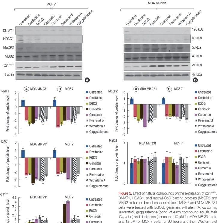
Effect of EGCG, genistein, curcumin, resveratrol, withaferin A, and guggulsterone on the p21WAF1, DNMT1, HDAC1, MeCP2, and MBD2 protein expression
To further evaluate the effects of the tested natural com- pounds on the expression of epigenetic regulators, the expres- sion of DNMT1, MeCP2, MBD2, HDAC1, and p21WAF1 pro-
Figure 4. Effect of natural compounds on the DNMT transcript levels, viz., DNMT1, DNMT3a, and DNMT3b in human breast cancer cell lines.
MCF 7 and MDA MB 231 cells were treated with EGCG, genistein, withaferin A, curcumin, resveratrol, guggulsterone (conc. of each com- pound used equals their IC50 value) and decitabine (at conc. of 10 μM for MDA MB 231 cells and 12 μM for MCF 7 cells) for 96 hours and then real time polymerase chain reaction analysis was performed for the transcript levels of DNMT1 (A), DNMT3a (B), and DNMT3b (C).
A DNMT1
Untreated Decitabine Curcumin EGCG Withaferin A Resveratrol Genistein Guggulsterone
Fold change
5 0 -5 -10 -15 -20 -25 -30 -35 -40 -45
MDA MB 231 MCF 7
C DNMT3b
Untreated Decitabine Curcumin EGCG Withaferin A Resveratrol Genistein Guggulsterone
Fold change
5 0 -5 -10 -15 -20 -25 -30 -35 -40 -45
MDA MB 231 MCF 7
B DNMT3a
Untreated Decitabine Curcumin EGCG Withaferin A Resveratrol Genistein Guggulsterone
Fold change
5 0 -5 -10 -15 -20 -25 -30 -35 -40 -45
MDA MB 231 MCF 7
DNMT1 HDAC1 MeCP2 MBD2
β actin p21WAF1
Untreated Decitabine
EGCG Genistein
Curcumin Resveratrol
Witheferin A Guggulsterone MCF 7
190 kDa 60 kDa 56kDa 49 kDa 21 kDa 42 kDa Untreated
Decitabine EGCG
Genistein Curcumin
Resveratrol Witheferin A
Guggulsterone MDA MB 231
Figure 5. Effect of natural compounds on the expression of p21WAF1, DNMT1, HDAC1, and methyl-CpG binding proteins (MeCP2 and MBD2) in human breast cancer cell lines. MCF 7 and MDA MB 231 cells were treated with EGCG, genistein, withaferin A, curcumin, resveratrol, guggulsterone (conc. of each compound equals their IC50 value) and decita bine (at conc. of 10 μM for MDA MB 231 cells and 12 μM for MCF 7 cells) for 96 hours and then Western blot analysis was performed for DNMT1, HDAC1, MeCP2, MBD2, and p21WAF1 in MCF 7 cells (A) and MDA MB 231 cells (B). The fold change values were calculated as a relative change in comparison to the control cells treated with DMSO (expression equals 1).
p21WAF1
Untreated Decitabine EGCG Genistein Curcumin Resveratrol Withaferin A Guggulsterone
Fold change of protein leve
l 4.5 4 3.5 3 2.5 2 1.5 1 0.5 0
MDA MB 231 MCF 7
MBD2
Untreated Decitabine EGCG Genistein Curcumin Resveratrol Withaferin A Guggulsterone
Fold change of protein level 2
1
0
MDA MB 231 MCF 7
HDAC1
Untreated Decitabine EGCG Genistein Curcumin Resveratrol Withaferin A Guggulsterone
Fold change of protein level 2
1 0 -1 -2 -3 -4
MDA MB 231 MCF 7
DNMT1 Untreated
Decitabine EGCG Genistein Curcumin Resveratrol Withaferin A Guggulsterone
Fold change of protein level 2
1 0 -1 -2 -3 -4
MDA MB 231 MCF 7
MeCP2 Untreated
Decitabine EGCG Genistein Curcumin Resveratrol Withaferin A Guggulsterone
Fold change of protein level 2
1 0 -1 -2 -3 -4 -5
MDA MB 231 MCF 7
teins were determined in MCF 7 and MDA MB 231 cells by Western blotting. The 2- to 3-folds decrease in the levels of DNMT1 and MeCP2 were observed in the MCF7 and MDA MB 231 cells. Down regulation was also observed in the expression of HDAC1 in the MDA MB 231 cells. However, no effect was observed on the expression levels of MBD2 in both cell lines (Figure 5). In addition to these proteins, the treat-
ment with all the compounds increased the level of p21WAF1 in both the cell lines.
DISCUSSION
Epigenetic gene regulation has been recognized to play a crucial role in the etiology of cancer. DNA methylation and
A B A B
A B
post translational histone modifications are important epigene- tic events in the regulation of gene expression and maintenance of cellular function which may contribute to cancer develop- ment [23]. Abnormal methylation in DNA is a hallmark of cancer and often leads to silencing of the tumor suppressor genes, which leads to cancer development and progression.
Dietary phytochemicals have been shown to be involved in the epigenetic modifications to regulate the cellular functions and to modify the risks of cancer [4-7,10,24].
In this study, we investigated whether the treatment of breast cancer cells with natural compounds, viz., EGCG, genistein, curcumin, resveratrol, withaferin A, and guggulsterone, resulted in a reversal of the epigenetic changes. We used decitabine, a well characterized DNA methyltransferase inhibitor, as control [4]. Our study reveals that EGCG completely demethylated RARβ2 and TMS1 in the MDA MB 231 cells and RARβ2 in the MCF 7 cells. Genistein treated MDA MB 231 cells showed complete demethylation of Cyclin D2. However, partial deme- thylation was observed in Cyclin D2 and MGMT on treatment with EGCG; and MGMT was partially demethylated on treat- ment with genistein. We also observed that the treatment of breast cancer cells at the lower concentrations of curcumin, resveratrol, withaferin A, and guggulsterone for longer time period (more than 96 hours) did not change the methylation status of our panel of genes (data not shown). All the natural compounds tested here downregulated the expression of DNMT1 in both of the breast cancer cell lines.
Histone modifications are epigenetic marks linked to the transcriptional activators and repression of genes [25]. Post- transcriptional modification of histone acetylation/methyla- tion may contribute to cancer development by modulation of the expression of tumor suppressor genes and oncogenes.
HDAC plays a key role in the regulation of histone deacety- lation. Our data demonstrate that all natural compounds test- ed, viz., EGCG, genistein, curcumin, resveratrol, withaferin A, and guggulsterone, significantly decreased the HDAC1 expres- sion.
Promoter hypermethylation mediated enhancement of the epigenetic gene silencing involves the activity of MBD pro- teins, which specifically bind to methylated DNA [26]. The physical association of MBDs with methylated DNA causes steric hindrance for transcription factor binding. Studies dem- onstrate that at least 2 members of the MBD family, viz., MBD2 and MeCP2 are expressed in the human breast cancer cells and may be involved in gene repression. MeCP2 is able to bind to a single symmetrically methylated CpG [21,22]. It interacts with a transcriptional repressor complex containing HDACs (histone deacetylases) and the transcriptional co- repressor Sin3a [26]. Hence, the protein expression of MeCP2
and MBD2 was also evaluated after treatment with EGCG, genistein, curcumin, resveratrol, withaferin A, and guggul- sterone. The expression of MeCP2 was reduced by 2 to 3 folds.
However, no effect was observed on the expression levels of MBD2.
In the cancer cells, the epigenetic changes and the coexist- ing genetic lesions often fundamentally cripple various bio- chemical pathways, e.g., cell cycle arrest and apoptosis. DNMT1 and p21WAF1 compete for the same binding site on PCNA; and an increase in the DNMT1 expression maypromote the disso- ciation of p21WAF1 from PCNA, perhaps making p21WAF1 more susceptible to ubiquitination and proteasomal degradation [27,28]. A decrease in the DNMT1 expression would then be expected to have an opposite effect on the p21WAF1 stability [27,28]. We also evaluated the effects of these natural com- pounds on p21WAF1 and observed that the treatments with EGCG, genistein, curcumin, resveratrol, withaferin A, and gug- gulsterone resulted in 2 to 4 folds increase in the p21WAF1 ex- pression.
In summary, similar to the previous reports, we demon- strated that EGCG and genistein can restore or reactivate the expression of the DNA hypermethylated silenced genes in breast cancer by the down regulation of DNMT1. Recently, the molecular docking of the interactions between curcumin and DNMT1 [8] have suggested that curcumin covalently blocks the catalytic thiolate of DNMT1 to exert its inhibitory effect. Some recent studies have suggested that other polyphe- nols, lacking a gallic/pyrogallic acid moiety, cannot form a similar strong coordination with the DNMT catalytic center, which, in turn, interferes with the activities of DNMTs inhibi- tion [24]. Hence, we presume that one of the plausible expla- nations for the curcumin and all other compounds tested not reversing the methylation of some genes may be due to their weak effects on the DNMT catalytic center. Another explana- tion may be indicating of the functional importance of MBD2.
Recent studies on the structure of MBD2 bound to a methyl- ated gene target sequence suggest that the sequence context may direct this preferential binding of MBD2 to certain CpG rich regions [29].
The findings of the current study are preliminary and indi- cate that the natural compounds, viz., EGCG, genistein, cur- cumin, resveratrol, withaferin A, and guggulsterone, may pro- vide cancer preventive activity through the modification of the epigenetic process of the gene silencing. However, in-depth future mechanistic studies aimed to elucidate how these com- pounds affect the gene transcription are warranted. It rein- forces the view that these agents can exert inhibitory activity and may be useful in the investigation of the effects of the dietary natural compounds as adjuvants in cancer therapies.
ACKNOWLEDGEMENTS
S.M. is thankful to UGC for providing him with the SRF Fellowship. The authors thank Dr. Fayaz Malik, the Indian Institute of Integrative Medicine (Council of Scientific and In- dustrial Research), for the kind gift of withaferin A. R.R.
gratefully acknowledges the financial support from Canadian Institutes of Health Research for the Chair in the Advanced Cancer Diagnostics.
CONFLICT OF INTEREST
The authors declare that they have no competing interests.
REFERENCES
1. Jones PA. DNA methylation and cancer. Oncogene 2002;21:5358-60.
2. Ptak C, Petronis A. Epigenetics and complex disease: from etiology to new therapeutics. Annu Rev Pharmacol Toxicol 2008;48:257-76.
3. Siedlecki P, Zielenkiewicz P. Mammalian DNA methyltransferases.
Acta Biochim Pol 2006;53:245-56.
4. Lyko F, Brown R. DNA methyltransferase inhibitors and the develop- ment of epigenetic cancer therapies. J Natl Cancer Inst 2005;97:1498-506.
5. Fang MZ, Wang Y, Ai N, Hou Z, Sun Y, Lu H, et al. Tea polyphenol (-)-epigallocatechin-3-gallate inhibits DNA methyltransferase and re- activates methylation-silenced genes in cancer cell lines. Cancer Res 2003;63:7563-70.
6. Fini L, Selgrad M, Fogliano V, Graziani G, Romano M, Hotchkiss E, et al. Annurca apple polyphenols have potent demethylating activity and can reactivate silenced tumor suppressor genes in colorectal cancer cells. J Nutr 2007;137:2622-8.
7. King-Batoon A, Leszczynska JM, Klein CB. Modulation of gene meth- ylation by genistein or lycopene in breast cancer cells. Environ Mol Mu- tagen 2008;49:36-45.
8. Liu Z, Xie Z, Jones W, Pavlovicz RE, Liu S, Yu J, et al. Curcumin is a po- tent DNA hypomethylation agent. Bioorg Med Chem Lett 2009;19:
706-9.
9. Majid S, Dar AA, Ahmad AE, Hirata H, Kawakami K, Shahryari V, et al. BTG3 tumor suppressor gene promoter demethylation, histone modification and cell cycle arrest by genistein in renal cancer. Carcino- genesis 2009;30:662-70.
10. Ramos S. Effects of dietary flavonoids on apoptotic pathways related to cancer chemoprevention. J Nutr Biochem 2007;18:427-42.
11. Choi JS, Choi YJ, Park SH, Kang JS, Kang YH. Flavones mitigate tumor necrosis factor-alpha-induced adhesion molecule upregulation in cul- tured human endothelial cells: role of nuclear factor-kappa B. J Nutr 2004;134:1013-9.
12. Sim GS, Lee BC, Cho HS, Lee JW, Kim JH, Lee DH, et al. Structure ac- tivity relationship of antioxidative property of flavonoids and inhibitory
effect on matrix metalloproteinase activity in UVA-irradiated human dermal fibroblast. Arch Pharm Res 2007;30:290-8.
13. Lee WJ, Shim JY, Zhu BT. Mechanisms for the inhibition of DNA meth- yltransferases by tea catechins and bioflavonoids. Mol Pharmacol 2005;
68:1018-30.
14. Lee WJ, Zhu BT. Inhibition of DNA methylation by caffeic acid and chlorogenic acid, two common catechol-containing coffee polyphenols.
Carcinogenesis 2006;27:269-77.
15. Chen D, Milacic V, Chen MS, Wan SB, Lam WH, Huo C, et al. Tea poly- phenols, their biological effects and potential molecular targets. Histol Histopathol 2008;23:487-96.
16. Chen D, Wang CY, Lambert JD, Ai N, Welsh WJ, Yang CS. Inhibition of human liver catechol-O-methyltransferase by tea catechins and their metabolites: structure-activity relationship and molecular-modeling studies. Biochem Pharmacol 2005;69:1523-31.
17. Moiseeva EP, Almeida GM, Jones GD, Manson MM. Extended treat- ment with physiologic concentrations of dietary phytochemicals results in altered gene expression, reduced growth, and apoptosis of cancer cells. Mol Cancer Ther 2007;6:3071-9.
18. Mirza S, Sharma G, Prasad CP, Parshad R, Srivastava A, Gupta SD, et al.
Promoter hypermethylation of TMS1, BRCA1, ERalpha and PRB in serum and tumor DNA of invasive ductal breast carcinoma patients.
Life Sci 2007;81:280-7.
19. Sharma G, Mirza S, Prasad CP, Srivastava A, Gupta SD, Ralhan R. Pro- moter hypermethylation of p16INK4A, p14ARF, CyclinD2 and Slit2 in serum and tumor DNA from breast cancer patients. Life Sci 2007;80:
1873-81.
20. Shukla S, Mirza S, Sharma G, Parshad R, Gupta SD, Ralhan R. Detec- tion of RASSF1A and RARbeta hypermethylation in serum DNA from breast cancer patients. Epigenetics 2006;1:88-93.
21. Garinis GA, Patrinos GP, Spanakis NE, Menounos PG. DNA hypermeth- ylation: when tumour suppressor genes go silent. Hum Genet 2002;111:
115-27.
22. Baylin SB, Herman JG. DNA hypermethylation in tumorigenesis: epi- genetics joins genetics. Trends Genet 2000;16:168-74.
23. Esteller M. Epigenetics in cancer. N Engl J Med 2008;358:1148-59.
24. Li Y, Tollefsbol TO. Impact on DNA methylation in cancer prevention and therapy by bioactive dietary components. Curr Med Chem 2010;
17:2141-51.
25. Rice JC, Allis CD. Histone methylation versus histone acetylation: new insights into epigenetic regulation. Curr Opin Cell Biol 2001;13:263-73.
26. Prokhortchouk E, Hendrich B. Methyl-CpG binding proteins and cancer:
are MeCpGs more important than MBDs? Oncogene 2002;21:5394-9.
27. Fang JY, Lu YY. Effects of histone acetylation and DNA methylation on p21( WAF1) regulation. World J Gastroenterol 2002;8:400-5.
28. Periyasamy S, Ammanamanchi S, Tillekeratne MP, Brattain MG. Re- pression of transforming growth factor-beta receptor type I promoter expression by Sp1 deficiency. Oncogene 2000;19:4660-7.
29. Berger J, Bird A. Role of MBD2 in gene regulation and tumorigenesis.
Biochem Soc Trans 2005;33(Pt 6):1537-40.