책임저자:이남준, 서울 종로구 대학로 101
서울대학교 의과대학 외과학교실, 110-744 Tel: 02-2072-2990, Fax: 02-766-3975 E-mail: [email protected]
접수일 : 2011년 9월 1일, 게재승인일 : 2011년 9월 5일
소아 간이식 술기
서울대학교 의과대학 외과학교실 간담췌외과
이남준ㆍ이광웅ㆍ서경석ㆍ이건욱ㆍ김수태
Transplantation Techniques Unique in Pediatric Liver Transplantation
Nam-Joon Yi, M.D., Kwang-Woong Lee, M.D., Kyung-Suk Suh, M.D., Kuhn Uk Lee, M.D. and Soo Tae Kim, M.D.
Division of HBP Surgery, Department of Surgery, Seoul National University College of Medicine, Seoul, Korea
In previous decades, pediatric liver transplantation has become a state-of-the-art operation with excellent success and limited mortality. Graft and patient survival have continued to improve as a result of proper selection criteria for both donors and recipients, improvement in medical, surgical and anesthetic management, organ availability, balanced immunosuppression, and early identification and treatment of postoperative complications. Most of all, refinements of the technique has directly related to good outcome. Therefore rapid establishment of surgical knowhow is mandatory. In pediatric liver transplantation, the utilization of split-liver grafts and grafts for living donors has provided more organs for pediatric patients and has had a significant impact on graft and patient survival. This has been one of the brilliant outcomes of surgical evolution. In addition, new surgical technique of minimal invasive live donor surgery has been recently widening the living donor liver transplantation for children. Although the recent outcome has been rapidly improved and the volume of living donor liver transplantation has been larger and larger in Korea, pediatric liver transplantation has been performed in a very limited large volume centers. Therefore, this review focuses on surgical technique in order to share the experiences and to improve the outcome of pediatric liver transplantation.
Key Words: Pediatric liver transplantation, Living donor liver transplantation, Left lateral section, Split liver transplantation, Reduced liver transplantation, Monosegment liver transplantation
중심 단어: 소아 간이식, 생체 간이식, 좌외구역, 분할 간이식, 축소 간이식, 단구역 간이식
서 론
간이식은 간부전 환자 뿐 아니라 대사성 간질환 환자 에서도 최상의 치료법으로서 질환 자체의 치료와 더불어 환자의 삶의 질을 향상시키는 역할을 해왔다. 소아에서 도 예외는 아니어서 세계적으로 첫 간이식은 1963년 피 츠버그에서 선천성 담도폐쇄증 환아에게(1), 국내에서는 1988년 서울대학교병원에서 윌슨씨병 환아에게 이루어졌 다(2). 1980년대까지만 해도 소아 수혜자에 대한 간이식은 비슷한 체중의 뇌사 공여자의 전간이식술만 이루어졌다.
하지만 소아 수혜자에게 적절한 뇌사자로 전간이식을 하 는 경우는 전세계적으로도 많지 않으며(3), 이식 장기의
부족은 간이식 적응증 확대에 가장 큰 걸림돌이 되어 왔 다. 이러한 이유로 50%의 환아들이 이식 대기 중 사망하 였다(4). 소아 뇌사 공여자가 드물기 때문에 소아 간이식 대기자에게는 성인 공여자로부터 적절한 크기의 부분 간 적출술과 부분 간이식술이 필요하게 되어 다양한 술기가 개발되었다. 따라서 축소 간이식, 분할 간이식, 생체 간 이식, 보조 간이식 등이 널리 행해졌고, 이외에도 혈액형 부적합 간이식, 심장사 공여자의 장기 이식, 간세포이식 등 다양하게 기증 형태를 넓혀 왔다.
국내 간이식은 연 1,000예 이상으로 그 중에서도 생체 간이식이 80%를 차지하며 이식 성적은 구미의 뇌사자 간 이식보다 오히려 우수할 정도로 괄목할 성장을 거듭하고 있다. 간이식 성적은 적절한 공여자와 수혜자의 선정으 로부터, 수술 성공 여부, 이식편 정착, 이식 후 감염증을 비롯한 심각한 합병증의 조기 발견과 적절한 처치, 최선 의 면역억제술 등 다양한 과정이 관여하지만 그 중에서 도 수술 자체의 성공 여부가 이식 성패에 매우 중요하다.
간이식 술기는 수혜자 뿐 아니라 공여자 수술도 성공적 으로 이루어져야 하며, 대부분의 수혜자가 간기능 부전으 로 문맥압 항진증과 출혈 경향이 있고, 소아 수혜자는 50% 이상에서 과거에 수술 기왕력을 가지고 있기 때문에 술기 자체의 어려움이 있어 경험과 요령이 필요하다.
그러나 연평균 국내 소아 간이식 건수는 40∼50예로 전체 간이식 건수의 5% 미만이다. 최근 5년간 국립장기 기증원(Korean Network for Organ Sharing, KONOS)의 보고에 따르면(5) 284건의 소아 간이식이 15개 센터에서 이루어졌는데, 이 중 87.3%에 해당하는 248건은 3개 센 터에서 이루어졌다. 따라서 국내에서는 소아 간이식의 다양한 술기를 익히기란 쉽지 않다. 이에 본고에서는 소 아 간이식을 위한 술기에 초점을 맞추어 기술하고자 한 다.
전간이식
전간이식술은 일반적인 성인 수술과 다르지 않다. 소 아 환자이기 때문에 발생하는 차이점만 기술하겠다.
1) 공여자 간적출 준비
먼저 담낭과 담도를 열어서 생리식염수로 충분히 씻어 주고, 대동맥과 문맥 관류를 준비한다. 소아 뇌사자의 간 적출 시에는 간동맥 박리에 주의를 요한다. 소아 공여자 는 맥박이 빠르고 혈압이 낮아 간동맥이 손상되기 쉽다.
특히 간동맥 분지에 해부학적인 변형이 있는 경우, 즉 우 간동맥이 상장간막동맥으로부터 분지되거나 좌간동맥이 좌위동맥으로부터 분지되는 경우, 좌우 간동맥이 조기 분 지되거나, 드물게 우간동맥이 위십이지장동맥으로부터 분지되거나 담도의 앞으로 지나는 경우에, 간동맥 박리에 유의하지 않으면 절리-결찰되어 재관류 후 발견한다고 해도 복원하기 쉽지 않다. 따라서 전간적출술 시에는 담 도절리 후 혹은 관류 전에 촉지하거나 도플러 초음파를 이용해 반드시 좌우간동맥의 간내 유입 여부를 확인하여 야 한다.
2) 공여자 관류술
관류용 헤파린을 전신 투여한 후 대동맥 결찰하고 대 정맥을 열어 주고 관류한다. 적절한 관류용 헤파린 용량 은 공여자 체중 1 kg 당 50 unit이다. 간 좌외구역을 젖 히고 복강동맥 상부의 대동맥은 박리한다. 하복부 대동 맥은 장골동맥 분지부로부터 신동맥 분지부까지의 길이 가 짧으므로 대동맥 관류 카테터의 삽관을 짧게 하여 신 동맥을 지나지 않도록 주의한다. 필요하다면 장골동맥에
삽관할 수 있다. 장간맥정맥이나 비장정맥이 가늘어 삽 관이 어려운 경우, 문맥 관류는 보류하고 적출 후 문맥에 직접 관류액을 넣기도 한다. 대동맥과 문맥에 각각 150 mL/kg으로 HTK 용액을 주입한다.
3) 수혜자 간적출술 (1) 간적출술
일반적인 주의점은 성인의 전간 적출술과 같다. 유출 로 재건이 고전적인 하대정맥치환술(Inferior Vena Cava, IVC replacement)인지 혹은 Piggyback 방법인지에 따라 수혜자의 하대정맥 보존 여부를 결정한다(6). 소아에서는 하대정맥치환술을 하더라도 적절한 수액요법만 가능하다 면 정맥우회술(veno-veno bypass)이 반드시 필요하지 않 다(4). 그러나 원인 질환이 대사성 질환이나 전격성간부 전으로 부대정맥이 발달하지 않아 장이 심하게 붓는 경 우에는 일시적인 문맥대정맥션트술(portocaval shunt)이 유용하다. 염증이나 과거 수술 때문에 유착이 심한 경우 에는 먼저 간문부 우외측 접근하여 유입혈류를 차단하고 하대정맥을 포함하여 일괄 적출하는 것이 수술 중 손상 을 줄일 수 있다(4).
(2) 간문부 박리술
이식 전 Kasai 수술을 비롯한 기존의 개복술로 유착이 있는 경우에는 가능하다면 Roux-en-Y limb을 먼저 절제 한 후 간문부를 일괄 처리하는 것이 문맥과 간동맥을 손 상 없이 박리할 수 있다. 문맥은 상장간정맥과 비장정맥 합류부까지, 간동맥은 위십이지장동맥이 분지부까지 확 보한다.
4) 전간이식술
간정맥 혹은 하대정맥, 문맥, 간동맥, 담도 순으로 재 건한다. 소아 혈관 문합시 저자들은 주로 흡수성 단봉합 사(polydioxanone suture, PDS)를 이용하여 연속봉합 (running suture)하고 간동맥문합의 경우만 비흡수성 단 봉합사(nylon)을 사용한다(7).
(1) 유출로 재건(5/0 vascular absorbable monofila- ment running suture)
하대정맥치환술을 하는 경우에는 하대정맥을 간 상부 에서 하부 순으로 문합한다. Piggyback 방법 역시 성인 에서의 술기와 동일하지만 넓은 유출로 재건을 위해서 수혜자의 하대정맥을 박리하여 하대정맥치환술과 같이 완전히 차단한 상태에서 재건하기도 한다. 하대정맥을 측측문합(side-to-side anastomosis) 할 때에는 이식편을 우측으로 젖힌 상태에서 수혜자의 하대정맥을 넓은 혈관 감자로 잡고 문합한다.
Fig. 1. Portal vein reconstruction. (A) Portal vein reconstruction without vein graft, (B) Portal vein reconstruction with vein conduit from donor. Reprinted from reference (7).
Fig. 2. Living donor liver transplantation. (A) Left lateral sectionectomy in a live donor. There is usually no ischemia and congestion in the graft, but the left medial section and a part of caudate lobe become ischemic in the remnant liver of the donor. (B) Hepatic vein anastomosis and the position of the graft in transplantation of a left lateral section. Reprinted from reference (12,13).
Abbreviations: LHV, left hepatic vein; LBD, left bile duct; LPV, left portal vein; MPV, main portal vein; PHA, proper hepatic artery.
(2) 유입로 재건
가. 문맥 재건(6/0 vascular absorbable monofilament running suture) (Fig. 1): 문맥대 문맥을 단단문합하는 것이 일반적이지만, 문맥이 가늘거나(4 mm 미만) 섬유 화가 진행된 경우에는 이식편 문맥을 상장간정맥과 비장 정맥 합류부에 문합하거나, 부분적인 혈관패치를 이용하 거나, 공여자의 장골정맥을 이용하여 conduit를 사용할 수 있다(8). 문맥무형성증일 때는 상장간정맥, 비신단락 을 가진 좌신정맥, 하대정맥, 기타 부대정맥 등에 공여자 로부터 구득한 혈관으로 점프문합술을 하기도 한다 (9,10). 문맥혈이 충분하지 않을 경우 부대정맥을 결찰하 여 문맥혈 스틸증후군(portal steal syndrome)을 예방한 다.
나. 동맥재건(7/0∼8/0 vascular nonabsorbable mon- ofilament running/interrupted suture): magnification loops (3.5×)를 사용한다. 수혜자의 좌우간동맥 분지나 총간동
맥과 위십이지장동맥 분지 등을 이용한 동맥분지 패치성 형술(branch patch technique)로, 적절한 길이와 구경의 공여자 동맥과 문합한다. 수혜자의 동맥이 너무 가늘거 나 손상되어 사용할 수 없는 경우에는 복강동맥 직상방 혹은 신동맥 직하부 대동맥을 이용할 수 있다.
(3) 담도 재건(6/0 vascular absorbable monofila- ment running/interrupted suture)
담도-담도 문합술은 담도 합병증 발생률이 높아서 흔 히 사용하지 않는다(11). 담도공장문합술을 하는 경우 Roux-en-Y limb 길이가 40∼60 cm 정도로 충분해야 역 행성 담도염을 예방할 수 있다.
부분간이식
부분 간이식은 술기의 발전을 토대로 장기의 부족을 해결하는 방법으로 고안되었고 소아 간이식 수혜 대기자
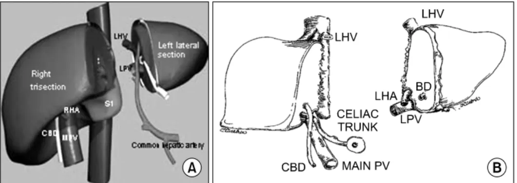
Fig. 3. Split liver transplantation of left lateral section and right trisection. (A) Classic split technique. The left graft composed of segment 2 and 3 includes the left hepatic vein, the left portal vein, the left hepatic artery from the common hepatic artery and the celiac triad, and the left hepatic duct. The right graft composed of segment 1 and 4∼8 includes the vena cava, the main portal vein, the right hepatic artery, and common bile duct. Reprinted from reference (3). Adapted split technique mimicking a graft from a live donor. The difference of the classical split liver graft is the arterial division level. Usually the left lateral graft includes only the left hepatic artery. Reprinted from reference (19). Abbreviations: BD, bile duct; CBD, common bile duct; PV, portal vein; LHV, left hepatic vein; LPV, left portal vein; S, segment.
의 대기 중 사망률을 감소시키고 이식 성적을 향상시키 는 방법으로 자리잡았다. 부분 간이식의 종류로는 아래 와 같은 방법이 있다.
1) 축소간이식
Bismuth 등에 의해 처음으로 기술되었는데(12), 성인 뇌사 공여자로부터 전간을 적출하여 벤치 수술 중 수혜 자의 복강 크기에 맞게 줄이는 방법이다. 이 방법을 통해 소아 수혜자의 대기 중 사망률을 감소시켰고, 수술 성적 은 전간이식과 비슷하였다. 공여장기 부족이 심각한 가 운데 남은 간을 폐기하는 수술이기 때문에 최근에는 잘 사용되지 않지만, 이식편이 커서 폐복이 어려운 경우 부 분적으로 이식편을 줄이기 위해 시행하기도 한다.
2) 생체 부분간이식(Fig. 2) (13,14)
세계적으로는 1989년(15)과 1990년(16)에, 국내에서는 1994년에(2) 처음으로 선천성 담도폐쇄증 환아에게 엄마 의 좌외구역을 이식하는 것이 보고되었다. 생체 간이식 은 이식 대기 시간을 줄이고, 이식 전 공여자와 수혜자가 철저한 수술 전 준비를 마친 후 정규 수술이 가능하며, 냉허혈 시간이 대개 1시간을 넘지 않고, 이식편의 질이 우수하다는 장점이 있다. 부분간을 사용하는 뇌사자 간 적출술과 다른 점은, 공여자 수술 중 간을 완전히 구동하 여 절리하고 공여자에게 전신 헤파린(3,000∼5,000 unit) 을 투여한 후 간유입혈과 간정맥을 단시간에 절리하고 벤치에서 체외 관류하게 된다. 최근 저자들은 공여자의 안전을 위해 관류액에 헤파린을 혼합하여 체외 관류 중 투입하고 있다. 좌간 이상의 이식술은 성인 수술에 준하
며, 좌외구역 이하의 이식술은 대개 소아에서 이용되는 수술이다.
3) 분할간이식(Fig. 3)
Pichlmayr 등에 의해 처음으로 기술되었는데(17), 성인 뇌사 공여자로부터 전간을 적출하여 벤치 수술 중 간원 인대를 중심으로 좌외구역(제2, 3구역)과 우삼구역(제1구 역 및 제4∼8구역)으로 나누어 각각 성인과 소아 수혜자 에게 이식하는 방법이다. 소아 수혜자의 대기 중 사망률 을 줄이면서 성인 수혜자도 동시에 이식 받을 수 있다는 점에서 이상적인 수술법이지만, 냉허혈시간이 길어져 초 기 이식 성적이 좋지 않았기 때문에 널리 이용되지 않았 다. 1996년 Rogiers 등(18)이 생체 간이식에서와 같이 in situ 방법으로 이식편을 절리하는 방법을 고안하였고 이 후 이식 성적이 향상되었다. 국내에서는 2000년에 처음 으로 in situ 방법의 분할간이식이 시행되었다(19). 분할 간이식 성적이 좋아지면서 최근에는 공여자와 수혜자 상 태에 따라서 좌간(제2∼4구역, 제1구역)과 우간(제5∼8 구역, 제1구역)으로 나누기도 한다. 고전적인 방법으로 좌외구역 이식편에 좌간정맥, 좌간문맥, 좌측 담도를 분 배하고 간동맥은 대동맥 패치를 가져오는 방법이다(Fig.
3A) (3). 생체간이식과 같은 방법으로 좌외구역을 가져오 기도 하는데(Fig. 3B) (19), 이는 좌외구역 적출을 우선적 으로 하여 적출 시간을 단축할 수 있는 장점이 있어 경 험이 많은 생체간이식센터에서 선호한다(6).
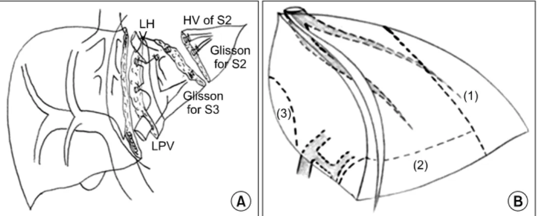
Fig. 4. Reduced left lateral section. (A) Monosegment (Segment 3). Reprinted from reference (22). (B) Hyperreduced left lateral section (LLS). Cutting lines to reduce a LLS; (1) the lateral part of the LLS is resected first while preserving the medial branches of the LHV.
(2) Further resection of the caudal part is performed without ligation of any portal branches of the S3. (3) The additional resection of the dorsal part is carried out, while preserving portal branches of the S2. Reprinted from reference (20). Abbreviations: HV, hepatic vein; LHV, left hepatic vein; LPV, left portal vein; S, segment.
부분 간공여자 수술의 주의점
부분 간이식에서는 수혜자의 체중 및 복강용적과 간용 적을 고려하여, 이식편 무게 대 수혜자 체중비(Graft ver- sus recipient weight ratio, GRWR)는 0.8% 이상, 3.5%
미만이 적절하다(20). 이에 따라서 공여자의 간절제술 범 위는 변형확대우간, 우간, 우후구역, 좌간, 확대좌간, 좌 외측구역, 단구역, 축소단구역 등 다양하다. 공여자 수술 준비 시, 간의 해부학적인 구조와 크기를 파악할 수 있도 록 컴퓨터단층촬영 및 담도 조영술(혹은 자기공명영상검 사)이 필요하다. 공여자 수술 시에는 간절리가 필요하므 로 출혈량이 최소화되도록 중심정맥압을 적절히 유지하 고 필요한 경우 선택적 간헐적 Pringle 술기를 사용할 수 있다. 간절리의 편의를 위해 견인기, Kelly 감자나 절리 기구(Cavitron Ultrasound Surgical Aspirator, CUSA 등) 을 이용하게 된다. 다양한 현수기법을 이용하면 간절리 가 용이하다. 간부분 적출술 중 좌간 이상의 이식술은 성 인간 부분 간이식과 동일하다(21). 좌외측우역 이하 단구 역 및 축소 간절제술은 소아에서 주로 활용되므로 이에 대해 기술하고자 한다.
1) 좌외구역 절제술
흔히 일반 간절제술 시 허용되는 좌외구역 절제술은 제2, 3구역 Glisson지를 결찰-절리하지만, 부분 간이식을 위한 공여자 간적출 시에는 좌간문맥과 좌간동맥, 좌간담 도를 절리한다(Fig. 2A). 우측간을 온전히 이식하거나 혹 은 생체 공여자가 합병증 없이 회복할 수 있도록 좌간정
맥, 좌간문맥, 좌간동맥, 좌간담도 절제부로 인하여 남은 우간 구조물에 변형이 오거나 좁아지지 않도록 주의한 다.
2) 단구역 절제술
주로 7 kg 미만의 소아나 신생아 간이식술, 전격성 간 부전으로 이식하는 경우에는 이식편에 비해 상대적인 복 강내 용적이 작아 좌외구역 보다 작은 이식편이 필요하 다(20,22-24). 이를 위해서는 제2 혹은 제3단구역을 절제 하게 된다. 단구역은 술기의 편의성을 고려하여 제2구역 보다 제3구역을 선호한다(Fig. 4A) (22).
3) 좌외구역 축소절제술
해부학적인 절제가 아니라 가능한 많은 좌외구역 축소 가 필요한 경우에는 주로 간의 끝단을 줄이기도 한다 (Fig. 4B) (20). 이식편 축소는 공여자 수술 중 시행하거 나 벤치 수술 중 시행하기도 하지만, 이식 재관류 후 폐 복이 어렵다고 생각될 때 추가적으로 시행할 수 있다.
4) 생체 간이식 공여자를 위한 최소 절개법
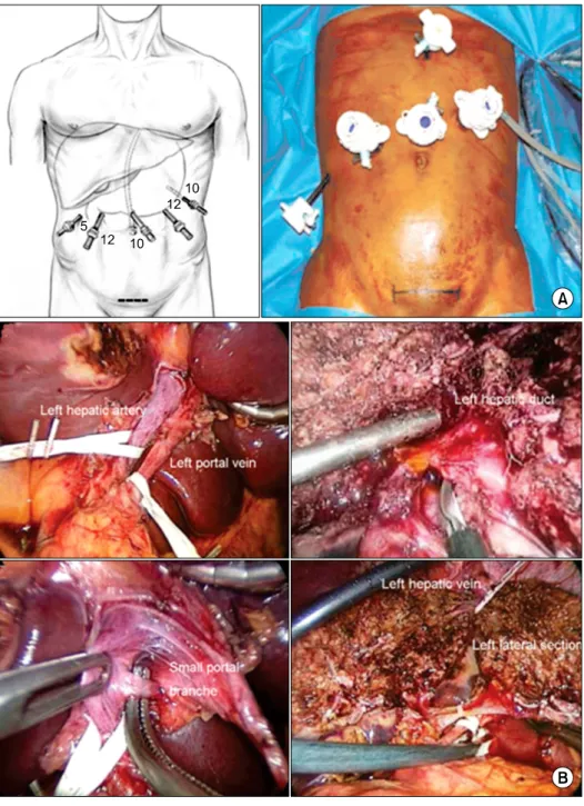
생체 공여자 간절제술 후 삶의 질에 관여하는 주요한 인자 중의 하나가 창상과 복강내 유착이다(25). Cherqui 등(26)은 2002년 처음으로 복강경 술식을 이용하여 좌외 구역을 절제하였다(Fig. 5). 개복술로 시행한 공여자들과 비교하였을 때 수술 시간이나 이식편 기능에 차이가 없 다고 보고하였다(27,28). 중앙절개만으로도 이식편 구득 이 가능하기 때문에 최소절개창으로 창상을 줄일 수도 있다(29).
Fig. 5. Laparoscopic left lateral sectionectomy in a live donor. (A) Port placement (n=port size, mm) for laparoscopic left lateral sectio- nectomy in a live donor. (B) Divi- sion of the left portal vein, hepatic artery, bile duct, and hepatic vein for harvest of a left lateral section.
Reprinted from reference (26,27).
부분 간이식 술기
좌외측우역 이식술 이하 단구역이식 및 축소 간이식은 소아 간이식에서 주로 활용되므로 이에 대해 기술하고자 한다.
1) 좌외측구역 이식술
수혜자 간적출술은 일반적인 전간 이식술에서와 다르 지 않지만 하대정맥을 보존하여야 한다(Fig. 2B). 다만 유착이 심하거나 종양절제를 위해 하대정맥 보존이 어렵
다면 동반절제 후 냉동보존혈관 혹은 인조혈관 등을 이 용하여 패치혈관성형술로 하대정맥을 복원할 수 있다 (Fig. 6) (3).
(1) 간정맥 문합술(5/0 vascular absorbable mono- filament running suture) (Fig. 2B,7) (3) 이식편의 좌간정맥은 수혜자의 좌간정맥보다 대개 크 다. 따라서 이식편의 간정맥은 수혜자 간정맥의 자연개 구부를 하대정맥에 연장하여 크게 열고 하대정맥의 중앙 부에 문합하게 된다(Fig. 7). 따라서 간정맥 문합 후 이식 편은 원래 위치보다 환자의 복부 쪽으로 이동하게 되고,
Fig. 6. Use of a left lateral section graft to a recipient affected hepatic malignancy with replacement of the recipient’s IVC us- ing a cryopreserved iliac vein. Reprinted from reference (3).
간문부는 자연히 우후측으로 이동하게 된다(Fig. 2B). 간 정맥의 문합은 좁아지지 않도록 하대정맥 유출로가 이식 편 간정맥보다 크게 되도록 문합한다. 단일 정맥일 때는
유출로 합병증이 적지만, 이식편 간정맥이 2개가 되면 합 병증이 증가하고, 이식편의 크기가 작은 경우(GRWR
<1.0%), 축소 간이식술을 한 경우 합병증이 증가하므로 (30) 이러한 경우 더욱 충분한 하대정맥 문합부를 확보하 도록 한다.
(2) 간문맥 문합술(6/0∼7/0 vascular absorbable mo- nofilament running suture) (Fig. 1)
전간이식술과 다르지 않지만, 이식편 문맥이 짧기 때 문에 주로 수혜자 간문맥의 분지부 패치를 이용하여 문 합한다. 이식편의 간문부가 우후측으로 돌아가 있기 때 문에 수혜자의 문맥 비틀림 현상에 주의한다. 수혜자 문 맥의 질이 좋지 않은 경우에는 앞서 언급한 문맥성형술 이 필요하다. 문맥 합병증은 8∼9% 정도 발생하는데 문 합 방법 보다는 수혜자가 6 kg 이하 소아에서 증가하므 로 주의를 요한다(8). 조기 합병증 보다는 후기에 증가하 며 후기 합병증인 경우에는 일차적인 수술 보다는 경피 경간 혹은 경피경비 문맥 성형술을 선호한다(8).
Fig. 7. Anastomosis between the left hepatic vein of the graft and the inferior vena cava of the recipient, performed with the triangulation technique. (A) Total clamp of the IVC for re- construction of hepatic vein. The bridge between the ostia of the right, middle, and left hepatic veins is cut to obtain a single opening. (B) Enlargement of IVC opening. The opening is further enlarged by cutting the anterior face of the vena cava to obtain a wide triangular orifice. (C) Anastomosis of the left hepatic vein to the IVC. Reprinted from reference (3).
Fig. 8. Microscopic technique of hepatic artery anastomosis. (A) Adventisectomoy of the end of the hepatic artery reducing thrombosis. ① Adventisectomoy of the end of the hepatic artery. ② Proper needling for hepatic artery anastomosis. (B) End-to-end anastomosis of hepatic artery. ① Carrel technique. ② Seidenberg technique. (C) Overcome size discrepancy of hepatic artery anastomosis. ① Fish mouth technique. ② Oblique technique. ③ Branch patch technique using the small right gastroepiploic artery of the recipient. Abbreviation: RGEA, right gastroepiploic artery. Reprinted from reference (31).
Fig. 9. Auxiliary parital orthotopic liver transplantation. (A) Schemat- ic figure of reconstruction. (B) Au- xiliary liver transplantation of the left lateral section. Abbreviations:
IVC, inferior vena cava; HV, hepatic vein; PV, portal vein.
(3) 간동맥 문합술(9/0~10/0 vascular nonabsorba- ble monofilament interrupted suture) (Fig. 8) (31)
간동맥 문합은 주로 미세현미경 하에서 단단 문합을 시행한다. 이식편의 동맥을 수혜자의 고유간동맥에 주로 문합하게 된다. 전간이식술과 달리 동맥이 짧고 가늘기 때문에 외번문합술(eversion anastomosis)이 어려우므로, 간동맥의 끝단 3 mm 정도를 외막절제하여 문합한다 (Fig. 8A). 단단문합의 방법(Fig. 8B)으로 Carrel 삼점법 과 Seidenberg 이점법이 선호된다. 이식편 대 수혜자 동 맥의 크기가 다른 경우(Fig. 8C)에는 내경이 작은 쪽 단 측에 절개창을 넣거나 비스듬히 잘라 문합직경을 늘리거 나 패치성형술을 사용한다. 간동맥 합병증 발생은 2∼
5% 정도이며 간이식 후 1주 이내에 폐색된 경우 응급재 이식 등록이 가능하다. 대개는 혈전제거 및 재문합술로 복원이 가능하다(7).
(4) 담도문합술(6/0∼7/0 vascular absorbable mon- ofilament running/interrupted suture) 담도대담도문합술이 담도공장문합술에 비해 담도합병 증이 증가하기 때문에 담도공장문합술을 선호한다(11).
Roux-limb의 길이는 역류성 담도염 예방을 위해 40∼60 cm 정도 둔다. 이식편 담도 내경이 2 mm보다 작은 경우 담도문합부를 지나는 내담도배액관(internal stent)을 설 치한다. 담즙 생성을 모니터하기 위해서 체외 담즙배액 관을 Roux-limb에 설치하기도 한다.
2) 단구역(monosegment) 및 축소간(reduced/hyperre- duced left lateral section) 이식술
제3구역을 이용하는 단구역 이식과 축소 간이식은 좌 외구역 이식 방법과 같다.
3) 보조간이식(Fig. 9)
전격성 간부전 혹은 이식 후 거부반응, 일차성 기능부 전 등 간기능이 일시적으로 나빠졌거나, 대사성 질환으로
이식편이 일시적으로만 필요하거나 작은 용적으로도 충 분한 수혜자의 간을 남기고 부분 이식편을 심을 수 있다 (32,33). 수혜자 간의 일부를 절제하고 원래 간의 위치에 이식하는 동소성 부분 보조간이식과 수혜자 간 전체를 남기고 다른 곳에 이식하는 이소성 보조간이식으로 구분 할 수 있다(33). 이소성 보조간이식은 술기상의 어려움으 로 잘 사용하지 않는다. 동소성 보조간이식을 위해서는 주로 수혜자 좌외측구역 혹은 좌간을 이식하게 된다. 이 식편의 간동맥, 문맥, 담도의 길이가 짧고 수혜자의 좌측 혈관과 담도의 길이가 짧기 때문에 수혜자 간적출 시부 터 주의해야 한다. 이식 방법은 좌간 혹은 좌외구역 이식 술과 같다.
폐 복
우간이식과 달리 좌외구역은 하대정맥 위에 현수되기 때문에 폐복 시 혹은 간재생이 일어나면서 이식편이 복 강내 우후측으로 비틀림 현상이 일어날 수 있다. 따라서 폐복 전에 적절한 위치에 이식편이 놓여 있는지 확인하 고 복막에 위치를 고정하도록 한다. 과용적 이식편을 심 은 경우에는 폐복 시 이식편이 눌리거나 복강 내압이 증 가되지 않는지 확인하여야 한다. 필요하다면 앞서 말한 이식편 축소술을 이용하거나, 단계적 폐복을 고려한다.
반복적인 수술로 근막층 손상이 있어 폐복이 어렵거나 복강내압 증가가 우려되면 인조 근막을 이용하여 폐복을 도울 수 있다. 폐복 전에는 좌우 횡경막 하에 폐쇄 배액 관을 삽입한다. 복수가 많았던 경우에는 추가적으로 골 반강 내에 배액관을 삽입할 수 있다.
결 론
서론에 언급했듯이 간이식의 단기 성패에는 다양한 과 정이 관여하는데, 소아 간이식은 이러한 여러 과정 중 외 과의사의 술기가 중요한 가장 손을 타는 분야이다. 수혜 자의 나이가 어리거나 체중이 적을수록 간이식의 성공률 이 낮고 수술합병증이 증가한다는 보고는 수혜자가 어릴 수록 기술 난이도가 높다는 반증이기도 하다. 다른 한편 으로는 수술이 성공적이라고 해도 아이들의 80년 여생을 끊임없이 감시하고 지켜주어야 하는 먼 여정이기도 하 다. 그래서 소아 간이식 분야는 외과의사의 자존심을 걸 만한 분야이기도 하다.
REFERENCES
1) Starzl TE. The puzzle people: memoirs of a transplant surgeon. Pittsburgh: University of Pittsburgh Press;
1992:145-54.
2) Lee SK. Outline and state of liver transplantation. In:
Park YH, Kim SH, et al. Hepato-biliaty-pancreatic surgery. 2nd ed. Seoul: Ŭihak Munhwasa; 2006:553-62.
(이승규. 간이식의 개요 및 현황. In: 박용현, 김선희, 등. 간 담췌외과학. 제2판. 서울: 의학문화사; 2006:553-62.) 3) Spada M, Riva S, Maggiore G, Cintorino D, Gridelli B.
Pediatric liver transplantation. World J Gastroenterol 2009;15:648-74.
4) Guiteau JJ, Cotton RT, Karpen SJ, O'Mahony CA, Goss JA. Pediatric liver transplantation for primary malignant liver tumors with a focus on hepatic epithelioid he- mangioendothelioma: the UNOS experience. Pediatr Transplant 2010;14:326-31.
5) Korean Network for Organ Sharing (KONOS). 2009 Annual Data Report [Internet]. Seoul: KONOS; 2010 [cited 2010 Sep 20]. Available from: http://www.konos.
go.kr.
6) Cho JW. Cadevaric liver transplantation. In: Park YH, Kim SH, et al. Hepato-biliaty-pancreatic surgery. 2nd ed.
Seoul: Ŭihak Munhwasa; 2006:563-72. (조재원. 사체간이 식. In: 박용현, 김선희, 등. 간담췌외과학. 제2판. 서울: 의학 문화사; 2006:563-72.)
7) Jin US, Chang H, Minn KW, Yi NJ, Suh KS. Microvas- cular anastomosis of hepatic artery in children under- going liver tTransplantation. J Korean Soc Plast Reconstr Surg 2006;33:454-7. (진웅식, 장학, 민경원, 이남준, 서경석.
소아 간이식에서 간동맥의 미세혈관 문합술. 대한성형외과 학회지 2006;33:454-7.)
8) Ueda M, Oike F, Kasahara M, Ogura Y, Ogawa K, Haga H, et al. Portal vein complications in pediatric living do- nor liver transplantation using left-side grafts. Am J Transplant 2008;8:2097-105.
9) Egawa H, Tanaka K, Kasahara M, Takada Y, Oike F, Ogawa K, et al. Single center experience of 39 patients with preoperative portal vein thrombosis among 404 adult living donor liver transplantations. Liver Transpl 2006;12:1512-8.
10) Lee SG, Moon DB, Ahn CS, Kim KH, Hwang S, Park KM, et al. Ligation of left renal vein for large sponta- neous splenorenal shunt to prevent portal flow steal in adult living donor liver transplantation. Transpl Int 2007;20:45-50.
11) Sakamoto S, Egawa H, Ogawa K, Ogura Y, Oike F, Ueda M, et al. The technical pitfalls of duct-to-duct biliary re- construction in pediatric living-donor left-lobe liver transplantation: the impact of stent placement. Pediatr Transplant 2008;12:661-5.
12) Bismuth H, Houssin D. Reduced-sized orthotopic liver graft in hepatic transplantation in children. Surgery
1984;95:367-70.
13) Yi NJ, Suh KS. Technical evolution in living donor liver transplantation. J Korean Soc Transplant 2006;20:149-59.
(이남준, 서경석. 생체 간이식 술기의 변화와 발전. 대한이식 학회지 2006;20:149-59.)
14) Suh KS. Technical variations in living donor liver transplantation. Curr Opin Organ Transpl 2004;9:90-8.
15) Raia S, Nery JR, Mies S. Liver transplantation from live donors. Lancet 1989;2:497.
16) Strong RW, Lynch SV, Ong TH, Matsunami H, Koido Y, Balderson GA. Successful liver transplantation from a living donor to her son. N Engl J Med 1990;322:1505-7.
17) Pichlmayr R, Ringe B, Gubernatis G, Hauss J, Bunzen- dahl H. [Transplantation of a donor liver to 2 recipients (splitting transplantation)--a new method in the further development of segmental liver transplantation]. Langen- becks Arch Chir 1988;373:127-30.
18) Rogiers X, Malago M, Gawad KA, Kuhlencordt R, Fröschle G, Sturm E, et al. One year of experience with extended application and modified techniques of split liv- er transplantation. Transplantation 1996;61:1059-61.
19) Suh KS, Lee KW, Koh YT, Roh HR, Chung JK, Minn KW, et al. First successful in situ split-liver trans- plantation in Korea. Transplant Proc 2000;32:2140.
20) Shirouzu Y, Ohya Y, Hayashida S, Yoshii T, Asonuma K, Inomata Y. Reduction of left-lateral segment from liv- ing donors for liver transplantation in infants weighing less than 7 kg: technical aspects and outcome. Pediatr Transplant 2010;14:709-14.
21) Lee SG. Living donor liver transplantation. In: Park YH, Kim SH, et al. Hepato-biliaty-pancreatic surgery. 2nd ed.
Seoul: Ŭihak Munhwasa; 2006:573-85. (이승규. 생체부분 간이식. In: 박용현, 김선희, 등. 간담췌외과학. 제2판. 서울:
의학문화사; 2006:573-85.)
22) Suh KS, Kim TH, Shin WY, Yi NJ, Minn KW, Lee KU.
Living donor liver transplantation (LDLT) using mono- segment graft for a small infant. J Korean Soc Transplant 2009;23:85-8. (서경석, 김태훈, 신우영, 이남준, 민경원, 이 건욱. 작은 유아에게 시행된 단일 분절 이식편을 이용한 생 체 간이식. 대한이식학회지 2009;23:85-8.)
23) Attia MS, Stringer MD, McClean P, Prasad KR. The re- duced left lateral segment in pediatric liver trans-
plantation: an alternative to the monosegment graft.
Pediatr Transplant 2008;12:696-700.
24) Thomas N, Thomas G, Verran D, Stormon M, O'Loughlin E, Shun A. Liver transplantation in children with hyper-reduced grafts - a single-center experience.
Pediatr Transplant 2010;14:426-30.
25) Yoo JY, Yi NJ, Suh KS, Kwon CH, Choi SH, Lee KU.
Donor quality of life in living donor liver transplantation.
J Korean Soc Transpl 2004;18:73-80. (유진영, 이남준, 서 경석, 권준혁, 이건욱. 생체 부분 간이식 공여자의 삶의 질에 관한 연구. 대한이식학회지 2004;18:73-80.)
26) Cherqui D, Soubrane O, Husson E, Barshasz E, Vignaux O, Ghimouz M, et al. Laparoscopic living donor hep- atectomy for liver transplantation in children. Lancet 2002;359:392-6.
27) Soubrane O, Cherqui D, Scatton O, Stenard F, Bernard D, Branchereau S, et al. Laparoscopic left lateral sectio- nectomy in living donors: safety and reproducibility of the technique in a single center. Ann Surg 2006;244:
815-20.
28) Kim KH, Jung DH, Park KM, Lee YJ, Kim DY, Kim KM, et al. Comparison of open and laparoscopic live do- nor left lateral sectionectomy. Br J Surg 2011;98:1302-8.
29) Lee KW, Kim SH, Han SS, Kim YK, Cho SY, You T, et al. Use of an upper midline incision for living donor partial hepatectomy: A series of 143 consecutive cases.
Liver Transpl 2011;17:969-75.
30) Sakamoto S, Egawa H, Kanazawa H, Shibata T, Miyaga- wa-Hayashino A, Haga H, et al. Hepatic venous outflow obstruction in pediatric living donor liver transplantation using left-sided lobe grafts: Kyoto University experience.
Liver Transpl 2010;16:1207-14.
31) Mathes SJ, Hentz VR. Plastic Surgery. 2nd ed. Philadel- phia, PA: Saunders Elsevier; 2006.
32) Cheong HI, Lee BS, Kang HG, Hahn H, Suh KS, Ha IS, et al. Attempted treatment of factor H deficiency by liver transplantation. Pediatr Nephrol 2004;19:454-8.
33) Suh KS. Auxiliary partial orthotopic liver transplantation.
In: Park YH, Kim SH, et al. Hepato-biliaty-pancreatic surgery. 2nd ed. Seoul: Ŭihak Munhwasa; 2006:586-9.
(서경석. 보조간이식. In: 박용현, 김선희, 등. 간담췌외과학.
제2판. 서울: 의학문화사; 2006:586-9.)