ISSN 1225-6552, eISSN 2287-7630 https://doi.org/10.7853/kjvs.2018.41.1.15
< Original Article >
Veterinary Service
Available online at http://kjves.org
*Corresponding author: Bae-Keun Park, Tel. +82-42-821-6785, Fax. +82-42-821-8903, E-mail. [email protected]
The first two authors contributed equally to this work.
Ocular setariasis by Setaria digitata in a horse in Korea
Hyeon-Cheol Kim1, Dong Choon Ahn2, Jin ho Park2, Do-Hyeon Yu3, Joon-Seok Chae4, Jae-Gyu Yoo5, Cheol ho Sim6, Kyoung-Seong Choi7, Young-Jae Park8, Bae-Keun Park9*
1College of Veterinary Medicine, Kangwon National University, Chuncheon 24341, Korea
2College of Veterinary Medicine, Chonbuk National University, Iksan 54596, Korea
3College of Veterinary Medicine, Chonnam National University, Gwangju 61186, Korea
4Laboratory of Veterinary Internal Medicine, Research Institute of Veterinary Science and College of Veterinary Medicine, Seoul National University, Seoul 08826, Korea
5Department of Animal Resources Development, National Institute of Animal Science, Wanju 55365, Korea
6Department of Biology, Baylor University, Waco, TX 76798, USA
7College of Animal Science, Kyungpook National University, Sangju 37224, Korea
8Jeonju Kijeon College, Jeonju 54989, Korea
9Research Institute of Veterinary Medicine, Chungnam National University, Daejeon 34134, Korea (Received 25 September 2017; revised 20 November 2017; accepted 30 November 2017)
Abstract
A three-year-old male Thoroughbred horse with corneal opacity in his left eye was donated to Chonbuk National University for anatomical study. Upon gross observation, two whitish parasites were moving swiftly within the vitreous chamber of the eyeball. The worms obtained from the eye after anatomical dissection were identified as Setaria digitata by morphological observation with light and scanning elec- tron microscopes; one male (43 mm in length) and one female (55 mm) were found. This aberrant ocu- lar infection by S. digitata is the first case reported in horses in Korea.
Key words : Horse, Korea, Ocular Infection, Setaria digitata
INTRODUCTION
Nematodes of the Setaria species (Filarioidea, Nema- toda), generally having a milkfish colour, are several centimetres long, are commonly found in the abdominal cavities of domestic ungulates (Soulsby, 1982). It has been previously reported that S. digitata was mainly found in cattle, buffaloes, and sheep in Far East and Asia including S. equine in equine, S. marshalli in cat- tle, and S. labiatopapillosa in cattle and buffaloes. In Korea, the two species S. digitata and S. marshalli have been found in domestic ruminants to date; such in- fections have presented mainly within cattle (Rhee et al, 1994). They are recognized by morphological ob- servation via light and electron microscopy (Soho and
Uni, 1977; Rhee et al, 1994; Shin et al, 2002; Marzok and Desouky, 2009; Kim et al, 2010).
S. digitata, S. marshalli, and S. labiatopapillosa are common nematodes found in cattle in the Far East and Asia (Rhee et al, 1994; Tung et al, 2003; Nakano et al, 2007; Bazargani et al, 2008). Adult worms are generally considered to be nonpathogenic, although they can cause mild fibrinous peritonitis. Meanwhile, Setaria spp. in- fections in unusual sites such as eyeball (Shin et al, 2002), central nervous system (Tung et al, 2003), and thoracic cavity (Kim et al, 2010) have been also reported.
In case of ocular infection, opacity of an eye is ob- served, eventually resulting in loss of visual function.
Lumbar paralysis is a major clinical sign of central nervous system infection. In the definitive host, these larvae migrate and mature to adults within the peritoneal cavity.
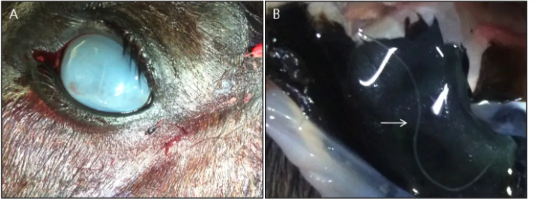
Fig. 1.The gross finding of worms in the eye of a horse. (A) Corneal opacity of the left eye. (B) Whitish parasite (arrow) moving in the aqueous humour of the left eye.
heart, lung, spleen, kidney, uterus, intestine, and eyes of the normal and unusual hosts (Mandal and Ray, 1994).
Ocular setariasis in equine is a disease resulting from erratic parasitism caused by Setaria spp., a genus of fi- laroid worms (Gangwar et al, 2008). The parasite ex- hibits migratory behaviour in unusual hosts such as horses, donkeys, or human beings, and immature worms can invade eyeballs through the vascular system (Sreedavi et al, 2002; Tuntivanich, 2011). In a study conducted by Tamilmahan et al (2013), occurrence of ocular setariasis in India was found in 138 of 242 (57.02%) eye defects surveyed in horses.
In the present study, ocular infection with S. digitata in a horse is described, and morphological character- istics of the nematodes are presented.
MATERIALS AND METHODS
In December 2013, a three-year-old male horse (B.W.
440 kg) was brought to the animal hospital of Chonbuk National University for anatomical study. Corneal opac- ity appeared in the left eye of the horse (Fig. 1A, B).
The examination of the eye revealed two worms ac- tively moving in the aqueous humour of the eye (Fig.
times with 0.2M cacodylate buffer (pH 7.3), fixed in 2.5% glutaraldehyde, and then post-fixed in 1% osmium tetroxide at 4°C. The specimens were dehydrated in a graded ethyl alcohol series, dried by CO2, osmium coat- ed, and then examined by SEM (S-4800, Hitachi) at 15 kV.
RESULTS
The worms were removed and measured; the male was 43 mm long, and the female was 55 mm long. No other parasites were found in the peritoneal cavity. The fine structure of Setaria spp. observed by light micro- scope and SEM enabled classification.
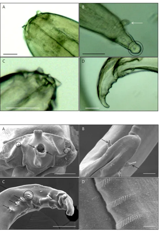
Anterior Part: The en face view of the head con- firmed that the mouth opening was surrounded by the peribuccal crown for each sex examined. The head of both male and female showed uniform roundness of the buccal opening and somewhat depressed dorso- and ven- tro-projections of the peribuccal crown at the tips (Fig.
2A, C). The buccal opening was round. The lateral lips were represented by one and two summits, respectively, of the mouth opening on both dorsal sides. The amphids were laterally situated, elevated, and had a dorsoventral
Fig. 2.Light micrographs show- ing fresh specimens of S. digitata for the lateral view from eye ball of horse. (A) Anterior part of the female. (B) Posterior part of the female. The tapering terminal end with a smooth knob (circle). Lateral appendage (arrow). (C) Anterior part of the male. (D) Posterior part of the male. Bar=50 m.
Fig. 3.Scanning electron micro- graphs of S. digitata. (A) Anterior part of the female. Depressed dor- so- and ventro-projection of the peribuccal crown (arrows). Amphids (circle). (B) Posterior part of the female. A pair of lateral appen- dages (arrows). (C) Posterior part of the male. Three pairs of post- cloacal papillae (arrows). A cloacal papilla (circle). (D) Latero-ventral view of the posterior part of the male. The transverse bands com- posed of longitudinal microstri- ations. Bar=5 m.
amphidal pore at the lower bottom (Fig. 3A).
Posterior Part: The lateral appendages were paired in both sexes (Figs. 2B, 3B, C). The posterior end of the female was a tapering terminus with a smooth knob (Fig. 2B). The male showed clear papillary arrangement;
three pairs of post-cloacal papillae, a pair of adcloacal papillae, and a central papilla just in front of the cloaca (Figs. 2D, 3C). The transverse bands were composed of longitudinal microstriations (Fig. 3D). Light microscopy
revealed the uteri in the female, but eggs were not identified.
DISCUSSION
In general, setariasis would be regarded as a benign and harmless parasitosis (Hornok et al, 2007). However, the condition can increase the risk of pathological
Many species of Setaria show no microfilarial perio- dicity, and only low numbers of microfilariae are pres- ent in the blood. The microfilariae are collected from the blood of the infected animal by mosquitoes as a vector. The development of infective larvae occurs in the thoracic muscles of mosquitoes such as Aedes, Culex, and Anopheles spp. (Soulsby, 1982). In horse and deer, the prepatent period is 7∼10 months, and that of S. marshalli is three months (Niimi et al, 1941;
Soulsby, 1982). In our study, eggs were not identified in the uterus of the identified female worm. In Korea, there is a lack of mosquitoes as vectors in winter.
Therefore, the parasitic infection likely occurred in summer of the same year.
The occurrence of equine intra-ocular parasite has been reported in some parts of the world. S. digitata is known to cause epizootic cerebrospinal nematodiasis in many domestic animals and can sometimes invade the eye (Jemelka, 1976; Soulsby, 1982; Radostits et al, 2000). Adult worms in the peritoneal cavity are gen- erally considered to be non-pathogenic in their natural hosts. However, the infection of microfilaria through mosquitoes can result in serious and fatal neuro- pathological disorders in animals such as sheep, goats, and horses when larvae of Setaria spp. migrate errati- cally to different parts of the body. During migration, the worms can accidentally get lodged in the eye chambers. Ocular setariasis is caused by the erratic mi- gration of S. digitata (Jemelka, 1976; Soulsby, 1982;
Nair et al, 1993; Shin et al, 2002). This infection is caused by an infected mosquito propagating the in- fective larvae into the horses’ eyes, where they enter and migrate in the aqueous humor. The adult S. digitata is commonly found in the peritoneal cavity of cattle.
However, the heterotrophic parasitism of the worm in our case caused detrimental changes to the cornea, lead-
This work was carried out with the support of
"Cooperative Research Program for Agriculture Science
& Technology Development (Project No. PJ010092)"
Rural Development Administration, Republic of Korea.
REFERENCES
Bazargani T, Eslami A, Gholami GR, Molai A, Ghafari Charati J, Dawoodi J, Ashrafi J. 2008. Cerebrospinal nem- atodiasis of cattle, sheep and goats in Iran. Iran J Parasitol 3: 16-20.
Gangwar AK, Sangeetha D, Singh HN, Singh A. 2008. Ocular fi- lariasis in equines. Indian Vet J 85: 547-548.
Hornok S, Genchi C, Bazzocchi C, Fok E, Farkas R. 2007.
Prevalence of Setaria equina microfilaraemia in horses in Hungary. Vet Record 161: 814-816.
Jemelka ED. 1976. Removal of Setaria digitata from the anterior chamber of the equine eye. Vet Med Small Anim Clin 71: 673-675.
Kim NS, Kim HC, Sim C, Ji JR, Kim NS, Park BK. 2010.
Congenital infection with Setaria digitata and Setaria marshalli in the thoracic cavity of a Korean calf: a case report. Veterinarni Medicina 55: 275-280.
Mandal SC, Ray S. 1994. Note on uncommon occurrence of Setaria labiatopapillosa. Indian Vet J 71: 726.
Marzok MA, Desouky AY. 2009. Ocular infection of donkeys (Equus asinus) with Setaria equine. Trop Anim Health Prod 41: 859-863.
Mohan K, Ananda KJ, Shridhar NB, Puttalakshmamma GC, D’Souza PE. 2009. Corneal opacity due to Setaria dig- itata in a Jersey cross-bred cow and its surgical mana- gement. Vet World 2: 69-70.
Nair KP, Pillai KM, George V. 1993. Setaria digitata as ectopic parasite in the cystic corpus luteum of a cow. J Vet Anim Sci 24: 92-93.
Nakano H, Tozuka M, Ikadai H, Ishida H, Goto R, Kudo N, Katayama Y, Muranaka M, Anzai T, Oyamada T. 2007.
Morphological survey of bovine Setaria in the abdominal cavities of cattle in Aomori and Kumamoto prefectures.
Jpn J Vet Med Sci 69: 413-415.
Niimi T, Nakamura N, Itagaki S. 1941. Chapter 2, parasitological
research, pp. 5-56. The 3rd Report the Sheep Cerebrospinal Filariosis Committee. Government Factory of Veterinary Serum in Korea (In Japanese).
Radostits OM, Gay CC, Blood DC, Hinchcliff KW. 2000. A text- book of the diseases of cattle, sheep, pigs, goats and horses. pp. 1378. Veterinary Medicine, 9th Edition:
Saunders Ltd, Philadelphia.
Rhee JK, Choi EY, Park BK, Jang BG. 1994. Application of scanning electron microscopy in assessing the prevalence of some Setaria species in Korean cattle. Korean J Parasitol 32: 1-6.
Shin SS, Cho KO, Wee SH. 2002. Ocular infection of cattle with Setaria digitata. J Vet Med Sci 64: 7-10.
Shoho C, Uni S. 1977. Scanning electron microscopy (SEM) of some Setaria species (Filarioidea, Nematoda). Z Parasitenk 53: 93-104.
Soulsby EJL. 1982. Helminthis, Arthropods and Protozoa of do- mesticated animals, pp: 316-319. 7th ed. Bailliere,
Tindall, London. 1982.
Sreedavi C, Sudhakar K, Murthy PR, Prasad V. 2002. Clinical microfilariasis in a horse: a case report. Indian Vet J 79:
487-488.
Tamilmahan P, Zama MMS, Pathak R, Muneeswaran NS, Karthik K. 2013. A retrospective study ocular occurrence in do- mestic animals: 799 cases. Vet World 6: 274-276.
Taylor MA, Coop RL, Wall RL. 2007. Veterinary parasitology, Blackwell, Oxford; 2007.
Tung KC, Lai CH, Ool HK, Yang CH, Wang JS. 2003.
Cerebrospinal setariosis with Setaria marshalli and Setaria digitata infection in cattle. J Vet Med Sci 65:
977-983.
Tuntivanich N, Tiawsirisup S, Tuntivanich P. 2011. Success of Anterior Chamber Paracentesis as a treatment for Ocular Setariasis in Equine Eye: Case Report. J Equine Vet Sci 31: 8-12.