서 론
전정유발전위(vestibular evoked potential)는 다른 유발전위들과는 다르게 사람에서 유도하기가 매우 어렵 다. 이는 미로( l a b y r i n t h )에서 유발되는 반응은 양측에서 기인하므로 병측이 어디인지 구분하기 어려운 한계가 있 고, 유발되는 반응이 전정기관에서 유래되는지에 대한 논 란이 있으며 사용되는 방법이나 기구가 매우 복잡하기 때 문에 임상에서 적용되기가 어렵기 때문이다.1 - 3
매우 큰 소리가 전정기관을 자극할 수 있는 것으로 알려 져 있으며, 이 자극을 통하여 유발되는 반응을 근육에서 기록할 수 있다는 것이 밝혀져 있다.4 - 8이 반응을 전정유 발근육전위(Vestibular Evoked Myogenic Potential:
V E M P )라고 부르며 비교적 쉽게 일관된 파형을 유도할 수 있기 때문에 임상의 각 분야에 응용되고 있다. 저자들 은 V E M P의 기원, 신경전달경로, 파형유도방법, 파형의 특성 및 그 임상적 유용성을 중심으로 서술하고자 한다.
V E M P의 기원 및 신경전달로
전정기관의 주머니(saccule) 부위는 내이에 있는 감각 기관의 하나로써 주로 중력 등의 선형 가속도에 반응하여 공간적인 지남력에 관여하는 기관으로 알려져 있다( F i g . 1). 전정 주머니 부위는 하등동물에서는 소리에 대한 감각 기이며,9 사람과 같은 포유류에서는 균형감각을 유지하는 것이 주된 기능이지만 어느 정도의 소리에 대한 감지 기 능도 존재한다고 알려져 있으며, 매우 큰 소리는 전정기 관을 자극할 수 있음이 확인되었다.1 0Bickford 등은 정상 인에서 큰 소리의 째깍음( c l i c k )에 의해 유발되고 뒷통수 점(inion) 부분 기록되는 파형을 처음으로 보고하였으 며,1 1초기에는 이 반응이 소뇌벌레에 존재하는 음성자극 에 반응하는 부위에서 유래하는 것으로 해석하였으나, 후 에 여러 연구를 통해 목근육의 긴장도가 파형형성에 매우 중요하다는 것이 밝혀졌고 그 기원이 뒤통수점 근처의 목 근육이라는 것이 확인되었다.1 1 - 1 3
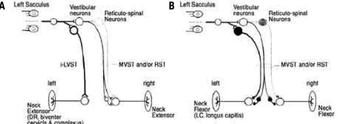
V E M P의 신경전달회로 구성은 주로 동물실험을 통한 연구되었다. 소리자극이 전정주머니를 자극하면 전정신경 및 신경절을 통하여 뇌줄기의 전정신경핵으로 전달되며 내측전정척수통로(medial vestibulospinal tract)을 통 하여 경부근육에 전달되고 외측전정척수신경통로( l a t e r a l vestibulospinal tract)을 통하여 하지의 근육으로 전달
된다.4 , 1 4 - 1 6 Fig .2는 고양이에서 연구된 주머니목반사
전정유발근육전위
강원대학교 의과대학 신경과학교실, 이비인후과학교실
김성훈・남의철
Vestibular Evoked Myogenic Potential
Sung Hun Kim, M.D., Eui Cheol Nam, M.D.*
Department of Neurology, Otolaryngology* College of Medicine, Kangwon National University
Loud click or tone burst sound can activate vestibular receptor and evoke reflex changes in tonic electromyographic activity within the stenocleidomastoid muscles. This reflex is assumed to originate in the saccule, the afferent pathways being the inferior vestibular nerve, and the efferent pathways the vestibulospinal tract. Averaging these muscular responses allows vestibular evoked myogenic potentials (VEMP) to be obtained. The earliest response ipsilateral to a loud click, p13n23, is dependent upon vestibular activation, specifically saccular afferents. These new techniques are beginning to be applied clinically in the patient of vestibular neuritis, Meniere’s disease, acoustic neuromas, Tullio phe- nomenon, etc. VEMP recording will provide both a straightforward non-invasive exploration of each vestibule indepen- dently and an attractive method by which to explore otolithic receptors and vestibulospinal pathways.
Key Words: Vestibular evoked myogenic potentials
Address for correspondence Kim Sung Hun, M.D.
Department of Neurology, College of Medicine, Kangwon National Univesity Dept. of Neurology, Kangwon National University Hospital, Chunchon, Kangwondo, 200-947
Tel: +82-33-258-2413 Fax: +82-33-257-4636 E-mail: [email protected]
(sacculocollic reflex)를 도시한 그림이다.
VEMP 유발 방법
표면 근전도 전극을 경부 앞쪽의 목빗근( s t e r n o c l e i- domastoid) 중앙부위에 위치시킨다. 환자의 자세는 앙와 위( s u p i n e )로 유지시키고 환자의 목을 앞쪽으로 굴곡 시 켜 목빗근의 긴장상태로 유지하게 한다. 목빗근을 긴장시 키는 다른 자세로는 환자를 않게 하고 턱을 반대쪽 어깨 쪽으로 돌리게 하는 것이다. 피검자는 소리자극이 들어가 는 동안에는 자세를 유지하게 하며 중간에는 휴식을 하게 한다. 중요한 것은 근근장이 유지되지 않으면 V E M P가 유발되지 않는 다는 점이며 V E M P의 크기는 근육의 긴장
도와 비례하게 된다.
큰 강도의 째깍음이나 음조터짐음(tone burst)이 사용 되며(90~100DB nHL이나 그 이상), 양측귀에 0 . 2초의 간격으로 자극한다.1 7 , 1 8음자극의 빈도는 500~1000Hz 정 도로 한다. 흉골부위에 기준전극을 붙이고 이마부위에 접 지전극을 위치시킨다. 근육유발전위는 증폭을 거치고 통 과거르개(band filter, 1~5Hz)를 통과하며 적어도 1 0 0번 의 평균화( a v e r a g i n g )를 거친다.
V E M P를 유발시키기 위해서는 고강도의 소리자극이 필요하기 때문에 음량조절기능을 가진 헤드폰을 사용하여 야 한다. VEMP가 확실히 유발되었는지를 확인하기 위해 서 적어도 3번의 검사가 필요하나 첫 번째, 두 번째 검사 에서 확실한 파형이 유발된다면 세 번째는 생략해도 무방
Figure 1. Schematic of the utricle and saccule. These sensory organs in the inner ear primarily respond to linear acceleration such as due to orientation to gravity, but the saccule is also somewhat sensitive to sound. This is the basis of the VEMP test
Figure 2. Schematic diagram of disynaptic and trisynaptic excitatory (A) and inhibitory (B) sacculo-neck motoneuron connections.
Saccular nerve contacts excitatory (s) and inhibitory (l) 2nd-order vestibular neurons. These project directly or indirectly (via spinal cord interneurons and/or reticulospinal neurons) to neck extensor and flexor moroneurons. Thick lines: strong connections. Thin lines: weaker connections. Locations of excitatory and inhibitory interneurons have not been identified, nor have midline crossing points within medial vestibulospinal tract (MVST) and/or reticulospinal tract. Only ipsilateral excitatory pathway to neck extensors projects through ipsilateral lateral vestibulospianal tract (i-LVST). Note qualitatively bilaterally symmetrical innervation pattern of sacculo-neck reflexes.
A B
하다. 일반적으로 V E M P는 1분 이내에 비교적 쉽게 파형 을 얻을 수 있다. 이명이 있는 환자는 검사에 상대적인 금 기이다.
VEMP 유발의 다른 방법
1. 골전도 V E M P
두개골두드림(skull tap)이나 골전도 음조도 V E M P를 유도할 수 있다. 두드림은 앞이마나 옆두개골 부위를 자 극할 수 있으며 자극 위치에 따라 유발되는 극성( p o l a r i- ty) 등이 변화할 수 있다. 골전도 음조터짐음 역시 V E M P 를 유발할 수 있고 200Hz 정도의 주파수를 이용한다. 임 상에서 흔히 쓰이는 골진동기는 충분한 강도를 얻기 위해 추가적인 증폭장치가 필요하다. 일반적으로 골전도에 의 해 유발되는 V E M P는 일반적인 V E M P에 비해 편향화 되 지는 않는다.1 9
2. 직류(galvanic) VEMP
직류자극도 V E M P를 생성한다.2 0이 방법은 전정주머니 를 우회하기 때문에 뇌간 및 전정신경 근위부의 병변을 평가하는데 유리하나 방법 및 유용성 등이 정립되어 있지 않기 때문에 연구가 더 필요하다.
정상 VEMP 파형 및 특징
1. 정상 VEMP 파형
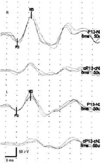
Colebatch 등은 째깍음을 이용하여 목빗근에서 기록한 VEMP 파형을 정립하였다.4 파형은 이중모양( b i p h a s i c ) 을 취하며 처음 13 ms 근처에서 기록되는 양성( p o s i t i v e ) 파형과 23 ms근처에서 기록되는 음성파형( p 1 3 n 2 3 )으로 이루어져 있다(Fig. 3). 파형은 음성자극의 동측 근육에서 기록된다.2 1VEMP 파형의 진폭은 기록 동안의 근육긴장 도와 비례하며 청각장애 정도와는 관련이 없으나 전정신 경이 손상된 경우에는 소실된다. 전정신경 구심로에 대한 직접기록 결과에 의하면 전정주머니 구심로가 가장 소리 에 민감한 것으로 확인되었다. 이는 등자뼈( s t a p e s )의 음 직임에 의해 유발된 속림프의 전류가 해부학적으로 가장 가까이에 위치한 전정주머니를 자극하기 때문인 것으로 보여진다.2 2 , 2 3단일운동단위(single motor unit) 기록 연 구에 따르면 VEMP 반응은 목빗근 운동신경( m o t o n e u- ron) 발사( f i r i n g )의 짧은 기간 동안의 억제에 의해 일어 나는 것으로 알려져 있으며, 이는 고양이에서 전정주머니 를 선택적으로 자극했을 때 목빗근 운동신경에서 억제시 냅후전위(inhibitory postsynaptic potential)가관찰되 는 결과와 일치한다.2 4 , 2 5
VEMP 검사가 완전히 청각이 소실된 사람에서도 유발 될 수 있으나 이는 감각신경성난청( s e n s o r i n e u r a l hearing loss) 환자에서다. 전음난청(conductive hear-
ing loss)이 있는 환자에서는 전정주머니에 소리 전달 과 정 자체에 문제가 있을 수 있기 때문에 VEMP 파형이 유 발되지 않을 수 있으므로 V E M P를 해석할 때 청력도 검 사 및 뼈전도, 공기전도 검사를 함께 고려하여야 한다.
2. 정상 기준
V E M P의 파형이 형성되지 않거나 p13-n23 진폭이 양 측에서 2배 이상의 차이가 날 때는 명백한 이상 소견이다.
연구에 따라서는 p 1 3의 잠복기 및 진폭을 각 실험실의 정 상치와 비교하여 이상 유무를 판단하기도 한다. VEMP 파형은 7 0세가 넘은 환자에서는 진폭이 작아지고 유발 역 치가 커지는 경향을 보인다.
임상적 유용성
1. 전정신경염
전정신경염의 경우 전정신경의 아래분할( i n f e r i o r d i v i s i o n )은 잘 침범되지 않고,2 6 전정주머니는 전정신경 의 아래분할과 연결되기 때문에 VEMP 파형의 이상이 관
Figure 3. 100dB(nHL) tone burst to each ear evoked normal p13n23 potentials in the ipisilateral sternocleidomastoid mus- cles.
찰되지 않을 것을 예측할 수 있다. 실제로 Ochi 등의 연구 에 따르면 8명의 급성 시기 전정신경염 환자에서 V E M P 를 시행한 결과 2명에서만 파형 형성이 관찰되지 않았으 며 1명에서는 1 0개월 정도를 추적관찰한 결과 VEMP 파 형이 점차적으로 회복됨을 보고하였다(Fig. 4).2 7 1 8명의 환자를 대상으로 골전도 V E M P와 째깍음 V E M P를 비교 한 연구에서는 골전도 V E M P에서는 1 0명의 환자에서 이 상 반응을 보였으나 째깍음 V E M P에서는 4명에서만 이상 반응을 보였다.2 8이 결과는 골전도 V E M P의 경우 전정주 머니의 기능을 평가 한 다기 보다 전정타원낭( u t r i c l e )의 기능을 반영한다는 것을 시사하는 것이다.
2. 양측성 전정신경 손상
아미노글라이코사이드 내이독성 등의 양측성 전정신경 장애 환자에서 VEMP 결과는 잘 알려져 있지는 않다. 7 명의 양측성 신경섬유종증 환자를 대상으로 한 연구에서 5명에서 칼로리 검사에서 이상이 발견되었지만 V E M P에 서는 1명에서만이 이상 소견을 보였다.2 9
3. Meniere 병
Rauch 등의 보고에 따르면 Meniere 병에서 V E M P의 자극 역치의 변화가 관찰되며, 이는 전정주머니의 수포확 장으로 인한 기능저하에 기인한다고 해석하였다. 또한 증 상이 없는 쪽 귀에서도 경미한 이상이 관찰되었다.3 0
Meniere 병 초기에는 전정주머니 수포확장으로 인하여 V E M P의 진폭이 증가할 수도 있지만 병이 진행되면서 파 형이 점차로 소실되게 되며 이는 전정주머니가 손상되었 음을 의미한다. 최근의 보고에 의하면 furosemide 투여 시 VEMP 파형이 커지는 것을 관찰함으로써 Meniere 병 의 진단에 도움이 될 수 있다는 것이 알려졌다.3 1
4. 전정신경종양
Murofish 등의 연구에 따르면 전전신경종양 환자의 8 0 %에서 VEMP 파형의 이상을 관찰할 수 있었고 그 중 몇 환자는 정상 칼로리 반응을 보이는 환자였다.3 2뇌줄기 청각유발전위(brainstem auditory evoked potential) 가 정상인 2명의 안뜰신경집종(acoustic neuroma) 환자 에서 VEMP 파형의 소실이 보고되었으며,3 3이는 V E M P 검사가 특히 전정신경 아래분할의 검사에 민감도가 높다 는 것을 시사하는 것이다.
5. Tullio 현상
Tullio 현상이란 소리에 의해 유발되는 전정계의 증상 및 증후군을 일컫는 것이다. 이 환자들은 큰 소리 등에 의 하여 현훈 등의 전정 증상이 유발될 수 있다. 현재까지 다 양한 원인이 알려져 있지만 대부분의 경우에는 상반고리 관(superior semicircular canal) 뼈지붕(bony roof)의 벌어짐( d e h i s c e n c e )이 원인인 것으로 알려져 있으며 이 벌어짐이 소리에너지를 전정신경에 전달하게 된다.3 4 Colebatch 등은 소리에 의해 유발되는 안구진탕 등의 증 상이 있는 Tullio 현상 환자들에서 째깍음에 의해 유발되 는 VEMP 파형의 역치가 증가함을 보고하였다(Fig. 5).3 5 Brantberg 등의 보고에서도 상반고리관 뼈지붕 벌어짐 환자에서 째깍음이나 음조터짐음 자극에 의해 VEMP 역 치가 감소하였다.3 6또한 방사선 검사 소견상 상반고리관 벌어짐을 보이는 4명의 Tullio 현상 환자에서 째깍음에 의해 유발되는 V E M P의 역치가 증상이 있는 쪽 귀에서 병적으로 감소된다는 것이 알려져 있다.3 7비록 방사선 소 견이 양측성의 이상을 보이지만, 역치의 감소는 증상이 있는 귀쪽에서만 관찰된다. VEMP 소견상의 역치의 감소 가 환자의 증상과 잘 연관되기 때문에 상반고리관 결손 진단이나 치료에 있어 V E M P검사는 매우 유용하며, 특히 수술적인 치료시 기능적인 측면을 평가하는 데 있어서 꼭 필요한 검사이다.
6. 기타질환
뇌줄기나 소뇌를 침범하는 임상적으로 명확한 다발성 경화증 환자에서 VEMP 잠복기가 연장되는 등의 이상 소 견이 관찰되었다.3 8 , 3 9 VEMP 파형은 또한 연축기운목 (spasmodic torticollis) 환자에서 비대칭 소견을 보인 다.4 07명의 뇌줄기 경색 환자에서 V E M P를 시행한 C h e n 등의 보고에 따르면 7 9 %에서 이상 소견을 얻을 수 있었 다.4 1 VEMP 전달로는 주로 연수(medulla) 등의 뇌줄기 아래쪽에 있고 칼로리 검사는 교뇌(pons) 등의 뇌줄기 위 Figure 4. VEMP waveforms were recorded at the initial stage
in the right ear (A) 10 months after the onset in the right ear (B) and in the left ear (C) The stimulus intensity is 105 dB.
A B
C
쪽에 전달로가 있기 때문에 두 검사는 뇌줄기의 기능을 평가하는 데 서로 보완적으로 작용한다.
결 론
대부분의 전정신경 검사들이 반고리관의 기능을 평가하 는 반면 VEMP 검사는 전정주머니 수용체의 기능을 반영 하여 전정 기능 검사에서 추가적인 정보를 얻을 수 있다.
V E M P의 작극 역치나 잠복기, 파형의 진폭이 이상 유무 를 판단하는 지표로써 이용된다. VEMP 검사는 목빗근의 수의적인 수축이 필요하므로 협조가 되지 않는 환자나 심 한 현훈을 호소하는 환자에서는 검사가 어려울 수도 있 다. 현재까지는 Tullio 현상 환자에서 V E M P의 유용성이 확립되어 있으며 다양한 전정계 질환 환자에서 유용성이 보고되고 있다.
전정 수용체에서 전정신경을 거쳐 척수로 내려가는 신경 전달로는 아직까지 잘 알려져 있지 않고 지속적인 연구가 필요한 분야이다. 그런 측면에서 VEMP 검사는 전정, 평형 수용체(otolithic receptor), 전정척수로의 기능을 연구하 고 평하가는 데 있어 매우 유망한 방법이라고 할 수 있다.
REFERENCES
01. Pirodda E, Ghdini S, Zemeti MA. Investigations into vestibular evoked responses. Acta Otolaryngol 1 9 8 7 ; 104:77-84.
02. Durrant JD, Furman JMR. Long-latency rotational evoked potentials in subjects with and without bilateral vestibular loss. Electroencephalogr Clin Neurophysiol 1988;71:251- 256.
03. Elidan J, Leibner E, Freeman S, Sela M, Nitzan M, Sohmer H. Short and middle latency vestibular evoked responses to acceleration in man. Electroencephalogr Clin Neurophysiol 1991;80:140-145.
04. Colebatch JG, Halmagyi GM, Skuse NF. Myogenic poten- tials generated by a click-evoked vestibulocolic reflex. J Neurol Neurosurg Psychiatry 1994;57:190-197.
05. Di Lazzaro V, Quartarone A, Higuchi K, Rothwell JC.
Short-latency trigemino-cervical reflexes in man. E x p Brain Res 1995;102:474-82.
06. Ferber-Viart C, Dubreuil C, Duclaux R, Collet L. Reflexe sonomoteur vestibulaire dans les neurinomes de l’a c o u s- tique. Rev Laryngol Otol Rhinol 1995;116:47-51.
07. Halmagyi GM, Colebatch JG. Vestibular evoked myo- genic potentials in the sternomastoid muscle are not of lat- eral canal origin. Acta Otolaryngol 1995;Suppl 520:1-3.
08. Robertson DD, Ireland DJ. Vestibular evoked myogenic potentials. J Otolaryngol 1995;24:3-7.
09. Lowenstein O, Roberts TDM. The localization and anam- ysis of the responses to vibration from the isolated elasmo- branch labyrinth. A contribution to the problem of the evoluation of hearing in vertebrates. J Physiol 1 9 5 1 ; 114:471-489.
10. Von Bekesy G. Uber akustishe Reizung des vestibularap- parates. Arch F D Ges Physiol 1935;236:59.
11. Bickford RG, Jacobson JL, Cody DTR. Averaged poten- tials to sound and other stimuli in man. Ann NY Acad Sci 1964;112:204-223.
12. Townsend GL, Cody DTR. The averaged inion response evoked by acoustic stimulation: its relation to the saccule.
Ann Otol 1971;80:121-132.
13. Lim CL, Clouston P, Sheean G, Yiannikas C. The influ- ence of voluntary EMG activity and click intensity on the vestibular click evoked potential. Muscle Nerve 1 9 9 5 ; 1 8 : 1210-1213.
14. Kushiro K, Zakir M, Sato H, et al. Saccular and utricular inputs to single vestibular neurons in cats. Exp Brain Res 2000;131:406-415.
15. Murofushi T, Halmagyi GM, Yavor RA, Colebatch JG.
Absent vestibular evoked myogenic potentials in vestibu- lar neurolabyrinthitis. An indicator of inferior vestibular nerve involvement? Arch Otolaryngol Head Neck Surg 1996;122:845-849.
16. Uchino Y. Sato H, Sasaki M, et al. Sacculocollic reflex arcs in cats J Neurophysiol 1997;77:3003-3012.
Figure 5. Click-evoked vestibular-evoked myogenic potentials for a patient with clinical evidence of the Tullio phenomenon (sound activation of the vestibular apparatus) on the left side. The two sides show similar responses with the loudest stimulus (100dB nHL) but the threshold is abnormally reduced (65dB) on the affected side, whereas the unaffected side had a (normal) threshold of 90dB. This patient had radiological evidence of dehiscence of the superior semicircular canals bilaterally, but the click threshold correctly identified the symptomatic side. SCM, Sternocleidomastoid muscles.
17. Cheng PW, Huang TW, Young YH. The influence of clicks versus short tone bursts on the vestibular evoked myogenic potentials Ear Hear 2003;24:195-197.
18. Murofushi T, Matsuzaki M, Wu CH. Short tone burst- evoked myogenic potentials on the sternocleidomastoid muscle: are these potentials also of vestibular origin? Arch Otolaryngol Head Neck Surg 1999;125:660-664.
19. Sheykholeslami K, Murofushi T, Kermany MH, Kaga K.
Bone-conducted evoked myogenic potentials from the sternocleidomastoid muscle. Acta Otolaryngol 2 0 0 0 ; 1 2 0 : 731-734.
20. Watson SR, Colebatch JG. Vestibulocollic reflexes evoked by short-duration galvanic stimulation in man. J Physiol 1998;513:587-597.
21. Li MW, Houlden D, Tomlinson RD. Click evoked EMG responses on sternocleidomastoid muscles: characteristics in normal subjects. J Vestib Res 1999; 9:327-334.
22. McCue MP, Guinan JJ. Acoustically responsive fibers in the vestibular nerve of the cat. J Neurosci 1994;14:6058- 6070.
23. Murofushi T, Curthoys IS, Topple AN, et al. Responses of guinea pig primary neurons to clicks. Exp Brain Res 1995;103:174-178.
24. Colebatch JG, Rothwell JC. Vestibular-evoked EMG responses in human neck muscles. J Physiol 1993;473:18.
25. Kushiro K, Zakir M, Ogawa Y, Sato H, Uchino Y.
Saccular and utricular inputs to sternocleidomastoid motoneurons of decerebrate cats. Exp Brain Res 1999;126:
410-416.
26. Fetter M, Dichgans J. Vestibular neuritis spares the inferi- or division of the vestibular nerve. B r a i n 1 9 9 6 ; 1 1 9 : 7 5 5 - 763.
27. OCHI K, Ohashi T, Watanabe S. Vestibular-evoked myo- genic potential in patients with unilateral vestibular neuri- tis: abnormal VEMP and its recovery. J Laryngol Otol 2003;117:104-108.
28. Brantberg K, Tribukait A, Fransson PA. Vestibular evoked myogenic potentials in response to skull taps for patients with vestibular neuritis. J Vestib Res 2003;13:121-130.
29. Wang CP, Hsu WC, Young YH. Vestibular evoked myo- genic potentials in neurofibromatosis 2. Ann Otol Rhinol Laryngol 2005;114:69-73.
30. Rauch SD, Zhou G, Kujawa SG, Guinan JJ, Herrmann BS.
Vestibular evoked myogenic potentials show altered tun- ing in patients with Meniere’s disease. Otol Neurotol 2004;25:333-338.
31. Seo T, Node M, Yukimasa A, Sakagami M. Furosemide loading vestibular evoked myogenic potential for unilater- al Meniere’s disease. Otol Neurotol 2003;24:283-288.
32. Murofushi T, Matsuzaki M, Mizuno M. Vestibular evoked myogenic potentials in patients with acoustic neuromas.
Arch Otolaryngol Head Neck Surg 1998;124:509-512.
33. Matsuzaki M, Murofushi T, Mizuno M. Vestibular evoked myogenic potentials in acoustic tumor patients with nor- mal auditory brainstem responses. Eur Arch Otorhinolaryngol 1999;256:1-4.
34. Minor LB, Solomon D, Zinreich JS, Zee DS. Sound- and/or pressure-induced vertigo due to bone dehiscence of the superior semicircular canal. Arch Otolaryngol Head Neck Surg 1998;124:249-258.
35. Colebatch JG, Day BL, Bronstein AM, et al. Vestibular hypersensitivity to clicks is characteristic of the Tullio p h e n o m e n o n . J Neurol Neurosurg Psychiatry 1 9 9 8 ; 65:670-678.
36. Brantberg K, Bergenius J, Tribukait A. Vestibular-evoked myogenic potentials in patients with dehiscence of the superior semicircular canal. Acta Otolaryngol 1 9 9 9 ; 119:633-640.
37. Watson SRD, Halmagyi GM, Colebatch JG. Vestibular hypersensitivity to sound (Tullio phenomenon): structural and functional assessment. Neurology 2000;54:722-728.
38. Shimizu K, Murofushi T, Sakurai M, Halmagyi M.
Vestibular evoked myogenic potentials in multiple sclero- sis. J Neurol Neurosurg Psychiatry 2000;69:276-277.
39. Versino M, Colnaghi S, Callieco R, Bergamaschi R, Romani, Cosi V. Vestibular evoked myogenic potentials in multiple sclerosis patients. Clin Neurophysiol 2003;113:1464-1469.
40. Colebatch JG, Di Lazzaro V, Quartarone A, Rothwell JC, Gresty M. Click-evoked vestibulocollic reflexes in torti- collis. Mov Disord 1995;10:455-459.
41. Chen CH, Young YH. Vestibular evoked myogenic poten- tials in brainstem stroke. Laryngoscope 2 0 0 3 ; 1 1 3 : 9 9 0 - 9 9 3 .