596 ORIGINAL ARTICLE
DOI 10.4070 / kcj.2008.38.11.596
Print ISSN 1738-5520 / On-line ISSN 1738-5555 Copyright ⓒ 2008 The Korean Society of Cardiology
Transient Left Ventricular Dysfunction After Percutaneous Patent Ductus Arteriosus Closure in Children
Yeo Hyang Kim, MD
1, Hee Joung Choi, MD
2, Yongkeun Cho, MD
3, Sang Bum Lee, MD
4and Myung Chul Hyun, MD
41
Department of Pediatrics, School of Medicine, Keimyung University, Daegu,
2
Department of Pediatrics, College of Medicine, Pochon CHA University, Gumi,
3
Department of Internal Medicine and
4Pediatrics, School of Medicine, Kyungpook National University, Daegu, Korea
ABSTRACT
Background and Objectives: The goal of this study was to assess changes in left ventricular (LV) function and to identify pre-closure factors associated with LV dysfunction {fractional shortening (FS) below 29%} after trans- catheter patent ductus arteriosus (PDA) closure. Subjects and Methods: Forty-three pediatric patients with PDAs underwent cardiac catheterization for hemodynamic studies and intervention. Doppler echocardiography was per- formed at pre-closure, post-closure, and follow-up. Results: S’ and A’ of the septum and mitral annulus were sig- nificantly decreased at post-closure and follow-up, respectively. In five of eight patients with Qp/Qs ratios over 1.60 and Pp/Ps ratios over 0.32 at pre-closure, the FS was decreased below 29% at post-closure. Qp/Qs ratio over 1.60 and Pp/Ps ratio over 0.32 at pre-closure had a sensitivity of 86% and a specificity of 84% for predicting FS to be below 29% at post-closure. Conclusion: Larger amounts of pre-closure left-to-right shunting and higher pulmonary artery pressure were associated with an increased likelihood of FS <29% after closure. The results of this study suggest that serial assessments of ventricular function are needed after PDA occlusion in patients with high Qp/Qs and Pp/Ps ratios. (Korean Circ J 2008;38:596-600)
KEY WORDS: Ductus arteriosus, patent; Catheter closure; Ventricular dysfunction.
Introduction
Patent ductus arteriosus (PDA) is a congenital heart disease that results in left-to-right shunting; it is asso- ciated with increased left ventricular (LV) preload.
1)2)Over the last 30 years, treatment of most PDAs has been accomplished by transcatheter interventional closure using various devices, such as detachable coils, Nit-Oc- cluders, and Amplatzer duct occluders,
3-8)although vid- eo-assisted thoracoscopic surgery has been introduced for large PDAs.
9)Previous studies have reported an im- mediate change in LV performance followed by recovery within a few months after successful transcatheter clo- sure of PDAs in children.
2)10)However, some cases with larger ductus size and higher pulmonary pressure had immediate LV dysfunction {fractional shortening (FS) below 29%} after interventional PDA closure.
Echocardiography is the most commonly used meth- od for evaluating LV function; such studies include 2- dimensional (2-D), M-mode, conventional Doppler, and tissue Doppler technology. The purpose of this study was to investigate changes in LV function before and af- ter percutaneous transcatheter PDA closure using con- ventional and tissue Doppler echocardiography (TDE).
In addition, we evaluated pre-closure factors associated with immediate LV dysfunction after the procedure.
Subjects and Methods
Study population
Forty-three pediatric patients were enrolled in this study. They were all diagnosed with PDAs, and they all underwent successful percutaneous transcatheter PDA closure between January 2005 and May 2007. Patients of unsuitable size for interventional closure or with ad- ditional congenital heart defects were excluded. Echo- cardiography was performed to detect combined cardiac defects and to analyze ventricular function. Study ap- proval was obtained from the institutional ethics com- mittee at the Kyungpook National University School of Medicine, and written informed consent was obtained
Received: July 9, 2008
Revision Received: September 1, 2008 Accepted: September 17, 2008
Correspondence: Myung Chul Hyun, MD, Department of Pediatrics, School of Medicine, Kyungpook National University, 200 Dongdeong-no, Jung- gu, Daegu 700-721, Korea
Tel: 82-53-420-5704, Fax: 82-53-425-6683
E-mail: mchyun@mail.knu.ac.kr
Yeo Hyang Kim, et al.
·597
from the patients’ parents in all cases.
Two-dimensional and tissue Doppler echocardiog- raphy
Echocardiography was performed with the patient in the supine position using an Acuson Sequoia (Siemens Medical Solution, Mountain View, CA, USA) with a 4 MHz transducer at pre-closure, post-closure (the next or second day after the procedure), and follow-up (3 months after the procedure). Some patients required sedation for echocardiography. The LV end-diastolic dimension (LVEDD) and LV end-systolic dimension (LVESD) were measured in the parasternal long-axis view. Fractional shortening (FS) was calculated as {(LVEDD-LVESD)/
LVEDD}×100. We defined FS below 29% as “Abnor- mal”
11)and considered FS changes 10% greater than the pre-closure level as “Change”.
2)For diastolic functional analysis, the mitral inflow signal was acquired from three cardiac cycles in the apical four-chamber view. The puls- ed Doppler sample volume was 2 mm, placed at the mi- tral valve tip. The early peak flow velocity (E) and atrial filling velocity (A) were measured three times and av- eraged, and the E/A ratio was calculated. In addition, the peak systolic S’, peak early diastolic E’, and peak late diastolic A’ velocities were obtained from the TDE using the pulsed wave Doppler method at the septal cor- ner, and the lateral portion of the mitral annulus and the E’/A’ and E/E’ ratios were calculated. The TDE was recorded from the apical four-chamber view, and the sample volume was 2 mm.
Cardiac catheterization
All patients received a first generation cephalosporin injection (50 mg/kg) 30 minutes before the procedure, were sedated with ketamine, midazolam, or propofol during the procedure, and also received a dose of 50- 100 unit/kg of heparin after vein and artery puncture.
Hemodynamic information was obtained from a stand- ard cardiac catheterization, and the pulmonary blood flow/systemic blood flow (Qp/Qs), pulmonary artery pressure/systemic artery pressure (Pp/Ps), and pulmo- nary artery resistance/systemic artery resistance (Rp/Rs) ratios were calculated before PDA closure using the Fick principle. A lateral aortogram was performed at the distal aortic arch before PDA closure, followed by PDA sizing in an appropriate image view. Percutaneous transcatheter closure of the PDA was performed using antegrade or retrograde technique, according to the device type. Near- ly complete or complete closure was confirmed by repeat aortogram after the procedure.
Statistical analysis
Statistical analysis was carried out using the statistical package for social science (SPSS) software program for Windows (version 12; SPSS, Chicago, Illinois, USA).
Continuous variables were compared using the Student t-test and are expressed as mean values±standard de- viation. Changes in echocardiographic parameters were compared using the paired t-test. Correlation between two continuous variables was assessed using linear re- gression analysis. Multiple stepwise linear regression analysis was used to identify pre-closure determinants of post-closure FS below 29%. Univariate analysis was performed first, and a p<0.05 was considered statisti- cally significant. Multivariate analysis was then perform- ed using variables that were significant on univariate analysis; a p<0.1 was considered significant. The best cutoff value for a FS below 29% post-closure was iden- tified based on the receiver operating characteristic curve analysis.
Results
Clinical characteristics
The clinical characteristics of the patients are summa- rized in Table 1. There were 14 boys and 29 girls, with a median age of 24 months at the time of the procedure (range 6-97 months). The mean narrowest PDA diam- eter measured by the aortogram was 2.0±1.3 mm (range 1.9-5.5 mm). All patients underwent percutaneous trans- catheter closure using devices such as Amplatzer duct occluders, Nit-Occluders, and detachable coils. PDA oc- clusions were performed through an antegrade approach using the Amplatzer duct occluder (AGA Medical Corp., Golden Valley, MN, USA) or Nit-Occluder in 26 pa- tients and a retrograde approach using a detachable coil in 13 patients. Both the Nit-Occluder and the detach- able coil were used in four patients.
Two-dimensional and tissue Doppler echocardiog- raphy
Compared to pre-closure values, the FS was signif-
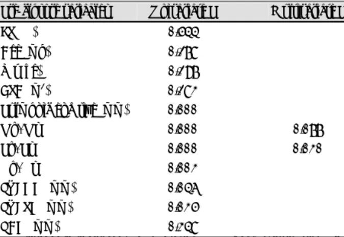
Table 1. Clinical characteristics of patients
PDA patients (n=43) Range
Age (mo) 24.1±19.1 6-97
Weight (kg) 11.7±4.0 5.7-17.8
BSA (m
2) 0.4±0.1 0.19-0.59
Pulmonic end size (mm) 2.0±1.3 1.9-5.9 Qp/Qs 1.43±0.61 1-3.30 Pp/Ps 0.30±0.18 0.11-0.91 Rp/Rs 0.13±0.10 0.02-0.44 Device for intervention
Amplatzer 15
Nit occluder 11 Detachable coil 13
Nit occluder+detachable coil 04
PDA: patent ductus arteriosus, BSA: body surface area, Qp/Qs:
pulmonary blood flow/systemic blood flow, Pp/Ps: pulmonary artery pressure/systemic artery pressure, Rp/Rs: pulmonary artery resistance/
systemic artery resistance
598 · Transient LV Dysfunction After PDA Closure
icantly decreased post-closure (p<0.01), but recovered at follow-up when it returned to pre-closure values (Ta- ble 2). The LVEDD was significantly decreased post- closure (p<0.01) and showed a persistent decrease at fol- low-up. However, there was no significant difference be-
tween the post-closure and follow-up values. The LVESD was no different in a comparison of pre- and post-clo- sure values, despite the significant decrease at follow- up. There was no significant interval change in the E, A, or E/A ratio based on the mitral inflow signal (p>
0.1). The TDE of the septum and mitral lateral annulus showed that the S’ was significantly decreased post-clo- sure and the A’ was significantly decreased at follow-up (Table 2). The S’ values at post-closure and follow-up, the E’ values at all examination times, and the A’ values at pre- and post-closure were all similar (p>0.05).
Parameters associated with left ventricular dysfunction
In order to identify the factors associated with a post- closure decrease in the FS below 29%, stepwise multiple linear regression analysis was performed (Table 3). Uni- variate stepwise linear regression analysis showed that the LVEDD and the pulmonic end size obtained from the lateral aortogram, Qp/Qs, Pp/Ps, and Rp/Rs ratios were associated with a FS below 29% at post-closure ex- amination (p<0.05). Among these parameters, the Qp/
Qs and Pp/Ps ratios were statistically significant factors on multivariate stepwise linear regression analysis for post-closure FS below 29% (p<0.1). The receiver oper- ating characteristic curve analysis showed that a Qp/
Qs ratio over 1.60 and a Pp/Ps ratio over 0.32 were the best cutoff values for post-closure FS below 29%, with a sensitivity of 86% and a specificity of 84%.
Table 4 lists the post-closure FS values, FS changes between pre- and post-closure, and Qp/Qs and Pp/Ps ratios. Changes in FS between pre- and post-closure were below 10%, 10-20%, and over 20% in 21 patients, 8 patients, and 14 patients, respectively. Changes in FS of over 10% were considered to be a “Change”, and 22 patients had such changes. A significant “Change” in FS was noted in 40% (14/35) of patients with post- closure FS equal to or greater than 29% and in 100%
(8/8) of patients with post-closure FS below 29%. The Qp/Qs ratio was over 1.60 in 12 of 43 patients and be- low 1.60 in 31 patients. Fifty-eight percent (7/12) of pa- tients with a Qp/Qs ratio over 1.60 had a post-closure FS below 29% compared with 3% (1/31) of patients with a Qp/Qs ratio below 1.60. In addition, the Pp/Ps ratio was over 0.32 in 12 of 43 patients and below 0.32 in 31 patients. Among patients with a Pp/Ps ratio over 0.32, the post-closure FS was below 29% in 50% (6/12)
Table 3. Multiple linear regression analysis for FS below 29% at post-closure
Pre-closure variables Univariate P Multivariate P
FS (%) 0.544
Age (mo) 0.278
Wt (kg) 0.297
BSA (m
2) 0.283
Pulmonic end size (mm) 0.000
Qp/Qs 0.000 0.077
Pp/Ps 0.000 0.030
Rp/Rs 0.003
LVEDD (mm) 0.046
LVESD (mm) 0.035
LAD (mm) 0.348
FS: fractional shortening, Wt: weight, BSA: body surface area, LV- EDD: left ventricular end-diastolic dimension, LVESD: left ventric- ular end-systolic dimension, LAD: left atrial dimension, Qp/Qs:
pulmonary blood flow/systemic blood flow, Pp/Ps: pulmonary artery pressure/systemic artery pressure, Rp/Rs: pulmonary artery resistance/
systemic artery resistance
Table 2. Changes in parameters on conventional and tissue Dop- pler echocardiography
Pre-closure Post-closure Follow-up FS (%) 37.6±4.6 33.3±5.9* 36.3±5.8
†LVEDD (cm) 3.30±0.48 3.07±0.49* 2.98±0.45*
LVESD (cm) 2.06±0.37 2.06±0.40 1.91±0.38
‡Septal S’ (cm/s) 7.86±2.15 7.43±1.55* 7.49±1.86*
E’ (cm/s) 11.85±2.79 12.12±3.52 11.77±2.51 A’ (cm/s) 8.46±2.95 7.79±2.84 7.78±2.29*
E/E’ 9.11±3.00 8.87±3.45 8.70±2.56 Mitral lateral
S’ (cm/s) 7.55±2.35 6.72±2.01* 7.03±1.55 E’ (cm/s) 13.79±4.07 12.79±4.40 13.71±4.42 A’ (cm/s) 8.59±3.72 7.54±2.95 7.10±2.30*
E/E’ 8.63±3.27 9.36±5.74 7.50±2.16
*p<0.01 versus pre-closure,
†p<0.01 versus post-closure,
‡p<0.01 versus pre- and post-closure. FS: fractional shortening, LVEDD: left ventricular end-diastolic dimension, LVESD: left ventricular end- systolic dimension. S’: peak systolic velocity in tissue Doppler im- aging, E’: peak early diastolic velocity in tissue Doppler imaging, A’:
peak late diastolic velocity in tissue Doppler imaging
Table 4. Patients according to post-closure changes in FS, Qp/Qs, and Pp/Ps
FS change Qp/Qs Pp/Ps
<10% 10-20% >20% >1.60 ≤1.60 >0.32 ≤ 0.32
>29% 21 7 7 5 30 6 29
Post-closure FS
≤29% 00 1 7 7 1 6 2
FS: fractional shortening, Qp/Qs: pulmonary blood flow/systemic blood flow, Pp/Ps: pulmonary artery pressure/systemic artery pressure, Rp/Rs:
pulmonary artery resistance/systemic artery resistance
Yeo Hyang Kim, et al.
·599
of them, compared with 6% (2/31) of the patients with a Qp/Qs ratio below 1.60.
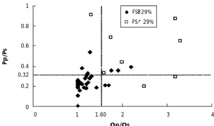
Eight patients had a Qp/Qs ratio over 1.60 and a Pp/
Ps ratio over 0.32 (Fig. 1). Five of eight patients had LV “dysfunction” with post-closure FS below 29% and FS change of over 20%. Post-closure FS below 29% was observed in one patient with a Qp/Qs ratio below 1.60 and a Pp/Ps ratio over 0.32, and in two patients with Qp/Qs ratios over 1.60 and Pp/Ps ratios below 0.32.
No patients with Qp/Qs ratios below 1.60 and Pp/Ps ratios below 0.32 had post-closure FS below 29%.
Discussion
Transcatheter interventional closure of PDAs has been practiced with various devices since 1967.
12)Re- cently, with the introduction of the Amplatzer ductal occluder into clinical practice, transcatheter closure has become possible for even very large PDAs.
13)Because a PDA is a congenital heart defect with a left-to-right shunt, it is associated with an increased LV preload. PDA closure causes an immediate reduction in ventricular preload.
1)2)14)Prior reports
15)have shown that patients with PDAs differ from normal control sub- jects in LV volume and function as measured by two- and three-dimensional echocardiography at baseline.
These changes caused by the PDA generally resolve by six months after percutaneous closure. Galal et al.
2)dem- onstrated that patients with large PDAs (larger than 3.1 mm) had significant deterioration in FS immediately after PDA closure, which was followed by FS recovery within six months.
Compared to the values before PDA closure, in this study, a significant decrease in the LVEDD was noted;
there was no difference in the LVESD immediately af- ter PDA closure. However, at follow-up, an additional
decrease in the LVEDD and a significant decrease in the LVESD were detected. An immediate decrease in the LVEDD and a late decrease in the LVESD resulted in a change in the FS when pre-closure, post-closure, and follow-up values were compared. Although FS is load-dependent and an acute change in LV volume load- ing after PDA closure is the primary underlying cause of this change in FS; FS below 29% after PDA closure was unusual and was considered to reflect LV dysfunction.
This study showed that LV dysfunction was present in eight patients immediately after PDA closure. Among these eight patients with LV dysfunction, seven patients had FS changes over 20%, five had Qp/Qs ratios over 1.60 and Pp/Ps ratios over 0.32, and two patients each had only Qp/Qs ratios over 1.60 or Pp/Ps ratios over 0.32. All of the patients were medicated with digitalis or angiotensin converting enzyme (ACE) inhibitors, but none showed clinical symptoms of ventricular dysfunc- tion. In one case with a post-closure FS below 29% and a FS change below 20%, the FS was 28% pre-closure, 23% post-closure, and 25% at follow-up, and the Qp/
Qs and Pp/Ps ratios were 1.60 and 0.33, respectively.
This patient may have had a cardiomyopathy concurrent with the PDA. However, further diagnostic evaluations such as a cardiac biopsy were not performed, and the patient has been undergoing regular follow-up.
The relationship of the Qp/Qs and Pp/Ps ratios with FS was thought to be explained by larger amounts of sh- unting, higher pulmonary artery pressure, greater changes in the FS, and significant LV dysfunction. Therefore, based on our findings, although the patients had nor- mal ventricular function before the procedure, patients with Qp/Qs ratios over 1.60 and Pp/Ps ratios over 0.32 at catheterization are predicted to be at risk for deterio- ration of ventricular function after the procedure. These patients require early evaluation of ventricular function using echocardiography.
Jeong et al.
16)reported persistent LV systolic dysfunc- tion (EF<50%) after PDA closure in adult PDA patients with low pre-closure LV EF. In contrast to Jeong’s study, our study showed recovery of the FS at follow-up, de- spite the immediate post-closure LV systolic dysfunction (FS<29%). This discrepancy might be due to differences in the populations studied (adults vs. children). A long- er duration of LV volume overload may cause changes in LV contractility and may lead to incomplete recovery after reduction in the loading condition. The ventric- ular remodeling status is different according to patient age. In this study, the tissue Doppler myocardial velocity was significantly decreased during the systolic and di- astolic phases at post-closure and follow-up compared to the pre-closure measurements. These findings suggest that an immediate reduction in the preload after PDA closure may lead to decreased myocardial velocity during the systolic phase after the procedure. After preload re-
Pp/Ps
1 0.8 0.6
0.4 0.32 0.2
0
0 1 1.60 2 3 4 Qp/Qs
FS>29%
FS≤29%