계명의대학술지 제34권 1호 Keimyung Med J Vol. 34, No. 1, June, 2015
© Copyright
Keimyung University School of Medicine 2015 Received: March 30, 2015
Accepted: April 22, 2015
Corresponding Author: Seok Jung Lee, M.D., Department of Orthopedic Surgery, Keimyung University School of Medicine, 56 Dalseong-ro, Jung-gu, Daegu 700-712, Korea Tel: +82-53-250-7739
E-mail: [email protected]
• The authors report no conflict of interest in this work.
Cauda equina syndrome (CES) is one of the typical symptoms condition requires emergency operation. This syndrome is always accompanied by sciatica. Because schwannomas rarely cause sciatica, it is seldom considered as a pain source in the patient with radiculopathy. A 74-year-old male patient presented with lumbar radiculopathy symptoms with, mild stenosis on lumbar MRI scan. All conservative treatment including medication failed to subside the symptoms. Thoracic spine MRI revealed a large tumor in the thoracic region. The symptoms disappeared after excision of the tumor. The tumor was diagnosed as a schwannoma.
Key Words :
Mimicking cauda equina syndrome, Schwannoma, Thoracic spine tumor서 론
척추관 협착은 척수를 압박하여 다양한 정도의 척수증이나 신경근증을 야기하는데, 이 중 마미증후군은 1934년 Mixter와 Barr[1]에 의해 처음 보고 되었으며, 척수 원추 이하 부위의 요천추 신경근인 마미에 포함된 신경 중 전부 또는 일부가 압박되어 나타나는 질환이다. 마미증후군은 발생 시 요통, 편측 또는 양측의 좌골 신경통, 안장 감각 소실, 하지의 근력 약화 및 감각 이상, 방광과 항문의 조절 기능 상실 등의 신경학적 증상이 나타나며 즉각적인 수술적 치료를 요하는 질환으로 알려져 있다. 저자들은 마미증후군으로 오인된 흉추의 종양으로 인한 흉추 척수증 1례를 경험하여 문헌고찰과 함께 보고하고자 한다.
Department of Orthopedic Surgery, Keimyung University School of Medicine, Daegu, Korea
Seok Jung Lee, M.D.
Thoracic Spine Tumor Mimicking Cauda Equina Syndrome
계명대학교 의과대학 정형외과학교실 이석중
마미증후군으로 오인된 흉추의 종양
71 마미증후군으로 오인된 흉추의 종양
증 례
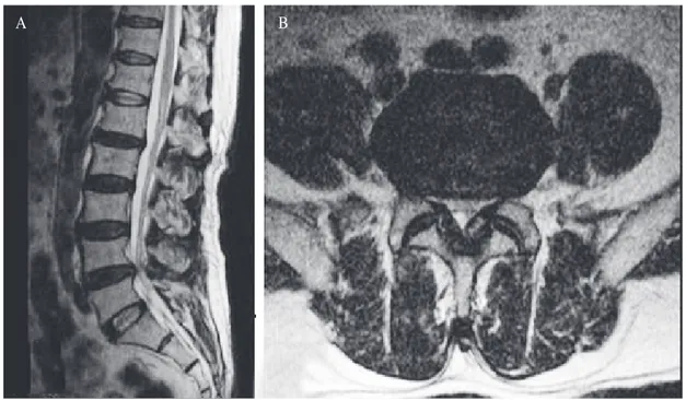
개인병원에서 요추 4/5번 간의 척추간 협착증 진단 하에 1년 동안 치료받았으나 내원 1개월여 전부터 심해진 하지 방사통과 신경인성 파행 및 내원 3일 전부터 대소변 보기 힘들고 엉치부위 감각 떨어지는 증상을 주소로 마미증후근 진단 하에 개인병원에서 응급수술을 권유받았으나 본원으로 전원 되신 74세 남자이다. 과거력상 15년 된 당뇨와 8년 된 고혈압, 4년 전에 간암으로 간 절제술 시행 받았던 기왕력이 있었다. 전원 당시 가져오신 개인병원의 자기공명 영상 상에서 요추 4/5번의 척추관 협착증은 관찰되나 마미증후근을 유발할 정도의 심각한 협착은 관찰되지 않았다(Fig. 1). 본원에서 시행한 이학적 검사상 양측 족부의 신전근력 3/5, 족배굴근력 3/5으로 감소되어 있었고 하지 및 항문주위 감각이 저하되어 있었으며, 항문 괄약근 긴장은 소실되어 있었다. 양측 하지 직 거 상 검 사 는 정 상 의 소 견 을 보 였 다 . 경 추 의 신경근증이나 척수병증으로 인한 증상은 관찰되지 않았으나, 검상돌기 밑으로 전반적인 감각저하와 함께
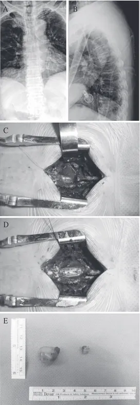
심부건반사는 대칭적으로 보존되어 있고, 양측에서 바빈스키(Babinski) 징후가 양성이었다. 하지 감각은 통증 및 위치각이 저명하게 소실되어 있었다. 이에 본원에서 시행한 흉추 자기공명 영상에서 흉추 5번에서 척수를 압박하는 종괴가 관찰되어(Fig. 2), 이에 대한 척추궁절제술 및 현미경하 종양 절제술 시행하였으며(Fig. 3), 술 후 하지 근력 및 대소변장애 또한 호전되었다. 조직학적 검사상 신경초종으로 진단되었다.
고 찰
흉추의 척수증은 대개 흉추에 외상이나 퇴행성 변화, 척추 전방 전위증 및 척추 종양에 의하여 척수가 압박을 받아 발생하며, 급성 외상이나 척추 종양으로 인하여 추체 골절이 동반되어 척수를 압박하는 경우는 급격한 통증과 신경 장애의 소견을 보이나 서서히 진행하는 종양이나 흉추 후관절에 발생한 골극은 점진적인 흉추 척수증의 소견을 보인다. 신경초종은
Fig. 1. Magnetic resonance images from the private hospital does not reveal stenotic lesion which can cause cauda equine syndrome. (A) T2 weighted sagittal image. (B) T2 weighted axial image at the level of the L4/5.
A B
72 계명의대학술지 제34권 1호 2015
척추에서 관찰되는 경막 내 척수외종양으로 대부분은 증상을 호소하지 않으나, 하지방사통과 신경인성 파행이 가장 빈번하게 호소하는 증상이다. 또한, 종양의 크기가 성장함에 따라 종양의 위치에 따라 다행한 근력저하와 감각저하를 나타내게 된다. 이학적 검사로는 종양의 위치 밑으로 이상감각, 운동의 제한, 바빈스키검사 양성, 경추의 경우에는 호프만 검사 양성, 강직, 대소변장애, 심부건반사 항진과 같은 척수증의 증상을 보인다[2,3].
척수증은 증상이 매우 다양하게 일어날 수 있으며, 본 예에서처럼 환자가 호소하는 증상이 하지방사통과 신 경 인 성 파 행 이 주 증 상 이 고 갑 자 기 심 해 진 하지방사통과 신경인성 파행 및 대소변 장애를 호소한다면 마미 증후군을 우선적으로 고려해 볼 수
있겠다[4].
하지만 흉추의 척수신경절에 기원하는 종양에 있어서는 흉추의 신경절로 인한 방사통이나 근력저하가 발행하지 않으며, 요추 신경 또한 흉추 내에 존재하기 때문에 하지방사통과 신경인성 파행과 같은 요추 신경근병증의 증상을 먼저 호소할 수 있다[5].
이후 종양이 크기가 증가하여 척수 압박이 진행할 경우 척수병증의 증상을 호소할 수 있다. 따라서 본 예에서처럼 요추 신경병증을 의심하였으나 영상소견에 비해서 심한 통증과 근력저하를 호소한다면 척수병증을 의심하고[6], 감각이상과 심부건반사, 바빈스키 징후 등의 척수병증에 대한 이학적 검사를 추가로 실시하고, 자기공명영상과 근전도검사를 시행하여 척수병증을 일으키는 원인에 대한 진단과 적절한 치료가 필요할 Fig. 2. Magnetic resonance images shows an intradural and an extradural mass at the level of the T5. (A) T1 weighted sagittal image, (B) T2 weighted sagittal image, (C) Enhanced sagittal image, (D) T1 weighted axial image at the level of T5, (E) T2 weighted axial image at the level of T5, (F) Enhanced axial image at the level of T5.
A
D E F
B C
73 마미증후군으로 오인된 흉추의 종양
것으로 사료된다.
참 고 문 헌
1. Mixter WJ, Barr JS. Rupture of the intervertebral disc with involvement of the spinal cord. N Engl J Med 1934;211:210–4.
2. Kim SB, Kim HS, Jang JS, Lee SH. Mobility of intradural extramedullary schwannoma at spine: report of three cases with literature review. J Korean Neurosurg Soc 2010;47:64–7.
3. Kothbauer KF. Neurosurgical management of intramedullary spinal cord tumors in children. Pediatr Neurosurg 2007;43:222–35.
4. Tay ECK, Chacha PB. Midline prolapse of lumbar intervertebral disc with compression of the cauda equina.
J Bone Joint Surg Br 1979;61:43-6.
5. Ukaigwe A, Olugbodi A, Alweis RL. Taking it to the next level: lumbar radiculopathy from thoracic nerve schwannoma. J Community Hosp Intern Med Perspect 2015;5:25744.
6. Goshgarian H. In: Lin VW, Cardenas DD, editors. Spinal Cord Medicine: Principles and Practice. New York:
Demos 2003, Chapter 2: 15-8.
A
C
D
E
B
Fig. 3. Post-OP x-ray & Clinical photo. (A) Anteroposterior view of thoracic spine, (B) Lateral view of thoracic spine, (C) Exposure of large extradural schwannoma with laminectomy, (D) The dura bulged out after extradural mass excision, (E) Tumor mass of large extradural schwannoma, small intradural schwannoma.