pISSN 2093-9043 eISSN 2233-6273
Purpose: Emergent intracranial occlusions causing acute ischemic stroke are often related to extracranial atherosclerotic stenosis. This study aimed to investigate the association between post-procedure intracerebral hemorrhage (ICH) and emergent extracranial artery stenting and assess their effects on clinical outcomes in patients with acute ischemic stroke.
Materials and Methods: We retrospectively analyzed patients undergoing hyperacute en- dovascular treatment for cervicocephalic vascular occlusion in three Korean hospitals between January 2011 and February 2016. Patients who had extracranial artery involvement and were treated from 24 hours of symptom onset to puncture were included in this study, and they were divided into the extracranial stenting (ES) and non-ES groups. Any type of petechial hem- orrhages and parenchymal hematoma was defined as ICH for the current study.
Results: In total, 76 patients were included in this study. Among them, 56 patients underwent ES, and 20 patients did not. Baseline characteristics, risk factors, laboratory data, treatment methods, successful reperfusion rates, and baseline stenotic degrees of extracranial internal carotid artery did not differ between these two groups. However, atrial fibrillation was more frequent in patients without than with ES (P=0.002), and post-procedure ICH was more fre- quent in patients with than without ES (P=0.035). Logistic regression models revealed that ES was independently associated with post-procedure ICH (odds ratio [OR], 7.807; 95% confidence interval [CI], 1.213–50.248; P=0.031), and ICH was independently associated with poor clinical outcomes (OR, 0.202; 95% CI, 0.054–0.759; P=0.018); however, ES itself was not associated with clinical outcomes (OR, 0.530; 95% CI, 0.117–2.395; P=0.409). Notably, ICH and ES had interaction for predicting good outcomes (P=0.041).
Conclusion: Post-procedure ICH was associated with ES and poor clinical outcomes. There- fore, ES should be cautiously considered in patients with hyperacute stroke.
Key Words: Carotid stenosis; Stents; Cerebral infarction; Endovascular procedures; Cerebral hemorrhage
Clinical Impact of Intracerebral Hemorrhage after Hyper- acute Extracranial Stenting in Patients with Ischemic Stroke
Je Hong Min, MD1, Seong-Joon Lee, MD, PhD1, Ji Man Hong, MD, PhD1, Jin Wook Choi, MD, PhD2, Dong-Hun Kang, MD3,4, Yong-Won Kim, MD4,5, Yong-Sun Kim, MD, PhD4, Jeong-Ho Hong, MD, PhD6, Joonsang Yoo, MD6, Chang-Hyun Kim, MD7, Sung-Il Sohn, MD, PhD7, Yang-Ha Hwang, MD, PhD5, Jin Soo Lee, MD, PhD1
1Department of Neurology, Ajou University School of Medicine, Ajou University Medical Center, Suwon, Korea
2Department of Radiology, Ajou University School of Medicine, Ajou University Medical Center, Suwon, Korea
3Department of Neurosurgery, School of Medicine, Kyungpook National University, Daegu, Korea
4Department of Radiology, School of Medicine, Kyungpook National University, Daegu, Korea
5Department of Neurology, School of Medicine, Kyungpook National University, Daegu, Korea
6Department of Neurology, Keimyung University Dongsan Medical Center, Daegu, Korea
7Department of Neurosurgery, Keimyung University Dongsan Medical Center, Daegu, Korea
Correspondence to:
Jin Soo Lee, MD, PhD
Department of Neurology, Ajou Uni- versity School of Medicine, 164 World Cup-ro, Yeongtong-gu, Suwon 16499, Korea
Tel: +82-31-219-5175 Fax: +82-31-219-5178 E-mail: [email protected] Received: May 31, 2019 Revised: June 26, 2019 Accepted: June 27, 2019
Copyright © 2019 Korean Society of Interventional Neuroradiology
This is an Open Access article distributed under the terms of the Creative Commons Attribution Non-Commercial License (http://creativecommons.org/licenses/by-nc/3.0) which permits unrestricted non-commercial use, distribution, and reproduction in any medium, provided the original work is properly cited.
INTRODUCTION
Extracranial atherosclerosis (ECAS) is observed in about 10–
20% of stroke patients and is not a rare cause of acute isch- emic stroke.1,2 Retrospective single-arm studies have shown that acute ischemic stroke patients undergoing emergent endovascular treatment (EVT) for acute extracranial internal carotid artery (ICA) occlusion show high recanalization rates and early neurologic improvement.3-5 In addition, several studies have demonstrated that patients with extracra- nial occlusion combined with intracranial artery tandem occlusion have better outcomes after EVT.6-8 Mechanical thrombectomy-based EVT is now considered a reasonable treatment especially for intracranial large artery occlusions.9 However, symptomatic intracranial hemorrhage (ICH) is observed in about 30–40% patients with acute large-artery occlusion after EVT,10,11 and it is thus considered a significant contributing factor for poor functional outcomes.
Carotid artery stenting is an EVT option for patients with extracranial carotid stenosis. Cases of extracranial stenting (ES) for ECAS-related occlusion in hyperacute stroke are increasing presently; however, the relationship between ES and the prevalence of post-procedural hemorrhagic transfor- mation (HT) remains unclear. Therefore, we investigated the association between ES and the prevalence of post-proce- dural HT and assessed the effect of ES on clinical outcomes in patients with hyperacute stroke.
MATERIALS AND METHODS
Study population and inclusion criteriaWe retrospectively analyzed patients in the Acute Stroke due to Intracranial Atherosclerotic occlusion and Neurointerven- tion-Korean Retrospective (ASIAN-KR) registry. This registry collected data on patients (aged ≥18 years) who underwent EVT for cervicocerebral artery occlusion causing ischemic stroke at three comprehensive stroke centers in Korea be- tween January 2011 and February 2016.12 Inclusion criteria were as follows: 1) onset-to-puncture time within 24 hours;
2) underlying etiology was proven as ECAS; or 3) patients un- derwent EVT for extracranial artery steno-occlusive disease.
All clinical data were de-identified and allocated study identification numbers. The protocol of data collection was approved by the Institutional Review Board of each hospital and was implemented in accordance with the ethical stan-
dards of the 1964 Declaration of Helsinki and its later amend- ments.
Evaluation methods
Premorbid and 3-month modified Rankin Scale (mRS) scores, National Institutes of Health Stroke Scale (NIHSS) scores, conventional vascular risk factors, and laboratory findings on admission were collected. A 3-month mRS score of 0 to 2 or no change compared with the premorbid mRS score was defined as a good outcome, and a 3-month mRS score of 3 to 6 or an increase compared with the premorbid mRS score was classified as a poor outcome.
The location of the initial occlusion site was determined using baseline computed tomography (CT) angiography, magnetic resonance angiography, or digital subtraction an- giography. Stenotic degree of extracranial artery associated with hyperacute stroke was assessed based on the North American Symptomatic Carotid Endarterectomy Trial crite- ria.13 Alberta Stroke Program Early CT Scores (ASPECTS) were determined using non-contrast CT in the anterior-circulation territory in patients with hyperacute stroke. Final reperfusion was evaluated using the modified thrombolysis in cerebral infarction (mTICI) scale,14 and mTICI grades 2b or 3 were defined as successful reperfusion. Post-procedural ICH was graded based on the criteria defined by the European Co- operative Acute Stroke Study, and was defined as presence of any grade of ICH for the current study.15 Subarachnoid hemorrhage was classified according to the modified Fisher scale.16
Endovascular procedures
Endovascular devices were selected at the discretion of neu- rointerventionists based on consensus within each stroke team. Contact aspiration and stent retrieval were routinely used as a frontline method. Contact aspiration is a meth- od of a forced arterial suction thrombectomy, which uses the Penumbra system (Penumbra Inc., Alameda, CA, USA).
Stent retrieval is a method of clot removal by capturing and removing the thrombus with a stent retriever, such as the Solitaire AB/FR (Medtronic, Irvine, CA, USA) or Trevo (Stryker, Kalamazoo, MI, USA). Balloon guide catheters, adjuvant local lytic infusion, intracranial or extracranial angioplasty, and/or stenting were implemented as needed.
Statistical analysis
Data are expressed as mean±standard deviations, median
(interquartile ranges), or numbers (percentages). Comparative analyses of clinical characteristics and treatment outcomes were performed between the ES and non-ES (NES) groups.
Differences between the groups were analyzed using χ2 tests for categorical variables, the Mann-Whitney U-test for ordinal variables, or t-tests for continuous variables. To evaluate the relation between ES and the prevalence of post-procedural ICH or further clinical outcomes, we performed multivariate logistic regression analyses with major confounders. A P of
<0.05 was considered significant. Statistical analyses were performed using the SPSS statistical package (version 25.0, IBM SPSS, Armonk, NY, USA).
RESULTS
Baseline characteristics
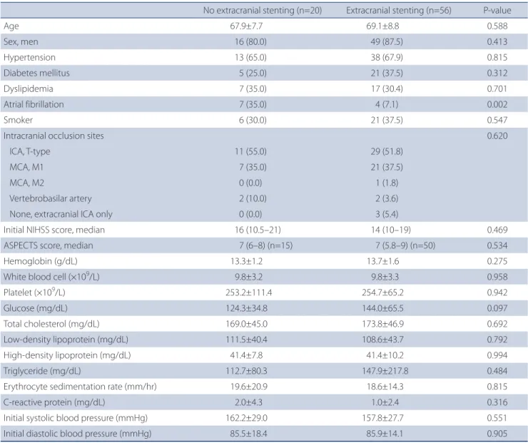
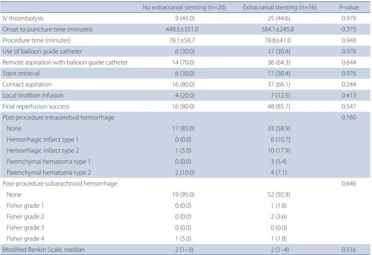
Among the 720 registered patients, 76 patients were eligible based on the inclusion criteria (Fig. 1). Baseline characteristics of patients are summarized in Table 1. Among the 76 eligible patients, 56 patients underwent ES, and 20 patients did not undergo any extracranial treatment or underwent other ex- tracranial treatments without stenting (such as balloon-an- gioplasty and contract aspiration). Atrial fibrillation was more prevalent in the NES group than in the ES group (35.0% vs.
7.1%, P=0.002); however, other variables including laboratory data; percentages of hypertension, diabetes mellitus, dyslip- idemia, or smoker; NIHSS or ASPECTS score on admission; or systolic or diastolic blood pressure did not differ between the two groups.
Treatment methods and outcomes associated with ES
Comparative analyses of treatment methods and outcomes between these two groups are summarized in Table 2. Over- all procedure time or treatment methods, except for the ES, were not different between the two groups. The rates of successful reperfusion were similar in these two groups, regardless of ES (80.0% vs. 85.7%, P=0.547). Functional out- comes, based on the mRS score, did not differ between the ES and NES groups. However, the prevalence of post-proce- dural cerebral hemorrhage after emergent extracranial EVT was associated with ES (odds ratio [OR], 7.807; 95% confi- dence interval [CI], 1.213 to 50.248; P=0.031) (Table 3). Notably, multivariate analyses with potential major confounders in a logistic regression model revealed that the presence of ES was the only independent predictor for the prevalence of hemorrhage after emergent extracranial EVT (OR, 7.807; 95%
CI, 1.213 to 50.248; P=0.031). Good outcomes at 3 months were more commonly observed in patients without than
Fig. 1. Flowchart of this study. ASIAN-KR, Acute Stroke due to Intracranial Atherosclerotic occlusion and Neurointervention-Korean Retrospective;
ECAS, Extracranial Atherosclerosis.
ASIAN-KR registry (n=720)
Onset-to-puncture time ≤1,440 minutes (n=711)
Onset-to-puncture time >1,440 minutes (n=9)
ECAS as a final etiology OR
presence of extracranial artery treatment (n=76)
Othere underlying mechanism (embolism, dissection and intractable) AND
absence of extracranial artery treatment (n=635)
Extracranial stenting (n=56)
Non-extracranial stenting (n=20)
Inclusion Exclusion
with any post-procedural cerebral hemorrhage (68.0% vs.
42.3%, P=0.031).
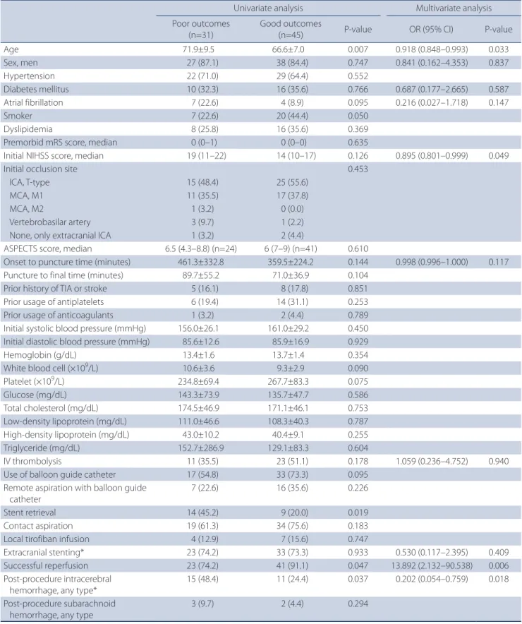
As observed in Table 4, the association between ES and better clinical outcomes was not demonstrated in multivar- iate logistic regression analyses (OR, 0.530; 95% CI, 0.117 to 2.395; P=0.409). However, good outcomes after EVT were the absence of any post-procedural intracerebral hemorrhage (OR, 0.202; 95% CI, 0.054 to 0.759; P=0.018). Because ES had an interaction with post-procedural intracerebral hemorrhage (P=0.041), we speculate that ES has an indirect impact on worse clinical outcomes.
DISCUSSION
In the present study, we found that the prevalence of post-procedural ICH was relatively higher in patients who underwent emergent ES than in those who did not. Good outcomes after hyperacute extracranial artery treatment were attributable to multiple factors such as younger age, lower initial NIHSS score, successful reperfusion, and the ab- sence of any ICH after EVT. Post-procedural ICH was related emergent extracranial artery stenting and associated with poor functional outcomes.
Table 1. Baseline characteristics of patients with emergent extracranial steno-occlusive disease
No extracranial stenting (n=20) Extracranial stenting (n=56) P-value
Age 67.9±7.7 69.1±8.8 0.588
Sex, men 16 (80.0) 49 (87.5) 0.413
Hypertension 13 (65.0) 38 (67.9) 0.815
Diabetes mellitus 5 (25.0) 21 (37.5) 0.312
Dyslipidemia 7 (35.0) 17 (30.4) 0.701
Atrial fibrillation 7 (35.0) 4 (7.1) 0.002
Smoker 6 (30.0) 21 (37.5) 0.547
Intracranial occlusion sites 0.620
ICA, T-type 11 (55.0) 29 (51.8)
MCA, M1 7 (35.0) 21 (37.5)
MCA, M2 0 (0.0) 1 (1.8)
Vertebrobasilar artery 2 (10.0) 2 (3.6)
None, extracranial ICA only 0 (0.0) 3 (5.4)
Initial NIHSS score, median 16 (10.5–21) 14 (10–19) 0.469
ASPECTS score, median 7 (6–8) (n=15) 7 (5.8–9) (n=50) 0.534
Hemoglobin (g/dL) 13.3±1.2 13.7±1.6 0.275
White blood cell (×109/L) 9.8±3.2 9.8±3.3 0.958
Platelet (×109/L) 253.2±111.4 254.7±65.2 0.942
Glucose (mg/dL) 124.3±34.8 144.0±65.5 0.097
Total cholesterol (mg/dL) 169.0±45.0 173.8±46.9 0.692
Low-density lipoprotein (mg/dL) 111.5±40.4 108.6±43.7 0.792
High-density lipoprotein (mg/dL) 41.4±7.8 41.4±10.2 0.994
Triglyceride (mg/dL) 112.7±80.3 147.9±217.8 0.484
Erythrocyte sedimentation rate (mm/hr) 19.6±20.9 18.6±14.3 0.815
C-reactive protein (mg/dL) 2.0±4.3 1.0±2.4 0.316
Initial systolic blood pressure (mmHg) 162.2±29.0 157.8±27.7 0.551
Initial diastolic blood pressure (mmHg) 85.5±18.4 85.9±14.1 0.905
Values are presented as mean±standard deviation or number (%).
ICA, internal carotid artery; MCA, middle cerebral artery; NIHSS, National Institutes of Health Stroke Scale; ASPECTS, Alberta Stroke Program Early CT Score.
It is well known that EVTs such as mechanical thrombecto- my are preferred to intravenous thrombolysis for intracranial large artery occlusions.9,17 Whereas EVTs for intracranial large artery occlusions have had sufficient evidences of efficacy and safety, evidences for emergent ES have barely been achieved. Nevertheless, accumulating evidence has indicated the safety and efficacy of emergent endovascular stenting in hyperacute stroke. It was reported that endovascular therapy of tandem occlusions with extracranial internal carotid artery revascularization as the first step had a high recanalization rate and led to an acceptable rate of good clinical outcomes.
However, little is known about the hemorrhagic risk after ES.
A retrospective study about the hemorrhagic risk after emer- gent EVT with intracranial or ES showed that patients under- going emergent stenting had higher grades of hemorrhagic subtypes, and acute phase stenting was an independent predictor of sICH after adjustment for potential confound-
ers and the procedure duration. However, this study had a single-center retrospective design, and the sample size was small (24 patients) in the emergent stenting group.18 Nota- bly, TITAN study did not find a definite association between extracranial ICA stenting and post-procedural hemorrhage, although procedure-related complications after 90 days of EVT, including any ICH, were lower in the intracranial throm- bectomy with ICA stenting group than in the intracranial thrombectomy only group.19 However, while final successful reperfusion rate was similar between ES and NES groups in the current study, reperfusion success rate was significantly higher in the ICA stenting group than thrombectomy only group in the TITAN study. Reperfusion failure may increase the final infarct volume, which, in turn, can cause larger area of breakdown of blood-brain barrier and the risk of hem- orrhagic transformation. Therefore, it is difficult to ascertain that ES-based EVT is safer than NES-based.
Table 2. Comparisons of treatment methods and outcomes
No extracranial stenting (n=20) Extracranial stenting (n=56) P-value
IV thrombolysis 9 (45.0) 25 (44.6) 0.978
Onset to puncture time (minutes) 448.5±351.0 384.1±245.8 0.375
Procedure time (minutes) 78.1±58.7 78.8±41.0 0.948
Use of balloon guide catheter 6 (30.0) 17 (30.4) 0.976
Remote aspiration with balloon guide catheter 14 (70.0) 36 (64.3) 0.644
Stent retrieval 6 (30.0) 17 (30.4) 0.976
Contact aspiration 16 (80.0) 37 (66.1) 0.244
Local tirofiban infusion 4 (20.0) 7 (12.5) 0.413
Final reperfusion success 16 (80.0) 48 (85.7) 0.547
Post-procedure intracerebral hemorrhage 0.160
None 17 (85.0) 33 (58.9)
Hemorrhagic infarct type 1 0 (0.0) 6 (10.7)
Hemorrhagic infarct type 2 1 (5.0) 10 (17.9)
Parenchymal hematoma type 1 0 (0.0) 3 (5.4)
Parenchymal hematoma type 2 2 (10.0) 4 (7.1)
Post-procedure subarachnoid hemorrhage 0.646
None 19 (95.0) 52 (92.9)
Fisher grade 1 0 (0.0) 1 (1.8)
Fisher grade 2 0 (0.0) 2 (3.6)
Fisher grade 3 0 (0.0) 0 (0.0)
Fisher grade 4 1 (5.0) 1 (1.8)
Modified Rankin Scale, median 2 (1–3) 2 (1–4) 0.516
Values are presented as mean±standard deviation or number (%).
IV, intravenous.
Table 3. Risk factors of intracerebral hemorrhage after EVT
Univariate analysis Multivariate analysis
No hemorrhage (n=50) Any hemorrhage (n=26) P-value OR (95% CI) P-value
Age 68.7±8.2 68.9±9.2 0.944 1.018 (0.947–1.094) 0.623
Sex, men 42 (84.0) 23 (88.5) 0.600 1.139 (0.165–7.878) 0.895
Hypertension 33 (70.2) 17 (68.0) 0.846
Diabetes mellitus 19 (40.4) 7 (28.0) 0.296 0.657 (0.149–2.896) 0.579
Atrial fibrillation 7 (14.9) 2 (8.0) 0.400
Smoker 15 (31.9) 10 (40.0) 0.493
Dyslipidemia 16 (34.0) 8 (32.0) 0.861
Prior history of TIA or stroke 7 (14.9) 4 (16.0) 0.901 3.083 (0.402–23.658) 0.279
Prior usage of antiplatelets 12 (25.5) 7 (28.0) 0.821 1.199 (0.263–5.466) 0.815
Prior usage of anticoagulants 1 (2.1) 1 (4.0) 0.645 15.776 (0.084–2975.334) 0.302
Initial NIHSS score, median 14 (10–19.3) 15.5 (10.8–20.3) 0.781 0.999 (0.904–1.104) 0.984
Intracranial occlusion location 0.405
ICA, T-type 27 (54.0) 13 (50.0)
MCA, M1 17 (34.0) 11 (42.3)
MCA, M2 0 (0.0) 1 (3.8)
Vertebrobasilar artery 3 (6.0) 0 (0.0)
None, only extracranial ICA 3 (6.0) 0 (0.0)
ASPECTS score, median 8 (5.5–9) (n=41) 6 (6–8) (n=24) 0.071
Hemoglobin (g/dL) 13.3±1.6 14.1±1.2 0.036
White blood cell (×109/L) 9.6±3.2 10.3±3.3 0.372
Platelet (×109/L) 253.7±88.5 255.6±58.4 0.921
Glucose (mg/dL) 135.2±44.4 145.7±81.5 0.545
Total cholesterol (mg/dL) 163.4±45.0 190.0±44.0 0.016
Low-density lipoprotein (mg/dL) 101.6±40.0 124.0±44.8 0.029
High-density lipoprotein (mg/dL) 39.1±9.1 45.8±9.1 0.003
Triglyceride (mg/dL) 149.0±231.3 118.7±68.8 0.517
Initial systolic blood pressure (mmHg) 160.2±28.4 156.6±27.3 0.593 Initial diastolic blood pressure (mmHg) 86.9±14.2 83.6±17.1 0.370
IV thrombolysis 23 (46.0) 11 (42.3) 0.759 1.735 (0.327–9.199) 0.518
Onset to puncture time (minutes) 388.0±270.5 426.1±290.8 0.571 1.001 (0.998–1.004) 0.414
Procedure time (minutes) 81.1±45.6 73.8±46.9 0.512 0.995 (0.980–1.010) 0.516
Use of balloon guide catheter 33 (66.0) 17 (65.4) 0.957
Remote aspiration with balloon guide catheter
17 (34.0) 6 (23.1) 0.325
Stent retrieval 12 (24.0) 11 (42.3) 0.099
Contact aspiration 35 (70.0) 18 (69.2) 0.945
Local tirofiban infusion 5 (10.0) 6 (23.1) 0.124
Extracranial stenting 33 (66.0) 23 (88.5) 0.035 7.807 (1.213–50.248) 0.031
Successful reperfusion 42 (84.0) 22 (84.6) 0.944
Good outcomes at 3 months 34 (68.0) 11 (42.3) 0.031
Values are presented as mean±standard deviation or number (%) unless otherwise indicated.
EVT, endovascular treatment; OR, odds ratio; CI, confidence interval; TIA, transient ischemic attack; NIHSS, National Institutes of Health Stroke Scale; ICA, internal carotid artery; MCA, middle cerebral artery; ASPECTS, Alberta Stroke Program Early CT Score; IV, intravenous.
Table 4. Predicting factors associated with good clinical outcomes
Univariate analysis Multivariate analysis
Poor outcomes (n=31)
Good outcomes
(n=45) P-value OR (95% CI) P-value
Age 71.9±9.5 66.6±7.0 0.007 0.918 (0.848–0.993) 0.033
Sex, men 27 (87.1) 38 (84.4) 0.747 0.841 (0.162–4.353) 0.837
Hypertension 22 (71.0) 29 (64.4) 0.552
Diabetes mellitus 10 (32.3) 16 (35.6) 0.766 0.687 (0.177–2.665) 0.587
Atrial fibrillation 7 (22.6) 4 (8.9) 0.095 0.216 (0.027–1.718) 0.147
Smoker 7 (22.6) 20 (44.4) 0.050
Dyslipidemia 8 (25.8) 16 (35.6) 0.369
Premorbid mRS score, median 0 (0–1) 0 (0–0) 0.635
Initial NIHSS score, median 19 (11–22) 14 (10–17) 0.126 0.895 (0.801–0.999) 0.049
Initial occlusion site 0.453
ICA, T-type 15 (48.4) 25 (55.6)
MCA, M1 11 (35.5) 17 (37.8)
MCA, M2 1 (3.2) 0 (0.0)
Vertebrobasilar artery 3 (9.7) 1 (2.2)
None, only extracranial ICA 1 (3.2) 2 (4.4)
ASPECTS score, median 6.5 (4.3–8.8) (n=24) 6 (7–9) (n=41) 0.610
Onset to puncture time (minutes) 461.3±332.8 359.5±224.2 0.144 0.998 (0.996–1.000) 0.117
Puncture to final time (minutes) 89.7±55.2 71.0±36.9 0.104
Prior history of TIA or stroke 5 (16.1) 8 (17.8) 0.851
Prior usage of antiplatelets 6 (19.4) 14 (31.1) 0.253
Prior usage of anticoagulants 1 (3.2) 2 (4.4) 0.789
Initial systolic blood pressure (mmHg) 156.0±26.1 161.0±29.2 0.450 Initial diastolic blood pressure (mmHg) 85.6±12.6 85.9±16.9 0.929
Hemoglobin (g/dL) 13.4±1.6 13.7±1.4 0.354
White blood cell (×109/L) 10.6±3.6 9.3±2.9 0.090
Platelet (×109/L) 234.8±69.4 267.7±83.3 0.075
Glucose (mg/dL) 143.3±73.9 135.7±47.7 0.586
Total cholesterol (mg/dL) 174.5±46.9 171.1±46.1 0.753
Low-density lipoprotein (mg/dL) 111.0±46.6 108.3±40.3 0.787
High-density lipoprotein (mg/dL) 43.0±10.2 40.4±9.1 0.255
Triglyceride (mg/dL) 152.7±286.9 129.1±83.3 0.604
IV thrombolysis 11 (35.5) 23 (51.1) 0.178 1.059 (0.236–4.752) 0.940
Use of balloon guide catheter 17 (54.8) 33 (73.3) 0.095
Remote aspiration with balloon guide catheter
7 (22.6) 16 (35.6) 0.226
Stent retrieval 14 (45.2) 9 (20.0) 0.019
Contact aspiration 19 (61.3) 34 (75.6) 0.183
Local tirofiban infusion 4 (12.9) 7 (15.6) 0.747
Extracranial stenting* 23 (74.2) 33 (73.3) 0.933 0.530 (0.117–2.395) 0.409
Successful reperfusion 23 (74.2) 41 (91.1) 0.047 13.892 (2.132–90.538) 0.006
Post-procedure intracerebral hemorrhage, any type*
15 (48.4) 11 (24.4) 0.037 0.202 (0.054–0.759) 0.018
Post-procedure subarachnoid hemorrhage, any type
3 (9.7) 2 (4.4) 0.294
Values are presented as mean±standard deviation or number (%) unless otherwise indicated.
OR, odds ratio; CI, confidence interval; mRS: modified Rankin Scale; NIHSS; National Institutes of Health Stroke Scale; ICA, internal carotid artery; MCA, middle cerebral artery; ASPECTS, Alberta Stroke Program Early CT Score; TIA, transient ischemic attack; IV, intravenous.
*Both variables had interaction (P=0.041).
Several reasons may explain the relative high risk of ICH fol- lowing ES in the current study. Many studies have suggested that hyperacute ICH after extracranial carotid artery stenting is mainly associated with abrupt cerebral hyperperfusion in the chronically hypoperfused areas.20-23 The high rate of cerebral hyperperfusion after stenting is associated with the underlying diseases causing microangiopathy, high degrees of stenosis with the poor collateral flow, high degrees of stenosis on the contralateral carotid artery, and recent stroke history.20,21 In addition, these post-procedural hemorrhages just after extracranial carotid artery stenting could result from rupture of small perforating arteries after rapidly normalized perfusion pressure in the chronically hypoperfused areas.24 Prior usage of antiplatelets or anticoagulants might also in- crease the risk of post-procedural hemorrhage. In our current study, no significant correlation was observed between prior antiplatelet/anticoagulant medication and post-procedural hemorrhage. However, peri- or post-procedural antiplatelet treatment might potentially affect the incidence of ICH in the current study. As large infarct volume or microbleeds are seen on CT or magnetic resonance imaging, physicians may skip antiplatelet medication to escape hemorrhagic transfor- mation. In contrast, physicians cannot skip the medication if ES is permanently deployed to escape in-stent thrombosis.
Therefore, the occurrence of ICH after stenting might be caused by sudden hyperemia/hyperperfusion combined with post-stent prevention, such as antiplatelets.
Many studies have improved the process of EVT for intra- cranial large artery occlusion (LAO) in patients with acute ischemic stroke and tandem occlusion from ECAS disease. It is suggested that sufficient dilation of stenosis cervical artery is important for the successful use in stent-retrieval throm- bectomy or spontaneous recanalization in cases of refractory LAO.25 Therefore, it is necessary to perform balloon angio- plasty or stenting for maintaining a good extracranial artery perfusion status. Our study showed that ES had an increased risk on post-procedure ICH; therefore, balloon angioplasty could be used as an alternative method, and cautious local administration of glycoprotein IIb/IIIa inhibitor, such as tiro- fiban, could be used to prevent re-occlusion of the extracra- nial artery after mechanical thrombectomy.
This study has several limitations. First, this study was a ret- rospective study; thus, the findings in the current study can- not be generalized. For escaping potential biases, we recruit- ed multicenter data. Second, ICH after ES in patients with posterior circulation stroke was not well evaluated. Therefore,
further studies must be implemented with a large amount of data related to extracranial vertebral artery stenting. Third, no exact data on pre- or post-stenting prevention were available; therefore, we could not exactly examine whether post-procedural ICH was related to stenting or/and antiplate- let medication during the stenting period. Therefore, further studies with a standardized dosage of antiplatelet medica- tions before and after stenting are needed.
CONCLUSION
Emergent extracranial artery stenting has been used as a treatment option for hyperacute stroke patients with ex- tracranial artery stenosis. This study found that ICH more frequently occurred in patients who underwent ES and was related to poor clinical outcomes in which ICH and ES had interaction. The findings indicate that ES must be cautiously considered in patients with ECAS-related acute ischemic stroke.
REFERENCES
1. Grau AJ, Weimar C, Buggle F, Heinrich A, Goertler M, Neumaier S, et al. Risk factors, outcome, and treatment in subtypes of isch- emic stroke: the German stroke data bank. Stroke 2001;32:2559- 2566
2. Rubiera M, Ribo M, Delgado-Mederos R, Santamarina E, Delga- do P, Montaner J, et al. Tandem internal carotid artery/middle cerebral artery occlusion: an independent predictor of poor outcome after systemic thrombolysis. Stroke 2006;37:2301-2305 3. Jovin TG, Gupta R, Uchino K, Jungreis CA, Wechsler LR, Hammer
MD, et al. Emergent stenting of extracranial internal carotid ar- tery occlusion in acute stroke has a high revascularization rate.
Stroke 2005;36:2426-2430
4. Papanagiotou P, Roth C, Walter S, Behnke S, Grunwald IQ, Viera J, et al. Carotid artery stenting in acute stroke. J Am Coll Cardiol 2011;58:2363-2369
5. Yoon W, Kim BM, Kim DJ, Kim DI, Kim SK. Outcomes and prog- nostic factors after emergent carotid artery stenting for hyper- acute stroke within 6 hours of symptom onset. Neurosurgery 2015;76:321-329
6. Grigoryan M, Haussen DC, Hassan AE, Lima A, Grossberg J, Re- bello LC, et al. Endovascular treatment of acute ischemic stroke due to tandem occlusions: large multicenter series and system-
atic review. Cerebrovasc Dis 2016;41:306-312
7. Maurer C, Joachimski F, Berlis A. Two in one: endovascular treat- ment of acute tandem occlusions in the anterior circulation.
Clin Neuroradiol 2015;25:397-402
8. Cohen JE, Gomori JM, Rajz G, Itshayek E, Eichel R, Leker RR. Ex- tracranial carotid artery stenting followed by intracranial stent- based thrombectomy for acute tandem occlusive disease. J Neurointerv Surg 2015;7:412-417
9. Powers WJ, Rabinstein AA, Ackerson T, Adeoye OM, Bambakidis NC, Becker K, et al. 2018 guidelines for the early management of patients with acute ischemic stroke: a guideline for health- care professionals from the American Heart Association/Ameri- can Stroke Association. Stroke 2018;49:e46-e99.
10. Kang DH, Yoon W, Kim SK, Baek BH, Lee YY, Kim YW, et al. En- dovascular treatment for emergent large vessel occlusion due to severe intracranial atherosclerotic stenosis. J Neurosurg 2019;130:1949-1956
11. Hao Y, Yang D, Wang H, Zi W, Zhang M, Geng Y, et al. Predictors for symptomatic intracranial hemorrhage after endovascular treatment of acute ischemic stroke. Stroke 2017;48:1203-1209 12. Lee JS, Lee SJ, Hong JM, Choi JW, Hong JH, Chang HW, et al.
Temporal changes in care processes and outcomes for endo- vascular treatment of acute ischemic stroke: retrospective regis- try data from three Korean centers. Neurointervention 2018;13:2- 12
13. North American Symptomatic Carotid Endarterectomy Trial.
Methods, patient characteristics, and progress. Stroke 1991;22:711- 720
14. Tomsick T, Broderick J, Carrozella J, Khatri P, Hill M, Palesch Y, et al. Revascularization results in the Interventional Management of Stroke II trial. AJNR Am J Neuroradiol 2008;29:582-587 15. Fiorelli M, Bastianello S, von Kummer R, del Zoppo GJ, Larrue V,
Lesaffre E, et al. Hemorrhagic transformation within 36 hours of a cerebral infarct: relationships with early clinical deterioration and 3-month outcome in the European Cooperative Acute Stroke Study I (ECASS I) cohort. Stroke 1999;30:2280-2284
16. Frontera JA, Claassen J, Schmidt JM, Wartenberg KE, Temes R, Connolly ES Jr, et al. Prediction of symptomatic vasospasm after subarachnoid hemorrhage: the modified fisher scale. Neurosur- gery 2006;59:21-27; discussion 21-27
17. Rha JH, Saver JL. The impact of recanalization on ischemic stroke outcome: a meta-analysis. Stroke 2007;38:967-973 18. Dorado L, Castaño C, Millán M, Aleu A, de la Ossa NP, Gomis M,
et al. Hemorrhagic risk of emergent endovascular treatment plus stenting in patients with acute ischemic stroke. J Stroke Cerebrovasc Dis 2013;22:1326-1331
19. Zhu F, Bracard S, Anxionnat R, Derelle AL, Tonnelet R, Liao L, et al. Impact of emergent cervical carotid stenting in tandem oc- clusion strokes treated by thrombectomy: a review of the TITAN collaboration. Front Neurol 2019;10:206
20. Zhang L, Dai D, Li Z, Duan G, Zhang YW, Yang P, et al. Risk fac- tors for hyperperfusion-induced intracranial hemorrhage after carotid artery stenting in patients with symptomatic severe carotid stenosis evaluation. J Neurointerv Surg 2019;11:474-478 21. Kozar S, Jeromel M. Hyperperfusion and intracranial haemor-
rhage after carotid angioplasty with stenting–latest review.
Signa Vitae 2014;9:9-14
22. Narita S, Aikawa H, Nagata S, Tsutsumi M, Nii K, Yoshida H, et al.
Intraprocedural prediction of hemorrhagic cerebral hyperper- fusion syndrome after carotid artery stenting. J Stroke Cerebro- vasc Dis 2013;22:615-619
23. Abou-Chebl A, Yadav JS, Reginelli JP, Bajzer C, Bhatt D, Krieger DW. Intracranial hemorrhage and hyperperfusion syndrome following carotid artery stenting: risk factors, prevention, and treatment. J Am Coll Cardiol 2004;43:1596-1601
24. Buhk JH, Cepek L, Knauth M. Hyperacute intracerebral hemor- rhage complicating carotid stenting should be distinguished from hyperperfusion syndrome. AJNR Am J Neuroradiol 2006;27:1508-1513
25. Kim BM. Causes and solutions of endovascular treatment fail- ure. J Stroke 2017;19:131-142