Cardioembolism and Involvement of the Insular Cortex in Patients with Ischemic Stroke
Jihoon Kang
1, Jeong-Ho Hong
2, Min Uk Jang
3, Beom Joon Kim
4, Hee-Joon Bae
4, Moon- Ku Han
4*
1 Department of Neurology, Samsung Changwon Hospital, Sungkyunkwan University School of Medicine, Changwon, Korea, 2 Department of Neurology, Dongsan Medical Center, Keimyung University School of Medicine, Daegu, Korea, 3 Department of Neurology, Chuncheon Sacred Heart Hospital, Hallym University School of Medicine, Chuncheon, Korea, 4 Department Neurology, Cerebrovascular Center, Seoul National University Bundang Hospital, Seoul National University, Seongnam, Korea
* [email protected]
Abstract
Background
To evaluate whether topographical characteristics of insular involvement in ischemic stroke are associated with cardioembolism.
Methods and Findings
A consecutive series of patients hospitalized for ischemic stroke within 7 days of symptom onset were identified. Based on diffusion-weighted imaging, we included those who had ischemic lesions in the middle cerebral artery (MCA) territory. Each patient was assigned to one of two groups based on the presence or absence of insular involvement. The primary outcome was the frequency of cardioembolism, which was compared based on insular involvement. Of 1,311 patients with ischemic stroke in the MCA territory, 112 had insular involvement (8.5%). The frequency of cardioembolism in patients with insular involvement (52.7%) was significantly higher than that in patients without insular involvement (30.4%, P < 0.001). Although insular involvement was associated with a severe baseline National Institutes of Health Stroke Scale score (13 vs. 4), it did not independently affect the 3-month functional outcome.
Conclusions
In cases of stroke in the MCA territory, involvement of the insular cortex may be associated with a risk of cardioembolism.
OPEN ACCESS
Citation: Kang J, Hong J-H, Jang MU, Kim BJ, Bae H-J, Han M-K (2015) Cardioembolism and Involvement of the Insular Cortex in Patients with Ischemic Stroke. PLoS ONE 10(10): e0139540.
doi:10.1371/journal.pone.0139540
Editor: Heng Zhao, Stanford University, UNITED STATES
Received: October 11, 2014 Accepted: August 13, 2015 Published: October 21, 2015
Copyright: © 2015 Kang et al. This is an open access article distributed under the terms of the Creative Commons Attribution License, which permits unrestricted use, distribution, and reproduction in any medium, provided the original author and source are credited.
Data Availability Statement: All relevant data are within the paper and its Supporting Information files.
Funding: The authors have no support or funding to report.
Competing Interests: The authors have declared
that no competing interests exist.
Introduction
The insula is located at the invaginated portion of the frontal cortex and is involved in various higher cortical functions, such as motor control, homeostasis, and interceptive awareness [1].
Damage to the insular cortex can result in several types of neurogenic signs and symptoms, including various systemic somatosensory symptoms, a swallowing disorder, and cerebrogenic arrhythmia [2, 3].
After stroke, patients with insular involvement demonstrate a higher risk of mortality and unfavorable outcomes compared to those without insular involvement [4, 5]. At the acute stage of ischemic stroke, insular involvement may indicate a higher risk of conversion of salvageable penumbra to irreversibly damaged tissue [6].
Insular involvement might be highly associated with the specific pathomechanism of ische- mic stroke [7, 8]. The insula is supplied directly by the proximal portions of the two main branches of the middle cerebral artery (MCA; M2), where they abruptly arise from the main stem (M1) at a right angle. This topographical characteristic of the insula could enable an embolism, especially a cardioembolism (CE), to occlude in the transition region between the M1 and M2 [9, 10]. Although this hypothesis may be useful for the early diagnosis and devel- opment of a therapeutic plan, it has rarely been examined.
In the present study, we aimed to determine whether CE may be the major cause of insular involvement in ischemic stroke in the MCA territory.
Methods
Approval of the standard protocol and patient consent
This study was approved by the local institutional review board (IRB) of Seoul National Uni- versity Bundang Hospital, Republic of Korea. The need for written informed consent was waived owing to the retrospective and observational design of the study, which posed no poten- tial harm to the enrolled patients. Patient records and information were anonymized and de- identified prior to analysis.
Subjects
A consecutive series of 2,770 patients who were hospitalized between December 2004 and July 2011 because of ischemic stroke within 7 days of symptom onset were identified in the institu- tional stroke registry [11]. Of these, 1,715 patients whose ischemic lesions were located in the MCA territory were selected by reviewing the stroke registry and formal imaging reports writ- ten by institutional neuroradiologists. We excluded patients whose stroke mechanism had been determined as small vessel occlusion and who had not been examined with diffusion- weighted imaging (DWI) and magnetic resonance angiography (MRA).
Data collection and outcome definitions
Insular involvement in the study cases was identified in two steps. First, formal imaging reports were collected from the institutional electronic medical records (EMR) and searched for the terms “insula,” “insular cortex,” “subinsula,” and “extreme capsule” with an automatic keyword searching software integrated in the EMR system.
Second, neurologists (J.K., H.H., and M.K.H.) independently reviewed brain MRIs of sub-
jects selected in the first step and made the final assessments of insular involvement based on
cerebral hemisphere templates [12]. We used DWI for ascertaining the insular involvement
and MRA for evaluating the stenosis or occlusion status of the cerebral artery.
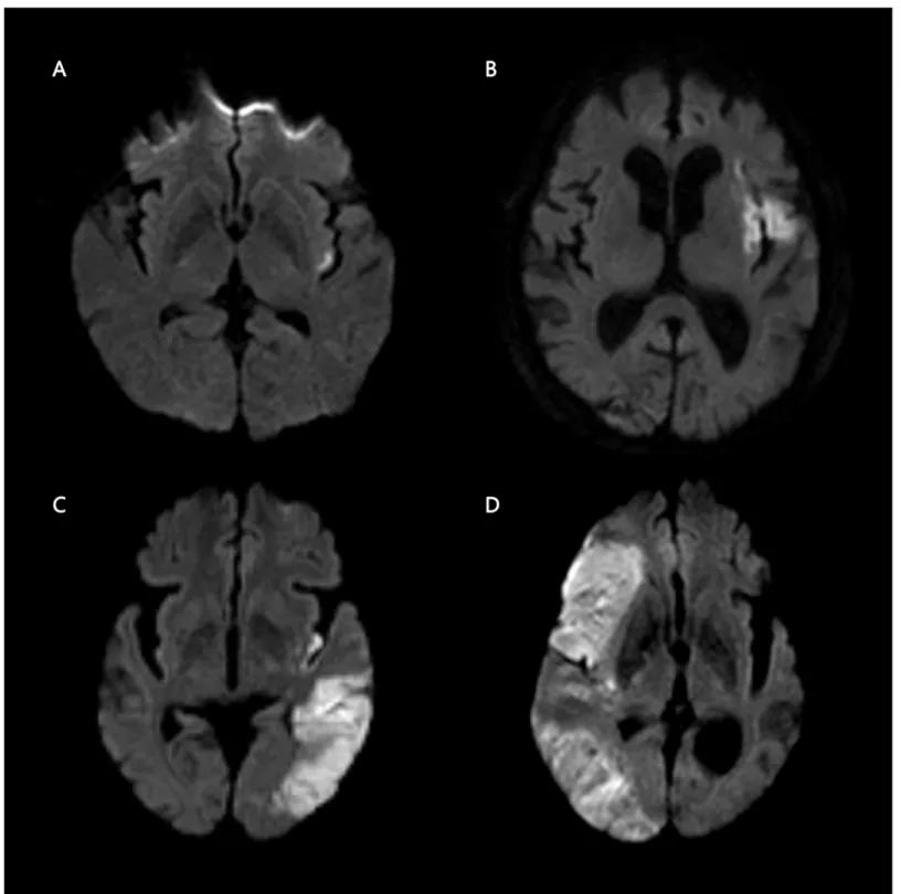
Patients with insular involvement were classified into 4 groups according to its pattern: iso- lated insula (IS), insula plus adjacent region (IA), insula plus remote region (IR), or large terri- torial infarction (more than one-third of the MCA territory; IL) (Fig 1). The adjacent region included the operculum, lenticulostriate nucleus, extreme capsule, and claustrum, whereas the remote region involved other areas within the MCA territory except for those assigned to the adjacent region.
With regard to the presence and location of symptomatic stenosis or occlusion of the cere- bral artery based on MRA results, the patients were categorized as follows: no vascular lesion, proximal vascular lesion, or distal vascular lesion. A proximal vascular lesion was defined as a symptomatic stenosis or occlusion located in the region from the proximal internal carotid artery to the proximal half of the main stem of the MCA, and these arteries were considered a possible embolic source. A distal vascular lesion was defined as a symptomatic stenosis or occlusion located in the region from the distal half of the M1 to the proximal portion of the M2, and these arteries were considered to be associated with the risk of in situ thrombosis.
Demographic, medical, and stroke data including age, baseline National Institutes of Health Stroke Scale (NIHSS) score, stroke mechanism, and presence of diabetes, hypertension, dyslipi- demia, and atrial fibrillation were extracted from the stroke registry. The primary outcome was the stroke mechanism, which was assigned by the neurologists based on the Trial of Org 10,172 in Acute Stroke Treatment (TOAST) classification [13]. CE was assigned when the ischemic lesions were from an embolism of cardiac origin in the absence of other embolic conditions and significant stenosis of the relevant cerebral artery on various angiographic studies.
Upon approval from the IRB, we also collected the 3-month modified Rankin scores (mRS) as the functional outcome. As a part of an institutional quality-of-care monitoring program for hospitalized stroke patients, the data on the 3-month functional status were prospectively recorded via direct observation, review of medical records, and telephone interviews. We defined the secondary outcome as a favorable functional status (mRS, 0–2).
Statistical analysis
Collected data are reported as number (percentage), mean ± standard deviation, or median (interquartile range, IQR) where appropriate. Baseline characteristics were compared between patients with and without insular involvement using a Pearson χ
2test for parametric variables, a Whitney-Mann U-test for non-parametric variables, and a t-test for continuous variables.
Comparison of primary and secondary outcomes with regard to insular involvement was per- formed using a Pearson χ
2test. For the secondary outcome, we estimated the adjusted odds ratio of insular involvement for a favorable functional status at 3 months. Based on potential variables that could affect the functional outcome that we identified from previous publications [13 – 15], we identified certain imbalanced variables with insular involvement in bivariate analy- sis (P < 0.2) and adjusted for these as confounders (P < 0.2). All probability values are 2-sided and considered statistically significant at <0.05. All analyses were performed using SPSS ver- sion 18.0 (SPSS Inc., Chicago, IL).
Results
A total of 1,311 patients with MCA territorial infarction were enrolled. The mean age was
67.7 ± 13.4 years, and men comprised 58.4% of the patients. The median baseline NIHSS score
was 5 (IQR, 2 –11), and 270 patients (20.6%) underwent some type of recanalization treatment
at the hyperacute stage. Stroke mechanisms were classified as follows: large artery atherosclero-
sis (LAA, 41.6%), CE (32.3%), other determined etiology (3.5%), and undetermined (UD,
22.6%).
Insular involvement was detected by DWI in 112 patients (8.5%). With regard to the pattern of insular involvement, IR was most commonly observed (32.7%), followed by IA (32.0%), IL (26.2%), and IS (4.1%) (Fig 1). Overall, patients with insular involvement demonstrated higher
Fig 1. Patterns of insular involvement in ischemic stroke. High signal intensity on diffusion-weighted images illustrates the patterns of insular involvement: isolated insula (A), insula plus adjacent region (B), insula plus remote region (C), and large territorial infarction (D).
doi:10.1371/journal.pone.0139540.g001
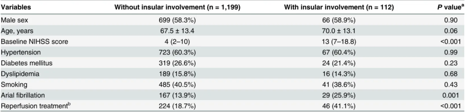
baseline stroke severity and atrial fibrillation rates, whereas some well-known atherosclerosis risk factors, such as hypertension, diabetes, and dyslipidemia, were not significantly different compared with patients without insular involvement (Table 1).
The distribution of stroke mechanisms was statistically different with regard to insular involvement (P < 0.001). In particular, the frequency of CE in patients with insular involve- ment (52.7%) was higher than that in patients without insular involvement (30.4%) (Table 2).
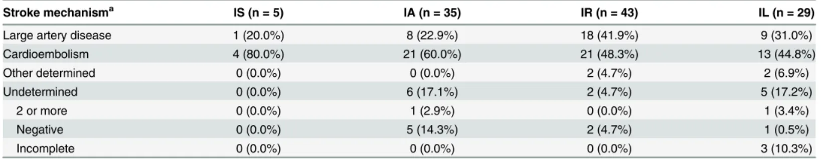
Depending on the pattern of insular involvement, the frequencies of CE ranged from 44.8% to 80.0%, but the difference between the patterns was not statistically significant (P = 0.18, Table 3).
A post-hoc analysis was used to investigate the location of symptomatic steno-occlusive lesions in patients who had insular involvement and were assigned to the LAA group of stroke mechanisms (n = 36). Of these patients, 75% had proximal vascular lesions located in the region from the proximal internal carotid artery to the proximal half of the M1, whereas the remaining 25% had vascular lesions between the distal half of the M1 and proximal part of the M2.
In the bivariate analysis, patients with insular involvement had significantly poorer func- tional status at 3 months compared with those without insular involvement (61.3% versus 37.4%, P < 0.001). However, after adjustment for age, baseline NIHSS score, atrial fibrillation, and recanalization treatment, insular involvement was not associated with the 3-month func- tional outcome (adjusted odds ratio, 0.79; 95% confidence interval, 0.47–1.32).
Table 1. Comparison of baseline characteristics of patients with and without insular involvement.
Variables Without insular involvement (n = 1,199) With insular involvement (n = 112) P value
aMale sex 699 (58.3%) 66 (58.9%) 0.90
Age, years 67.5 ± 13.4 70.0 ± 13.1 0.06
Baseline NIHSS score 4 (2 –10) 13 (7 –18.8) <0.001
Hypertension 723 (60.3%) 67 (60.4%) 0.99
Diabetes mellitus 319 (26.6%) 24 (21.4%) 0.23
Dyslipidemia 189 (15.8%) 16 (14.3%) 0.68
Smoking 485 (40.5%) 41 (38.6%) 0.43
Arial fibrillation 167 (13.9%) 29 (25.9%) 0.001
Reperfusion treatment
b224 (18.7%) 46 (41.1%) <0.001
Data are presented as number of patients (percentage), mean ± SD, or median (interquartile range).
a
P values were calculated using Pearson χ
2test, Whitney Mann U test, or t-test (see Methods).
b
Reperfusion treatment consisted of intravenous thrombolysis, intra-arterial treatment, and combined treatment.
doi:10.1371/journal.pone.0139540.t001
Table 2. Comparison of stroke mechanisms with regard to insular involvement.
Stroke mechanism Without insular involvement (n = 1,199) With insular involvement (n = 112)
Large artery disease 510 (42.5%) 36 (32.1%)
Cardioembolism 364 (30.4%) 59 (52.7%)
Other determined 42 (3.5%) 4 (3.6%)
Undetermined
Two or more 50 (4.2%) 2 (1.8%)
Negative 201 (16.8%) 8 (7.1%)
Incomplete 32 (2.7%) 3 (2.7%)
Data are presented as number of patients (percentage).
doi:10.1371/journal.pone.0139540.t002
Discussion
The results suggest that CE is the primary stroke mechanism in patients with insular involve- ment (approximately 50% of cases), while LAA constitutes the most common cause of ischemic stroke in the MCA territory. In addition, the data also indicate that arterial embolism may be responsible for insular involvement in most patients with LAA.
A previous study of 150 subjects with MCA territory ischemic stroke conducted in the United States described the clinical features of patients with insular involvement and proposed a greater association between insular involvement and CE (55.6% with insular lesions vs. 37.2%
without insular lesions) [7]. This higher frequency of CE in patients with insular involvement was somewhat surprising, given that a Korean epidemiologic study found that CE explained only 11% of total ischemic stroke cases [16]. Of note, a more recent statistical report demon- strated a greater frequency of CE in Korean stroke subjects (20%) [17], suggesting a stronger association between insular involvement and this stroke mechanism.
CE was consistently the most common cause of ischemic stroke for all insular lesion pat- terns. In particular, CE was detected in 80% and 60% of patients with IS and IA patterns, respectively, which supports the study hypothesis because the IS and IA patterns are more likely to result from occlusion of the transitional segment from the distal part of M1 to the proximal part of M2 by an embolus from the heart.
With regard to clinical practice, the association between CE and insular involvement may be useful for developing diagnostic approaches and deciding treatment strategies at the acute stage of ischemic stroke. Rapid detection of CE through the use of intensive studies, such as transthoracic echocardiography, transesophageal echocardiography, and multi-direction com- puted tomography, is important for determining the stroke etiology [18], and the results of the present study might help to select patients who would benefit from these extensive examina- tions. Moreover, our data is potentially useful for planning secondary prevention therapy, which can reduce the high stroke recurrence rate within the first month [19]. Finally, an associ- ation might exist with arterial embolism in patients with symptomatic steno-occlusive vascular lesions. Accordingly, intensive monitoring with transcranial sonography [20, 21] could be used to detect micro-emboli, and dual antiplatelet therapy could be utilized [22].
In the present study, patients with insular involvement showed high baseline neurologic impairment compared with the patients without insular involvement (NIHSS score, 13 vs. 4), which is consistent with a previous report (NIHSS score, 13.5 vs. 6) [7]. However, in contrast to other published data, an independent association between insular involvement and the
Table 3. Distribution of stroke mechanisms with regard to patterns of insular involvement.
Stroke mechanism
aIS (n = 5) IA (n = 35) IR (n = 43) IL (n = 29)
Large artery disease 1 (20.0%) 8 (22.9%) 18 (41.9%) 9 (31.0%)
Cardioembolism 4 (80.0%) 21 (60.0%) 21 (48.3%) 13 (44.8%)
Other determined 0 (0.0%) 0 (0.0%) 2 (4.7%) 2 (6.9%)
Undetermined 0 (0.0%) 6 (17.1%) 2 (4.7%) 5 (17.2%)
2 or more 0 (0.0%) 1 (2.9%) 0 (0.0%) 1 (3.4%)
Negative 0 (0.0%) 5 (14.3%) 2 (4.7%) 1 (0.5%)
Incomplete 0 (0.0%) 0 (0.0%) 0 (0.0%) 3 (10.3%)
Data are presented as number of patients (percentage). IS, insular isolated; IA, insular plus adjacent region; IR, insular plus remote region; IL, insular plus large territorial region
a