저작자표시-비영리-변경금지 2.0 대한민국 이용자는 아래의 조건을 따르는 경우에 한하여 자유롭게 l 이 저작물을 복제, 배포, 전송, 전시, 공연 및 방송할 수 있습니다. 다음과 같은 조건을 따라야 합니다: l 귀하는, 이 저작물의 재이용이나 배포의 경우, 이 저작물에 적용된 이용허락조건 을 명확하게 나타내어야 합니다. l 저작권자로부터 별도의 허가를 받으면 이러한 조건들은 적용되지 않습니다. 저작권법에 따른 이용자의 권리는 위의 내용에 의하여 영향을 받지 않습니다. 이것은 이용허락규약(Legal Code)을 이해하기 쉽게 요약한 것입니다. Disclaimer 저작자표시. 귀하는 원저작자를 표시하여야 합니다. 비영리. 귀하는 이 저작물을 영리 목적으로 이용할 수 없습니다. 변경금지. 귀하는 이 저작물을 개작, 변형 또는 가공할 수 없습니다.
Master's Thesis of Science in Medicine
Clinical characteristics with or without
retrieved thrombus in hyperacute ischemic stroke:
a histopathological evaluation
Ajou University Graduate School
Science in Medicine Major
Clinical characteristics with or without
retrieved thrombus in hyperacute ischemic stroke:
a histopathological evaluation
Ji Man Hong, Advisor
I submit this thesis as the
Master's thesis of Science in Medicine.
Feb, 2017
Ajou University Graduate School
Science in Medicine Major
The Master's thesis of Mun Hee Choi in Medicine is hereby approved.
Thesis Defense Committee President
홍 지 만 Seal
Member 김 장 희 Seal
Member 이 진 수 Seal
Ajou University Graduate School
i
-ABSTRACT-
Clinical Characteristics with or without Retrieved Thrombus in Hyperacute
Ischemic Stroke: a Histopathological Evaluation
With the advance of endovascular retrieval device for the cerebral thrombosis, it enables us to obtain a fresh thrombus of cerebral occlusion-site even in stroke survivors. We investigated the histological composition of retrieved thrombus and different effect of intravenous thrombolysis (IVT) according to the histological compositions of clots. We reviewed clinical, radiological, and interventional data of acute ischemic stroke patients from prospectively collected stroke and neurointervention registry at a stroke referral center from July 2014 to Jan 2016. The response to IVT was assessed by the change of clot burden score from initial CT angiography to digital subtraction angiography. The proportion of red blood cells (RBC), congregated fibrin/platelet, and white blood cells of the thrombus was analyzed by semi-automated color-based segmentation method (Positive Pixel Count Algorithm, Aperio scanscope). Patients were classified into RBC-dominant and fibrin-dominant or mixed group using receiver operating characteristic (ROC) curve analysis. Sixty patients were enrolled including 33 thrombus-available and 27 thrombus-unavailable patients. Thrombus-available group more likely to have cardioembolism and vessel sign, worse clot burden score, and use of retrieval stent. All 60 patients who received IVT, 35 patients showed IVT response and 25 patients did not show. The IVT-responsive group had more severe stroke (baseline NIHSS; 16.8±4.0 vs. 13.8±5.1, p=0.012), and showed more RBC-dominance (50.0% vs. 5.9%, p=0.007). Multivariate logistic regression analysis model revealed RBC dominance was an only variable influencing the good response to IVT (OR 15.816; 95% confidence interval 1.368-182.897, p=0.027). In analysis of characteristics according RBC ratio, the vessel signs and symptomatic hemorrhage according to the RBC ratio was more prevalent in patients with higher tertile than in those with lower levels. This study shows the histological composition of thrombus might lead to the different clinical characteristics, especially the response to IVT. The RBC dominance can be an important factor affecting the response to IVT, comparable with clinical or radiological findings in this study. The response to IVT appears to be
ii
influenced by histopathological features of retrieved thrombi and further studies are need to evaluate clinical implication.
iii
TABLE OF CONTENTS
ABSTRACT --- i
TABEL OF CONTENTS --- iii
LIST OF FIGURES --- iv
LIST OF TABLES --- - v
I. INTRODUCTION --- 1
II. PATIENTS AND METHODS --- -- 3
A. Patients and protocols --- 3
B. Endovascular treatment --- 3
C. Clinical and radiological evaluation --- 5
D. Histopathologic analysis --- 5
E. Statistical analysis --- 6
III. RESULTS --- 8
A. Comparison between the thrombus-available and thrombus-unavailable groups --- 8
B. Comparison between the IVT-responsive and IVT-unresponsive groups --- 11
C. Predictors of the response to IVT --- 14
D. Patient characteristics according to RBC ratio --- 16
IV. DISCUSSION --- 20
V. CONCLUSION --- 22
REFERENCES --- 23
iv
LIST OF FIGURES
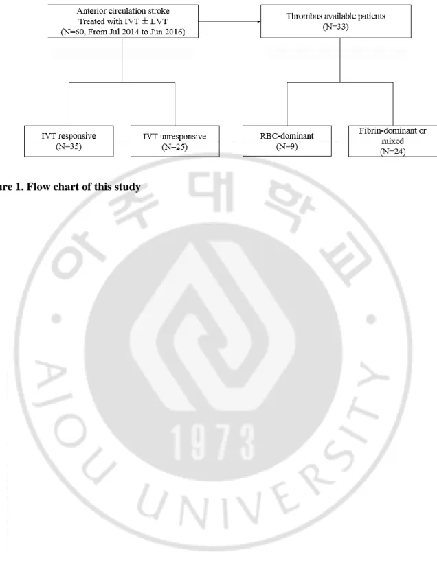
Fig 1.Flow chart of this study --- 4
Fig 2. The receiver operating characteristic (ROC) curve for development of thrombus component group
--- 7
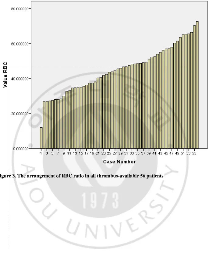
Fig 3. The arrangement of RBC ratio in all thrombus-available 56 patients --- 17
v
LIST OF TABLES
Table 1.Comparison of demographics and baseline characteristics between the thrombus-available and
thrombus-unavailable groups --- 9
Table 2. Comparison of radiological and treatment parameters between the thrombus-available and thrombus-unavailable groups --- 10
Table 3Comparison of demographics and baseline characteristics between the responsive and
IVT-unresponsive groups --- --- 12
Table 4. Comparison of radiological and treatment parameters between the responsive and IVT-unresponsive groups --- --- 13
Table 5. Logistic regression analyses of the relations between clinical covariates and the response to IVT
--- ---- 15
Table 6. Patient characteristics stratified by RBC ratio in all thrombus-available 56 patients --- --- 18
1
I.
INTRODUCTION
Endovascular treatment (EVT) is recently regarded as mandatory treatment option in acute ischemic stroke (AIS) with a proximal vessel occlusion. (Powers et al., 2015; Goyal et al., 2016) The serial success of large-scale, multicenter trials has led to changes in the treatment of AIS. (Berkhemer et al., 2015; Goyal et al., 2015; Saver et al., 2015; Campbell et al., 2015; Jovin et al., 2015) In real-world practice, however, all EVT candidates are not eligible for intra-arterial treatment. Substantial patients with acute ischemic stroke did not arrive at the appropriate hospital within the time available for EVT. Hong et al., 2013) Therefore, intravenous thrombolysis (IVT) with alteplase was still an important therapeutic modality.
The response to IVT has been assessed by various methods. Clinically, serial assessment of neurologic deficits using National Institutes of Health Stroke Scale (NIHSS) scores can be regarded as surrogate for the response to IVT. (Mikulik et al., 2007) However, due to the urgency of the treatment processes, serial neurologic examinations were largely limited. Considering the imaging parameters for recanalization, various modalities and protocols were used for evaluation of the response to IVT.
Non-invasive imaging studies-magnetic resonance angiography (MRA) (Kimura et al., 2011;Koga et al., 2013)
(Ritzenthaler et al., 2016) and transcranial Doppler (TCD) (Wunderlich et al., 2005; Wunderlich et al., 2007) were also used to evaluate recanalization after the IVT. Invasive but more accurate digital subtracted angiography (DSA) was also applied in several studies regarding angiographic grading system, such as modified thrombolysis in cerebral infarction (mTICI). (Zaidat et al., 2013) However, suboptimal
patients treated with IVT were recanalized to a certain level-mTICI 2b or 3, (Bhatia et al., 2010) and in
turn patients with mTICI 2b of 3 were primarily limited to obtain entirety of thrombi. TICI grading system is focused on reperfusion status after treatment, it does not detect subtle change of the occluded vessels. Because of this, it is difficult to apply mTICI grade for assessing the response to IVT. For the detailed evaluation on the response to IVT, another grading system is needed to address these questions.
On the other side, the response to IVT is influenced by numerous factors including occlusion site, clot burden, collateral status and vessel signs.(Seners et al., 2016) Proximal vessel occlusion showed
2
strong relationship with poor response to IVT.(Mishra et al., 2014) The length or burden of thrombi also was known to affect the response to IVT.(Mishra et al., 2014; Behrens et al., 2014) Additionally, poor collaterals was predictive of poor response to IVT in several studies.(von Kummer and Hacke, 1992; Nicoli et al., 2013) Hyperdense artery sign (HAS) on CT scan and susceptibility vessel sign (SVS) on
T2*-MR are well established early markers of acute arterial occlusion. (Leys et al., 1992; Cho et al., 2005)
These vessel signs are known to be related with poor clinical outcome. (Kharitonova et al., 2009)
However, the response to IVT regarding the vessel signs have not drawn definite conclusion. Qi ri et al. demonstrated poor neurological recovery in patients with proximal hyperdense middle cerebral artery
sign (HMCAS) after thrombolysis. (Li et al., 2014) Whereas D. Georgiadis et al. revealed that the
patients treated with IVT had better clinical outcome in positive HMCAS group. (Georgiadis et al., 2009)
In the aspect of thrombus histology, platelet-rich thrombus is known to be much more resistant to IVT compared to erythrocyte-rich clot in experimental study.(Jang et al., 1989) Until now, research on the effect of IVT according to histopathological feature of thrombus in stroke patients was not available. With the use of retrievable stent and aspiration catheter for acute large-vessel occlusion, it enable physicians to obtain fresh thrombi from cranial arteries even in stroke survivors. Furthermore, substantial AIS patients received IV thrombolysis (IVT) and endovascular treatment sequentially, the response to IVT can be evaluated angiographically in hyperacute stage of ischemic stroke. Recent studies on histological analysis of retrieved clots presented the relationship of stroke etiology, imaging features, and clinical outcome.(Niesten et al., 2014; Kim et al., 2015; Simons et al., 2015; Boeckh-Behrens et al., 2016) However, due to contradictory results and limited study design, the histological composition of thrombi still does not have definite clinical implications. Prior studies have suggested that studying thrombus composition can provide insights into stroke etiology, predict recanalization success following both intravenous thrombolysis
In this context, focused on the patients who were available for histologic analysis of retrieved thrombi, we aimed to investigate the different effect of IVT according to the histological compositions of clots.
3
II.
PATIENTS AND METHOD
A. Patients and protocols
Between July 2014 and June 2016, 89 consecutive acute stroke patients presenting with anterior circulation syndrome who received endovascular treatment enrolled in this study at a tertiary university hospital. All patients revealed acute neurologic symptoms attributable to cervical artery occlusion confirmed by CT angiography (CTA). Among the 89 patients, 60 patients received IVT treatment. We reviewed clinical, radiological and interventional data of 60 patients from prospectively collected stroke and neurointervention registry. Additionally, histopathological features of thrombus-available patients with or without IVT treatment were analyzed. Figure 1 showed a flow chart of this study. This study was approved by the institutional review board at Ajou University Medical Center.
B. Endovascular treatment
The baseline neurologic assessment was performed by stroke neurologists, including the National Institutes of Health Stroke Scale (NIHSS) score. Nonenhanced brain CT and CTA were conducted to evaluate eligibility for endovascular treatment. If candidate, the use of IV-rtPA was allowed before endovascular treatment. The inclusion criteria for endovascular treatment were as follows: 1) presentation within 8 h of symptom onset, 2) a baseline NIHSS score of ≥ 4, 3) the presence of anterior circulation artery occlusion on CTA, 4) no intracerebral hemorrhage on brain CT, 5) Alberta Stroke program early
CT (ASPECT) score (Barber et al., 2000) on initial nonenhanced CT ≥ 6, and 6) premobid modified
Rankin Scale (mRS) score of ≤ 2.
Cerebral angiography and endovascular treatment were performed by experienced neurointerventionalists. First, a guide catheter was positioned from the femoral artery to the proximal site of occluded vessels. According to the decision of examiners, a stent retriever (Solitaire AB/FR, Covidien, Irvine, California and Trevo, Concentric Medical Inc, Mountain View, California) or a Penumbra reperfusion catheter (Penumbra, Alameda, California) was selected for the first-line endovascular treatment. When first-line treatment modality was unsuccessful, alternative mechanical approaches were performed including switching strategy, angioplasty, and intra-art
4
5
rial tirofiban infusion. The details of the techniques were followed by previously described methods.(Lee
et al., 2013; Kim et al., 2016) Reperfusion status was assessed on the final angiogram 10-15 m after recanalization.
C. Clinical and radiological evaluation
Clinical data were comprised of demographic features, stroke risk factors, severity and clinical outcomes, subtypes of stroke, and use of IVT. Subtypes of stroke were determined according to the classification of the Trial of Org 10172 in Acute Stroke Treatment (Adams et al., 1993) by stroke neurologists. Stroke severity was assessed by NIHSS scores on admission, and clinical outcomes were evaluated by modified Rankin scale (mRS) at 3 months. The mRS score 0-2 at 3 months were classified as good outcome. Radiological data included ASPECT score HAS in baseline CT scans, the locations of the occlusion on CTA, susceptibility vessel sign (SVS) on gradient-recalled-echo (GRE) imaging of initial MR, and the response to IVT. The response to IVT was assessed by the change of clot burden score (CBS)(Tan et al., 2009) from initial CT angiography to digital subtraction angiography. Any increase of CBS defined as presence of IVT-responsiveness. Interventional data were time parameters-onset to puncture time, CT to puncture time, median procedure time, onset to reperfusion time. The reperfusion status was evaluated by the modified TICI score, (Zaidat et al., 2013) and the presence of symptomatic hemorrhage was investigated. Successful reperfusion was defined as a modified TICI score 2b or 3. Symptomatic hemorrhage was confined to any hemorrhage associated with a worsening of NIHSS score ≥ 4 within 24 h. (Saver et al., 2012)
D. Histopathologic analysis
After retrieval, thrombus material was fixed in 10% neutralized buffered formalin, and then embedded in paraffin. The formalin-fixed, paraffin-embedded tissue was cut into 4-μm thickness, followed by hematoxylin-eosin staining of slices. Stained slides were scanned at high resolution (× 400) by using a Scanscope XT digital scanner (Apergio, Vista, California). Histopathologic analysis of thrombi was conducted keeping blind to clinical and radiological findings. For assay of thrombi component, we used semi-automated color-based segmentation method (Positive Pixel Count Algorithm, Aperio
6
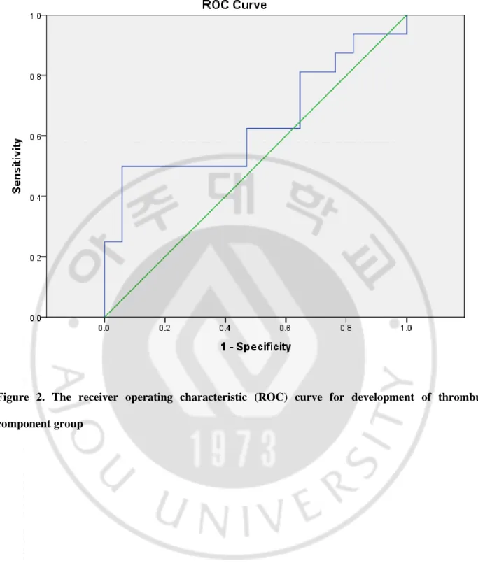
scanscope, Version 12.2, Leica Biosystems, Aperio, Vista, California) of the scanned slides. The percentage of red blood cells (RBCs), congregated fibrin/platelet, and white blood cells (WBCs) by area was used to thrombus analysis. Accordingly, thrombi were dichotomized into RBC-dominant and fibrin-dominant or mixed group using receiver operating characteristic (ROC) curve analysis. The cut-off value of RBC components related to the response to IVT was 12% (Figure 2; area under the curve, 0.643; sensitivity, 0.500; specificity, 0.941).
E. Statistical analysis
Baseline characteristics were presented as means (with standard deviation) or medians (with range) in numeric variables, and frequencies and percentage in categorical variables. To test for significant differences between groups, the χ2 or Fisher exact test was used for categorical variables, and the independent t test and Mann-Whitney U test were used for numeric variables. To identify factors associated with the response to IVT, univariate analyses were executed on age, sex, risk factors, stroke subtypes, baseline NIHSS, imaging parameters, time parameters, and thrombi histopathology. A multivariate logistic regression analysis model was built to assess the influence of thrombi histopathology on the IVT response. Variables were selected based on the results of univariate analyses (p<0.05). For analyses of histophatologic findings, we arranged the thombus-available 56 patients according to the RBC ratios and divided into the three groups. The lower, middle, and higher tertile groups were compared using χ2 or Fisher exact test for categorical variables and oneway Analysis of Variance (ANOVA) test for numeric variables. Statistical analyses were performed by using SPSS for Windows (Version 18.0; IBM, Armonk, New York). P<0.005 was considered statistical significant.
7
Figure 2. The receiver operating characteristic (ROC) curve for development of thrombus component group
8
III. RESULTS
A. Comparison between the thrombus-available and thrombus-unavailable groups
Sixty patients with acute ischemic stroke fulfilled the selection criteria and were included in this study. Retrieved clots were available in 33 patients, whereas 27 patients did not obtain thrombus— disintegration during retrieval or histological processing in 17 patients, and recanalization after IVT in 10 patients (Figure 1). Table 1 showed demographics and clinical characteristics between the thrombus-available and the thrombus-unthrombus-available groups. Age and sex were balanced between the two groups.
Hypertension was significantly morefrequent in patients with thrombus-unavailable patients
(thrombus-available; n=16, 48.5% vs. thrombus-un(thrombus-available; n=21, 77.8%; p=0.020). Other risk factors and the ratio of patients with prior antiplatelet use were similar in the two groups. The M1 segment of the middle cerebral artery was most commonly involved vessel in both groups. In thrombus-available group, cardioembolism (CE) was most common subtype of stroke, followed by large artery atherosclerosis (LAA) and cryptogenic embolism. The thrombus-unavailable group showed significantly different distribution of stroke subtype (p=0.010), 10 LAA patients (37.0%), 12 CE patients (44.4%), and 5 cryptogenic patients (18.5%). Baseline severity and clinical outcome after 3 months showed no difference between the two groups.
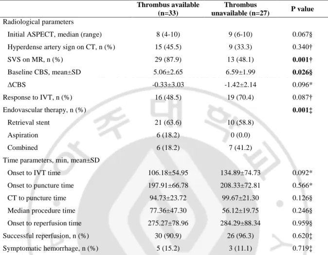
Radiological and treatment related parameters were presented in Table 2. ASPECT score and HAS on CT scan were balanced, whereas SVS on MR were more prevalent in the thrombus-available group (n=29, 87.9% vs. n=13, 48.1%; p=0.001). Clot burden was higher in the thrombus-available group (5.06±2.65 vs. 6.59±1.99; p=0.026). The response to IVT showed a better tendency in unavailable group without statistical significance (48.5% vs. 70.4%; p=0.087). In the thrombus-unavailable group, combined EVT methods were used simultaneously (n=9, 16.1% vs. n=11, 47.8%; p=0.000), resulting the disintegration of clots. Time parameters of EVT were balanced, successful reperfusion and symptomatic hemorrhage were achieved similar ratio in the two groups.
9
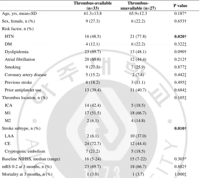
Table 1. Comparison of demographics and baseline characteristics between the thrombus-available and thrombus-unavailable groups
HTN = hypertension, DM = diabetes mellitus, ICA = internal carotid artery, LAA = large artery atherosclerosis, CE = cardioembolism, NIHSS = National Institutes of Health Stroke Scale, mRS = modified Rankin scale; *independent t-test, †Pearson’s chi-square test, ‡Fisher’s exact test
Thrombus-available (n=33)
Thrombus-unavailable (n=27) P value
Age, yrs, mean±SD 61.3±13.8 65.9±12.3 0.187*
Sex, female, n (%) 9 (27.3) 6 (22.2) 0.653† Risk factor, n (%) HTN 16 (48.5) 21 (77.8) 0.020† DM 4 (12.1) 6 (22.2) 0.322‡ Dyslipidemia 23 (69.7) 13 (48.1) 0.090† Atrial fibrillation 20 (60.6) 12 (44.4) 0.212† Smoking 9 (27.3) 7 (25.9) 0.877‡
Coronary artery disease 5 (15.2) 2 (7.4) 0.442‡
Previous stroke 6 (18.2) 3 (11.1) 0.495‡
Prior antiplatelet use 13 (39.4) 11 (40.7) 0.684‡
Thrombus location, n (%) 0.105‡ ICA 14 (42.4) 5 (18.5) M1 17 (51.5) 18 (66.7) M2 2 (6.1) 4 (14.8) Stroke subtype, n (%) 0.010† LAA 2 (6.1) 10 (37.0) CE 24 (72.7) 12 (44.4) Cryptogenic embolism 7 (21.2) 5 (18.5)
Baseline NIHSS, median (range) 16 (5-24) 15 (7-22) 0.303*
mRS 0-2 at 3 months, n (%) 23 (69.7) 18 (66.7) 0.802†
10
Table 2. Comparison of radiological and treatment parameters between the thrombus-available and thrombus-unavailable groups
ASPECT = Alberta Stroke program early CT score, SVS = susceptibility vessel sign, CBS = clot burden score, IVT = intravenous thrombolysis; *independent t-test, †Pearson’s chi-square test, ‡Fisher’s exact test, §Mann-Whitney U test Thrombus available (n=33) Thrombus unavailable (n=27) P value Radiological parameters
Initial ASPECT, median (range) 8 (4-10) 9 (6-10) 0.067§
Hyperdense artery sign on CT, n (%) 15 (45.5) 9 (33.3) 0.340†
SVS on MR, n (%) 29 (87.9) 13 (48.1) 0.001† Baseline CBS, mean±SD ΔCBS 5.06±2.65 -0.33±3.03 6.59±1.99 -1.42±2.14 0.026§ 0.096* Response to IVT, n (%) 16 (48.5) 19 (70.4) 0.087† Endovascular therapy, n (%) 0.001‡ Retrieval stent 21 (63.6) 10 (58.8) Aspiration 6 (18.2) 0 (0.0) Combined 6 (18.2) 7 (41.2)
Time parameters, min, mean±SD
Onset to IVT time 106.18±54.95 134.89±74.73 0.092*
Onset to puncture time 197.91±66.78 208.33±72.81 0.566*
CT to puncture time 94.73±23.72 99.67±21.30 0.126§
Median procedure time 77.36±47.30 56.12±19.75 0.246§
Onset to reperfusion time 275.27±78.96 284.29±88.34 0.959§
Successful reperfusion, n (%) 30 (90.9) 26 (96.3) 0.620‡
11
B. Comparison between the IVT-responsive and IVT-unresponsive groups
In all 60 patients, the responses to IVT were shown in 35 patients classified into the IVT-responsive and the rest 25 patients into IVT-unIVT-responsive groups. The comparison between the two groups were presented in Table 3 and Table 4. In general demographics, age were significantly younger in the IVT-unresponsive group (IVT-responsive; 66.2±11.9 vs. IVT-unresponsive; 59.3±14.2, p=0.045). Risk factor for ischemic stroke and prior antiplatelet use did not affect the response to IVT. M2 were more common in the IVT-responsive group (n=5, 14.3% vs. n=1, 4.0%), however, a statistical significance on thrombus location was not shown between the two groups. Subtype of stroke was not different in the two groups. Baseline stroke severity, represented with NIHSS, was higher in the IVT-responsive group (16.8±4.0 vs. 13.8±5.1, p=0.012). Consequently, mRS at 3 months was worse on the IVT-responsive group (n=18, 51.4% vs. n=21, 84.0%, p=0.012).
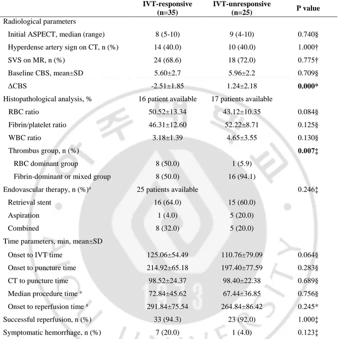
Table 4 presented with radiological, histopathological and treatment parameters between the IVT-responsive and IVT-unIVT-responsive groups. The vessel signs on CT and MR scan were not different between the two groups (HAS; n=14, 40.0% vs. n=10, 40.0%, p=1.000, SVS on MR; n=24, 68.6% vs. n=18, 72.0%, p=0.775), and the clot burden score also did not show difference (5.60±2.7 vs. 5.96±2.2, p=0.709). Thirty-three patients were thrombus-available—16 in the IVT-responsive group and 17 in the IVT-unresponsive group. In histopathological analysis, RBC-dominant patients were more common in IVT-responsive group (n=8, 50.0% vs. n=1, 5.9%) and fibrin-dominant or mixed patients were more common in IVT-unresponsive group (n=8, 50.0% vs. n=16, 94.1%; p=0.007), however, the ratio of component did not differ between the two groups. Treatment parameters related with EVT were balanced in the two groups.
12
Table 3. Comparison of demographics and baseline characteristics between the IVT-responsive and IVT-unresponsive groups
HTN = hypertension, DM = diabetes mellitus, ICA = internal carotid artery, LAA = large artery atherosclerosis, CE = cardioembolism, NIHSS = National Institutes of Health Stroke Scale, mRS = modified Rankin scale; *independent t-test, †Pearson’s chi-square test, ‡Fisher’s exact test
IVT-responsive (n=35)
IVT-unresponsive
(n=25) P value
Age, yrs, mean±SD 66.2±11.9 59.3±14.2 0.045*
Sex, female, n (%) 10 (28.6) 5 (20.0) 0.450† Risk factor, n (%) HTN 25 (71.4) 12 (48.0) 0.066† DM 7 (20.0) 3 (12.0) 0.499‡ Dyslipidemia 22 (63.0) 14 (56.0) 0.498† Atrial fibrillation 21 (60.0) 11 (44.0) 0.221† Smoking 9 (25.7) 7 (28.0) 0.773‡
Coronary artery disease 3 (8.6) 4 (16.0) 0.436‡
Previous stroke 5 (14.3) 4 (16.0) 1.000‡
Prior antiplatelet use 14 (40.0) 10 (40.0) 1.000†
Thrombus location, n (%) 0.507‡ ICA 11 (31.4) 8 (32.0) M1 19 (54.3) 16 (64.0) M2 5 (14.3) 1 (4.0) Stroke subtype, n (%) 0.710† LAA 6 (17.1) 6 (24.0) CE 21 (60.0) 15 (60.0) Cryptogenic embolism 8 (22.9) 4 (16.0)
Baseline NIHSS, median (range) 17 (7-24) 12 (5-22) 0.012*
mRS 0-2 at 3 months, n (%) 18 (51.4) 21 (84.0) 0.012†
13
Table 4. Comparison of radiological and treatment parameters between the IVT-responsive and IVT-unresponsive groups
ASPECT = Alberta Stroke program early CT score, SVS = susceptibility vessel sign, CBS = clot burden score, IVT = intravenous thrombolysis; *independent t-test, †Pearson’s chi-square test, ‡Fisher’s exact test, §Mann-Whitney U test; a: exclusion of 10 IVT-responsive patients due to recanalization after IVT use (only diagnostic angiography)
IVT-responsive (n=35)
IVT-unresponsive
(n=25) P value
Radiological parameters
Initial ASPECT, median (range) 8 (5-10) 9 (4-10) 0.740§
Hyperdense artery sign on CT, n (%) 14 (40.0) 10 (40.0) 1.000†
SVS on MR, n (%) 24 (68.6) 18 (72.0) 0.775†
Baseline CBS, mean±SD 5.60±2.7 5.96±2.2 0.709§
ΔCBS -2.51±1.85 1.24±2.18 0.000*
Histopathological analysis, % 16 patient available 17 patients available
RBC ratio 50.52±13.34 43.12±10.35 0.084§
Fibrin/platelet ratio 46.31±12.60 52.22±8.71 0.125§
WBC ratio 3.18±1.39 4.65±3.55 0.130§
Thrombus group, n (%) 0.007‡
RBC dominant group 8 (50.0) 1 (5.9)
Fibrin-dominant or mixed group 8 (50.0) 16 (94.1)
Endovascular therapy, n (%)a 25 patients available 0.246‡
Retrieval stent 16 (64.0) 15 (60.0)
Aspiration 1 (4.0) 5 (20.0)
Combined 8 (32.0) 5 (20.0)
Time parameters, min, mean±SD
Onset to IVT time 125.06±54.49 110.76±79.09 0.064§
Onset to puncture time 214.92±65.18 197.40±77.59 0.283§
CT to puncture time 98.52±24.37 98.40±22.38 0.689§
Median procedure time a 72.84±45.62 67.44±36.85 0.756§
Onset to reperfusion time a 291.84±75.54 264.84±86.42 0.245*
Successful reperfusion, n (%) 33 (94.3) 23 (92.0) 1.000‡
14
C. Predictors of the response to IVT
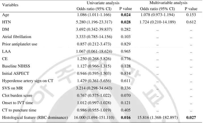
Logistic regression analysis was performed to detect influential factors on the response to IVT (Table 5). Multiple logistic regression was executed in thrombus-available and IVT-received 33 patients. In univariate analysis, age, hypertension, and RBC dominance had a significant effect on outcome (age; OR=1.086, 95% confidence interval, 1.011-1.166, p=0.024, hypertension; 5.280, 1.196-23.317, p=0.028, RBC dominance; 16.000, 1.694-151.110, p=0.027). Baseline NIHSS and clot burden score did not show significant relationship. After adjustment of age and hypertension, RBC dominance was an only variable influencing the good response to IVT (15.816; 1.368-182.897, p=0.027).
15
Table 5. Logistic regression analyses of the relations between clinical covariates and the response to IVT (n=33)
Variables Univariate analysis Multivariable analysis
Odds ratio (95% CI) P value Odds ratio (95% CI) P value
Age 1.086 (1.011-1.166) 0.024 1.078 (0.973-1.194) 0.153
HTN 5.280 (1.196-23.317) 0.028 1.724 (0.210-14.189) 0.612
DM 3.692 (0.342-39.837) 0.282 .. ..
Atrial fibrillation 3.333 (0.785-14.156) 0.103 .. ..
Prior antiplatelet use 0.857 (0.212-3.473) 0.829 .. ..
LAA 1.067 (0.061-18.624) 0.965 .. ..
CE 1.250 (0.268-5.826) 0.776 .. ..
Baseline NIHSS 1.127 (0.966-1.315) 0.128 .. ..
Initial ASPECT 0.946 (0.595-1.503) 0.814 .. ..
Hyperdense artery sign on CT 1.429 (0.361-5.656) 0.611 .. ..
SVS on MR 3.214 (0.298-34.643) 0.336 .. ..
Clot burden score 0.767 (0.575-1.022) 0.070 .. ..
Onset to IVT time 1.012 (0.997-1.028) 0.121 .. ..
CT to puncture time 0.986 (0.955-1.019) 0.405 .. ..
Histological feature (RBC dominance) 16.000 (1.694-151.110) 0.016 15.816 (1.368-182.897) 0.027 HTN = hypertension, DM = diabetes mellitus, LAA = large artery atherosclerosis, CE = cardioembolism, NIHSS = National Institutes of Health Stroke Scale, ASPECT = Alberta Stroke program early CT score, SVS = susceptibility vessel sign
16
D. Patient characteristics according to RBC ratio
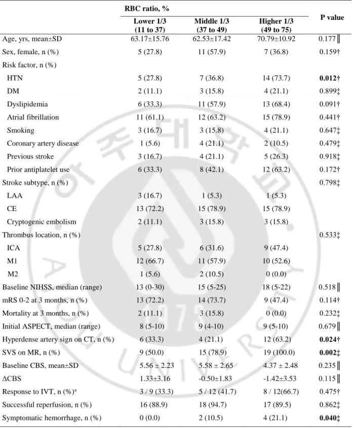
We analyzed the characteristics of all thrombus-available 56 patients, including who were did not receive IVT treatment. The patients were divided into the three groups according to the RBC ratio. The cut-off value for lower tertile group was 37%, and the value for middle tertile group was 49% (Figure 3). The patient characteristics according to the RBC ratio are shown in Table 6. Age, sex, risk factor profiles, subtype of stroke, and stroke severity were not different among the groups except hypertension (lower 1/3; n=5, 27.8% vs. middle 1/3; n=7, 36.8% vs. higher 1/3; n=14, 73.7%, p=0.012). However, vessel signs and symptomatic hemorrhage was more prevalent in patients with higher tertile than in those with lower levels (HAS; n=6, 33.3% vs. n=4, 21.1% vs. n=12, 63.2%; p=0.024, SVS; n=9, 50.0% vs. n=15, 78.9% vs. n=19, 100.0%; p=0.002, symptomatic hemorrhage; n=0, 0.0% vs. n=2, 10.5% vs. n=4, 21.1%; p=0.040). Figure 4 presented radiological and histopathological findings of the representative cases from RBC-dominant and fibrin-RBC-dominant patients.
17
18
Table 6. Patient characteristics stratified by RBC ratio in all thrombus-available 56 patients
HTN = hypertension, DM = diabetes mellitus, LAA = large artery atherosclerosis, CE = cardioembolism, ICA = internal carotid artery, NIHSS = National Institutes of Health Stroke Scale, ASPECT = Alberta Stroke program early CT score, SVS = susceptibility vessel sign, CBS = Clot burden score, IVT = intravenous thrombolysis;
†Pearson’s chi-square test, ‡Fisher’s exact test,║Oneway Analysis of Variance; a: only in patients received IVT RBC ratio, % PP value Lower 1/3 (11 to 37) Middle 1/3 (37 to 49) Higher 1/3 (49 to 75)
Age, yrs, mean±SD 63.17±15.76 62.53±17.42 70.79±10.92 0.177║
Sex, female, n (%) 5 (27.8) 11 (57.9) 7 (36.8) 0.159† Risk factor, n (%) HTN 5 (27.8) 7 (36.8) 14 (73.7) 0.012† DM 2 (11.1) 3 (15.8) 4 (21.1) 0.899‡ Dyslipidemia 6 (33.3) 11 (57.9) 13 (68.4) 0.091† Atrial fibrillation 11 (61.1) 12 (63.2) 15 (78.9) 0.441† Smoking 3 (16.7) 3 (15.8) 4 (21.1) 0.647‡
Coronary artery disease 1 (5.6) 4 (21.1) 2 (10.5) 0.479‡
Previous stroke 3 (16.7) 4 (21.1) 5 (26.3) 0.918‡
Prior antiplatelet use 6 (33.3) 8 (42.1) 12 (63.2) 0.172†
Stroke subtype, n (%) 0.798‡ LAA 3 (16.7) 1 (5.3) 1 (5.3) CE 13 (72.2) 15 (78.9) 15 (78.9) Cryptogenic embolism 2 (11.1) 3 (15.8) 3 (15.8) Thrombus location, n (%) 0.533‡ ICA 5 (27.8) 6 (31.6) 9 (47.4) M1 12 (66.7) 11 (57.9) 10 (52.6) M2 1 (5.6) 2 (10.5) 0 (0.0)
Baseline NIHSS, median (range) 13 (0-30) 15 (5-25) 18 (5-22) 0.518║ mRS 0-2 at 3 months, n (%) 13 (72.2) 14 (73.7) 9 (47.4) 0.114†
Mortality at 3 months, n (%) 2 (11.1) 3 (15.8) 0 (0.0) 0.232‡
Initial ASPECT, median (range) 8 (5-10) 9 (4-10) 9 (5-10) 0.679║ Hyperdense artery sign on CT, n (%) 6 (33.3) 4 (21.1) 12 (63.2) 0.024†
SVS on MR, n (%) 9 (50.0) 15 (78.9) 19 (100.0) 0.002‡ Baseline CBS, mean±SD 5.56 ± 2.23 5.58 ± 2.65 4.37 ± 2.48 0.235║ ΔCBS 1.33±3.16 -0.50±1.83 -1.42±3.53 0.115║ Response to IVT, n (%)a 3 / 9 (33.3) 5 / 12 (41.7) 8 / 12(66.7) 0.475† Successful reperfusion, n (%) 16 (88.9) 18 (94.7) 17 (89.5) 0.862‡ Symptomatic hemorrhage, n (%) 0 (0.0) 2 (10.5) 4 (21.1) 0.040‡
19
Figure 4. Representative cases from RBC-dominant (A) and fibrin-dominant or mixed groups (B)
(A) Early vessel signs are seen on nonenhance CT and gradient echo image of MR scan (red arrows). Initial CT angiography and digital subtraction angiography showed improvement of clot burden score (2 6 points). The color of gross specimen was categorized into dark red visually, and histological compositions were analyzed into RBC 65.0%, fibrin 33.7%, and WBC 1.3%.
(B) Patient from fibrin-dominant or mixed group did not show early vessel signs and response to IVT (clot burden score 4 3 points). The color of gross specimen was categorized into scarlet, and histological compositions were comprised in RBC 27.2%, fibrin 69.2%, and WBC 3.6%.
20
IV.
DISCUSSION
This study shows the histological composition of thrombus might lead to the different clinical characteristics, especially the response to IVT. In RBC dominant group, early vessel signs and the good response to IVT were more prevalent compared with fibrin-dominant or mixed group. After adjustment of other factors, the response to IVT was influenced by RBC dominance, most importantly. According to the RBC ratio, the vessel signs and symptomatic hemorrhage were more prevalent in patients with higher tertile RBC ratio group.
We could obtain entire thrombi from patients with cardioembolism, higher clot burden, and SVS on MR. Atrial fibrillation and the involvement of internal carotid artery (ICA) were more prevalent in thrombus-available group without significance. In comparison between the thrombus-available and thrombus-unavailable groups, various and combined EVT modalities were used in thrombus-unavailable patients. These process could be affect the integrity of thrombi, consequently, we could not obtain intact thrombi. The response to IVT did not show significance, it might be resulted from mixed characteristics of the patients. The presence or absence of intact thrombi did not affect to clinical and interventional outcome. We could scientifically confirm the experiences from the clinical field from this study.
In our study, the most relevant factor influencing the response to IVT was RBC dominance from the histopathological analysis. Although there was no statistical difference, IVT-responsive group was more prevalent M2 occlusion and symptomatic hemorrhage. In IVT-unresponsive group, the patients were younger and have more fibrin-dominant thrombi. Compared with previous studies, vessel signs and clot burden were not different according to IVT responsiveness. It may be due to small sample size of our data. Traditionally, thrombi were categorized into white—platelet-fibrin rich and red—RBC-rich clots. Red clots tend to form in low flow circumstance, whereas white clots adhere to roughened places in fast flow. (Caplan, 2006) During the formation of thrombus, incorporation of RBCs deemed to be mediated through a loose entrapment than that of platelets.(Brown et al., 1977) Therefore, thrombolytic agents may act by lysing the fibrin bridges in red thrombi, and do not have a lytic effect on white clots. In myocardial infarction, usually attributed to platelet-rich clots, the effect of IVT was limited in contrast to an acute
21
ischemic stroke. (O'Gara et al., 2013) Therefore, primary percutaneous coronary intervention was recommended than IVT in patients with acute myocardial infarction.
To find the factors related with RBC ratio, we arranged the patients with available thrombi according to the RBC ratio. Consequently, we analyzed clinical and radiological characteristics among the patients divided into three groups according to the RBC ratio. As the RBC ratio increased, the proportion of patients with vessel signs and symptomatic hemorrhage increased. There was a tendency of good response to IVT related with increase of RBC ratio. Clinical outcome at 3 months was worse with increase of RBC ratio, related with symptomatic hemorrhage. RBC dominance was related with vessel signs on imaging studies and might be related with symptomatic hemorrhage. This is line with previous studies, and worse outcome in patients with vessel signs is generally accepted. (Qureshi et al., 2006) Even the response to IVT was more prevalent in RBC-dominant group, considering clinical outcome and symptomatic hemorrhage, the use of IVT should be cautious in patients with vessel signs.
This study has several limitations. This is a retrospective study with small sample size. We try to reduce bias using prospectively collected registry and following a flow chart. Although single center study, in our best knowledge, this is a first study to evaluate the response of IVT focused on the patients who were available for histologic analysis of retrieved thrombi. Inhomogeneity due to the use of various EVT modalities could affect the results of our study. In future, prospective multi-center study are needed.
22
V.
CONCLUSION
This study shows the histological composition of thrombus might lead to the different clinical characteristics, especially the response to IVT. The RBC dominance can be an important factor affecting the response to IVT comparable with clinical or radiological findings in this study. These findings might affect clinical outcome through increased symptomatic hemorrhage related with increased RBC ratio. The response to IVT appears to be influenced by histopathological features of retrieved thrombi and clinical implication will be further evaluated.
23
REFERENCES
1. Adams HP, Jr., Bendixen BH, Kappelle LJ, Biller J, Love BB, Gordon DL, Marsh EE, 3rd:
Classification of subtype of acute ischemic stroke. Definitions for use in a multicenter clinical trial. TOAST. Trial of Org 10172 in Acute Stroke Treatment. Stroke 24: 35-41, 1993
2. Barber PA, Demchuk AM, Zhang J, Buchan AM: Validity and reliability of a quantitative
computed tomography score in predicting outcome of hyperacute stroke before thrombolytic therapy. ASPECTS Study Group. Alberta Stroke Programme Early CT Score. Lancet 355: 1670-1674, 2000
3. Behrens L, Mohlenbruch M, Stampfl S, Ringleb PA, Hametner C, Kellert L, Pham M, Herweh C,
Bendszus M, Rohde S: Effect of thrombus size on recanalization by bridging intravenous thrombolysis.
Eur J Neurol 21: 1406-1410, 2014
4. Berkhemer OA, Fransen PS, Beumer D, van den Berg LA, Lingsma HF, Yoo AJ, Schonewille
WJ, Vos JA, Nederkoorn PJ, Wermer MJ, van Walderveen MA, Staals J, Hofmeijer J, van Oostayen JA, Lycklama a Nijeholt GJ, Boiten J, Brouwer PA, Emmer BJ, de Bruijn SF, van Dijk LC, Kappelle LJ, Lo RH, van Dijk EJ, de Vries J, de Kort PL, van Rooij WJ, van den Berg JS, van Hasselt BA, Aerden LA, Dallinga RJ, Visser MC, Bot JC, Vroomen PC, Eshghi O, Schreuder TH, Heijboer RJ, Keizer K, Tielbeek AV, den Hertog HM, Gerrits DG, van den Berg-Vos RM, Karas GB, Steyerberg EW, Flach HZ, Marquering HA, Sprengers ME, Jenniskens SF, Beenen LF, van den Berg R, Koudstaal PJ, van Zwam WH, Roos YB, van der Lugt A, van Oostenbrugge RJ, Majoie CB, Dippel DW, Investigators MC: A randomized trial of intraarterial treatment for acute ischemic stroke. N Engl J Med 372: 11-20, 2015
5. Bhatia R, Hill MD, Shobha N, Menon B, Bal S, Kochar P, Watson T, Goyal M, Demchuk AM:
Low rates of acute recanalization with intravenous recombinant tissue plasminogen activator in ischemic stroke: real-world experience and a call for action. Stroke 41: 2254-2258, 2010
6. Boeckh-Behrens T, Schubert M, Forschler A, Prothmann S, Kreiser K, Zimmer C, Riegger J,
Bauer J, Neff F, Kehl V, Pelisek J, Schirmer L, Mehr M, Poppert H: The Impact of Histological Clot Composition in Embolic Stroke. Clin Neuroradiol 26: 189-197, 2016
7. Brown RS, Niewiarowski S, Stewart GJ, Millman M: A double-isotope study on incorporation
24
8. Campbell BC, Mitchell PJ, Kleinig TJ, Dewey HM, Churilov L, Yassi N, Yan B, Dowling RJ,
Parsons MW, Oxley TJ, Wu TY, Brooks M, Simpson MA, Miteff F, Levi CR, Krause M, Harrington TJ, Faulder KC, Steinfort BS, Priglinger M, Ang T, Scroop R, Barber PA, McGuinness B, Wijeratne T, Phan TG, Chong W, Chandra RV, Bladin CF, Badve M, Rice H, de Villiers L, Ma H, Desmond PM, Donnan GA, Davis SM, Investigators E-I: Endovascular therapy for ischemic stroke with perfusion-imaging selection. N Engl J Med 372: 1009-1018, 2015
9. Caplan LR: Antiplatelet therapy in stroke prevention: present and future. Cerebrovasc Dis 21
Suppl 1: 1-6, 2006
10. Cho KH, Kim JS, Kwon SU, Cho AH, Kang DW: Significance of susceptibility vessel sign on
T2*-weighted gradient echo imaging for identification of stroke subtypes. Stroke 36: 2379-2383, 2005
11. Georgiadis D, Wirz F, von Budingen HC, Valko P, Hund-Georgiadis M, Nedeltchev K, Rousson
V, Baumgartner RW: Intravenous thrombolysis in stroke patients with hyperdense middle cerebral artery sign. Eur J Neurol 16: 162-167, 2009
12. Goyal M, Demchuk AM, Menon BK, Eesa M, Rempel JL, Thornton J, Roy D, Jovin TG,
Willinsky RA, Sapkota BL, Dowlatshahi D, Frei DF, Kamal NR, Montanera WJ, Poppe AY, Ryckborst KJ, Silver FL, Shuaib A, Tampieri D, Williams D, Bang OY, Baxter BW, Burns PA, Choe H, Heo JH, Holmstedt CA, Jankowitz B, Kelly M, Linares G, Mandzia JL, Shankar J, Sohn SI, Swartz RH, Barber PA, Coutts SB, Smith EE, Morrish WF, Weill A, Subramaniam S, Mitha AP, Wong JH, Lowerison MW, Sajobi TT, Hill MD, Investigators ET: Randomized assessment of rapid endovascular treatment of ischemic stroke. N Engl J Med 372: 1019-1030, 2015
13. Goyal M, Menon BK, van Zwam WH, Dippel DW, Mitchell PJ, Demchuk AM, Davalos A,
Majoie CB, van der Lugt A, de Miquel MA, Donnan GA, Roos YB, Bonafe A, Jahan R, Diener HC, van den Berg LA, Levy EI, Berkhemer OA, Pereira VM, Rempel J, Millan M, Davis SM, Roy D, Thornton J, Roman LS, Ribo M, Beumer D, Stouch B, Brown S, Campbell BC, van Oostenbrugge RJ, Saver JL, Hill MD, Jovin TG, collaborators H: Endovascular thrombectomy after large-vessel ischaemic stroke: a meta-analysis of individual patient data from five randomised trials. Lancet 387: 1723-1731, 2016
25
Kim JS, Yoon BW: Stroke statistics in Korea: part I. Epidemiology and risk factors: a report from the korean stroke society and clinical research center for stroke. J Stroke 15: 2-20, 2013
15. Jang IK, Gold HK, Ziskind AA, Fallon JT, Holt RE, Leinbach RC, May JW, Collen D:
Differential sensitivity of erythrocyte-rich and platelet-rich arterial thrombi to lysis with recombinant tissue-type plasminogen activator. A possible explanation for resistance to coronary thrombolysis.
Circulation 79: 920-928, 1989
16. Jovin TG, Chamorro A, Cobo E, de Miquel MA, Molina CA, Rovira A, San Roman L, Serena J,
Abilleira S, Ribo M, Millan M, Urra X, Cardona P, Lopez-Cancio E, Tomasello A, Castano C, Blasco J, Aja L, Dorado L, Quesada H, Rubiera M, Hernandez-Perez M, Goyal M, Demchuk AM, von Kummer R, Gallofre M, Davalos A, Investigators RT: Thrombectomy within 8 hours after symptom onset in ischemic stroke. N Engl J Med 372: 2296-2306, 2015
17. Kharitonova T, Ahmed N, Thoren M, Wardlaw JM, von Kummer R, Glahn J, Wahlgren N:
Hyperdense middle cerebral artery sign on admission CT scan--prognostic significance for ischaemic stroke patients treated with intravenous thrombolysis in the safe implementation of thrombolysis in Stroke International Stroke Thrombolysis Register. Cerebrovasc Dis 27: 51-59, 2009
18. Kim SK, Yoon W, Kim TS, Kim HS, Heo TW, Park MS: Histologic Analysis of Retrieved Clots
in Acute Ischemic Stroke: Correlation with Stroke Etiology and Gradient-Echo MRI. AJNR Am J
Neuroradiol 36: 1756-1762, 2015
19. Kim YW, Hong JM, Park DG, Choi JW, Kang DH, Kim YS, Zaidat OO, Demchuk AM, Hwang
YH, Lee JS: Effect of Intracranial Atherosclerotic Disease on Endovascular Treatment for Patients with Acute Vertebrobasilar Occlusion. AJNR Am J Neuroradiol, 2016
20. Kimura K, Sakamoto Y, Aoki J, Iguchi Y, Shibazaki K, Inoue T: Clinical and MRI predictors of
no early recanalization within 1 hour after tissue-type plasminogen activator administration. Stroke 42: 3150-3155, 2011
21. Koga M, Arihiro S, Miyashita F, Yamamoto H, Yamada N, Nagatsuka K, Minematsu K, Toyoda
K: Factors associated with early recanalization failure following intravenous rt-PA therapy for ischemic stroke. Cerebrovasc Dis 36: 299-305, 2013
26
22. Lee JS, Hong JM, Lee SJ, Joo IS, Lim YC, Kim SY: The combined use of mechanical
thrombectomy devices is feasible for treating acute carotid terminus occlusion. Acta Neurochir (Wien) 155: 635-641, 2013
23. Leys D, Pruvo JP, Godefroy O, Rondepierre P, Leclerc X: Prevalence and significance of
hyperdense middle cerebral artery in acute stroke. Stroke 23: 317-324, 1992
24. Li Q, Davis S, Mitchell P, Dowling R, Yan B: Proximal hyperdense middle cerebral artery sign
predicts poor response to thrombolysis. PLoS One 9: e96123, 2014
25. Mikulik R, Ribo M, Hill MD, Grotta JC, Malkoff M, Molina C, Rubiera M, Delgado-Mederos R,
Alvarez-Sabin J, Alexandrov AV, Investigators C: Accuracy of serial National Institutes of Health Stroke Scale scores to identify artery status in acute ischemic stroke. Circulation 115: 2660-2665, 2007
26. Mishra SM, Dykeman J, Sajobi TT, Trivedi A, Almekhlafi M, Sohn SI, Bal S, Qazi E, Calleja A,
Eesa M, Goyal M, Demchuk AM, Menon BK: Early reperfusion rates with IV tPA are determined by CTA clot characteristics. AJNR Am J Neuroradiol 35: 2265-2272, 2014
27. Nicoli F, Lafaye de Micheaux P, Girard N: Perfusion-weighted imaging-derived collateral flow
index is a predictor of MCA M1 recanalization after i.v. thrombolysis. AJNR Am J Neuroradiol 34: 107-114, 2013
28. Niesten JM, van der Schaaf IC, van Dam L, Vink A, Vos JA, Schonewille WJ, de Bruin PC,
Mali WP, Velthuis BK: Histopathologic composition of cerebral thrombi of acute stroke patients is correlated with stroke subtype and thrombus attenuation. PLoS One 9: e88882, 2014
29. O'Gara PT, Kushner FG, Ascheim DD, Casey DE, Jr., Chung MK, de Lemos JA, Ettinger SM,
Fang JC, Fesmire FM, Franklin BA, Granger CB, Krumholz HM, Linderbaum JA, Morrow DA, Newby LK, Ornato JP, Ou N, Radford MJ, Tamis-Holland JE, Tommaso CL, Tracy CM, Woo YJ, Zhao DX, Anderson JL, Jacobs AK, Halperin JL, Albert NM, Brindis RG, Creager MA, DeMets D, Guyton RA, Hochman JS, Kovacs RJ, Kushner FG, Ohman EM, Stevenson WG, Yancy CW, American College of Cardiology Foundation/American Heart Association Task Force on Practice G: 2013 ACCF/AHA guideline for the management of ST-elevation myocardial infarction: a report of the American College of Cardiology Foundation/American Heart Association Task Force on Practice Guidelines. Circulation 127:
27
e362-425, 2013
30. Powers WJ, Derdeyn CP, Biller J, Coffey CS, Hoh BL, Jauch EC, Johnston KC, Johnston SC,
Khalessi AA, Kidwell CS, Meschia JF, Ovbiagele B, Yavagal DR, American Heart Association Stroke C: 2015 American Heart Association/American Stroke Association Focused Update of the 2013 Guidelines for the Early Management of Patients With Acute Ischemic Stroke Regarding Endovascular Treatment: A Guideline for Healthcare Professionals From the American Heart Association/American Stroke Association. Stroke 46: 3020-3035, 2015
31. Qureshi AI, Ezzeddine MA, Nasar A, Suri MF, Kirmani JF, Janjua N, Divani AA: Is IV tissue
plasminogen activator beneficial in patients with hyperdense artery sign? Neurology 66: 1171-1174, 2006
32. Ritzenthaler T, Lacalm A, Cho TH, Maucort-Boulch D, Klaerke Mikkelsen I, Ribe L,
Ostergaard L, Hjort N, Fiehler J, Pedraza S, Louis Tisserand G, Baron JC, Berthezene Y, Nighoghossian N: Sequential MR Assessment of the Susceptibility Vessel Sign and Arterial Occlusion in Acute Stroke. J
Neuroimaging 26: 355-359, 2016
33. Saver JL, Goyal M, Bonafe A, Diener HC, Levy EI, Pereira VM, Albers GW, Cognard C, Cohen
DJ, Hacke W, Jansen O, Jovin TG, Mattle HP, Nogueira RG, Siddiqui AH, Yavagal DR, Baxter BW, Devlin TG, Lopes DK, Reddy VK, du Mesnil de Rochemont R, Singer OC, Jahan R, Investigators SP: Stent-retriever thrombectomy after intravenous t-PA vs. t-PA alone in stroke. N Engl J Med 372: 2285-2295, 2015
34. Saver JL, Jahan R, Levy EI, Jovin TG, Baxter B, Nogueira RG, Clark W, Budzik R, Zaidat OO,
Trialists S: Solitaire flow restoration device versus the Merci Retriever in patients with acute ischaemic stroke (SWIFT): a randomised, parallel-group, non-inferiority trial. Lancet 380: 1241-1249, 2012
35. Seners P, Turc G, Maier B, Mas JL, Oppenheim C, Baron JC: Incidence and Predictors of Early
Recanalization After Intravenous Thrombolysis: A Systematic Review and Meta-Analysis. Stroke 47: 2409-2412, 2016
36. Simons N, Mitchell P, Dowling R, Gonzales M, Yan B: Thrombus composition in acute
ischemic stroke: a histopathological study of thrombus extracted by endovascular retrieval. J Neuroradiol 42: 86-92, 2015
28
37. Tan IY, Demchuk AM, Hopyan J, Zhang L, Gladstone D, Wong K, Martin M, Symons SP, Fox
AJ, Aviv RI: CT angiography clot burden score and collateral score: correlation with clinical and radiologic outcomes in acute middle cerebral artery infarct. AJNR Am J Neuroradiol 30: 525-531, 2009
38. von Kummer R, Hacke W: Safety and efficacy of intravenous tissue plasminogen activator and
heparin in acute middle cerebral artery stroke. Stroke 23: 646-652, 1992
39. Wunderlich MT, Goertler M, Postert T, Schmitt E, Seidel G, Gahn G, Samii C, Stolz E, Duplex
Sonography in Acute Stroke Study G, Competence Network S: Recanalization after intravenous thrombolysis: does a recanalization time window exist? Neurology 68: 1364-1368, 2007
40. Wunderlich MT, Stolz E, Seidel G, Postert T, Gahn G, Sliwka U, Goertler M, Duplex
Sonography in Acute Stroke Study G: Conservative medical treatment and intravenous thrombolysis in acute stroke from carotid T occlusion. Cerebrovasc Dis 20: 355-361, 2005
41. Zaidat OO, Yoo AJ, Khatri P, Tomsick TA, von Kummer R, Saver JL, Marks MP, Prabhakaran
S, Kallmes DF, Fitzsimmons BF, Mocco J, Wardlaw JM, Barnwell SL, Jovin TG, Linfante I, Siddiqui AH, Alexander MJ, Hirsch JA, Wintermark M, Albers G, Woo HH, Heck DV, Lev M, Aviv R, Hacke W, Warach S, Broderick J, Derdeyn CP, Furlan A, Nogueira RG, Yavagal DR, Goyal M, Demchuk AM, Bendszus M, Liebeskind DS, Cerebral Angiographic Revascularization Grading C, group SRw, Force STiCIT: Recommendations on angiographic revascularization grading standards for acute ischemic stroke: a consensus statement. Stroke 44: 2650-2663, 2013
29 -국문요약-