B16 흑색종세포에서 로바스타틴에 의한 멜라닌 합성 촉진효과에 미치는 세포내 칼슘의 역할
이 용 수# 덕성여자대학교 약학대학
(Received January 22, 2013; Revised February 18, 2013; Accepted February 20, 2013)
Role of Intracellular Ca
2+in the Lovastatin-Induced Stimulation of Melanin Synthesis in B16 Melanoma Cells
Yong Soo Lee#
College of Pharmacy, Duksung Women’s University, Seoul 132-714, Korea
Abstract — Although statins, inhibitors of 3-hydroxy-3-methylglutaryl-coenzyme A (HMG-CoA) reductase, have been shown to increase melanin synthesis, the exact mechanism of this action is not fully understood. In this study we inves- tigated the possible involvement of intracellular Ca2+ signal in the mechanism of stimulation of melanin synthesis induced by lovastatin in B16 cells. Lovastatin stimulated the production of melanin in a dose-dependent manner in the cells. Treat- ment with mevalonate, FPP and GGPP, precursors of cholesterol, did not significantly suppress the lovastatin-induced mel- anin production, suggesting that inhibition of cholesterol synthesis may not be involved in the mechanism of the action of lovastatin. In addition, lovastatin did not significantly alter the cAMP concentration and the stimulated production of melanin by lovastatin was not significantly changed by treatment with H89, a potent inhibitor of protein kinase A, which dem- onstrates that cAMP pathway may not be involved. However, lovastatin increased intracellular Ca2+ concentration in a dose- related fashion. Treatment with EGTA, an extracellular Ca2+ chelator did not significantly alter the lovastatin-induced intra- cellular Ca2+ increase and melanin synthesis, whereas intracellular Ca2+ reduction with BAPTA/AM and intracellular Ca2+
release blockers (dantrolene and TMB-8) completely blunted these actions of lovastatin. Taken together, these results sug- gest that the intracellular Ca2+ release may play an important role in the lovastatin-induced stimulation of melanin synthesis in B16 cells. These results further suggest that lovastatin may be useful for the treatment of hypopigmentation disorders, such as vitiligo.
Keywords □ lovastatin, melanogenesis, cAMP, Ca2+ signal, hypopigmentation, B16 melanoma cell
사람 피부색은 그 안에 존재하는 멜라닌의 농도에 의해 결정 된다. 피부 기저층의 멜라닌생성세포가 합성한 멜라닌은 각질형 성세포의 각화과정에 의해 세포질 돌기를 통하여 각질층으로 이 동된다.1) 멜라닌은 태양광선으로부터 나오는 자외선의 투과를 방 해하여 피부를 보호하는 기능을 하고 있지만 국소적으로 너무 많 이 생성되거나, 노화 등에 의해 피부의 기능이 감소하게 되면 멜 라닌이 피부 표면에 침착되어 기미, 주근깨 등의 다양한 과색소 질환을 일으키게 된다.2)멜라닌생성세포에서 멜라닌 합성은 멜라 노좀에 존재하는 복합적인 cascade 효소 반응에 의해 일어난다.3)
이 중 멜라닌합성의 속도를 결정하는 효소로서 tyrosinase는 두 가지 활성을 가지고 있는데 tyrosine을 3,4-dihydroxyphenylalanin (DOPA)로 전환하는 tyrosine hydroxylase 활성과 DOPA를 DOPA quinone으로 산화하는 DOPA oxidase 활성이 그것이다.
또한 5,6-dihydroxyindole-2-carboxylic acid(DHICA)는 tyrosinase- related protein(TRP)-1이라는 효소에 의해 indole-5,6-quinone- 2-carboxylic acid로 산화되며 TRP-2로도 불리는 dopachrome tautomerase(DCT) 효소는 dopachrome을 DHICA로 이성화한다.
멜라닌은 흑·갈색의 유멜라닌(eumelanin)과 적·노랑색의 피 어멜라닌(pheomelanin)으로 나뉘는데 tyrosinase는 이들 두 가 지 타입의 멜라닌 합성에 필요하며, TRP-1과 DCT는 유멜라닌 의 합성에 더 많이 관여하는 것으로 알려져 있다. 최근 다양한 식물추출물을 활용하여 tyrosinase 활성 및 멜라닌 합성 억제 작
#본 논문에 관한 문의는 저자에게로
(전화) 02-901-8396 (팩스) 02-901-8386 (E-mail) [email protected]
종설
용을 스크리닝하여 미백효과를 나타내는 기능성 화장품의 원료 물질로 개발하려는 시도가 많이 이루어지고 있다.4)
스타틴 계열 약물은 3-hydroxy-3-glutaryl-coenzyme A(HMG- CoA) 환원 효소를 억제하여 콜레스테롤의 합성을 저해한다. 최 근 연구에서 스타틴 계열 약물은 고콜레스테롤혈증의 치료뿐만 아니라 관상 혈관 질환이나5)암,6)골다공증7)및 뇌졸중8)등의 예방과 치료에 도움을 준다는 사실이 밝혀졌다. 또한 스타틴 약 물은 세포사멸(apoptosis)과 세포 주기 정지(cell cycle arrest)를 통하여 세포 주기의 진행을 억제하는 효과와9)흑색종 세포의 침 윤을 방해하는 효과10)를 가지고 있음이 보고되었다. 본 연구실 에서도 로바스타틴이 간암세포의 세포사멸을 유도한다는 사실을 밝힌 바 있다.11)
최근 사람 표피 멜라닌 세포에서 HMG-CoA 환원효소를 통하 여 콜레스테롤이 합성되고 콜레스테롤은 cAMP 경로를 통하여 멜라닌 합성을 촉진한다는 연구결과가 발표되었다.12)흥미롭게 도 HMG-CoA 환원효소를 억제하여 콜레스테롤 합성을 방해하 는 약물인 플루바스타틴에 의해 멜라닌 합성이 촉진된다는 상반 된 결과도 발표되었다.13)이처럼 아직까지 멜라닌 합성에 미치 는 콜레스테롤 및 스타틴 계열의 효과와 자세한 작용기전에 대 한 연구는 매우 부족한 상태이다.
따라서 본 연구에서는 스타틴 계열의 약물인 로바스타틴을 사 용하여 이 약물이 멜라닌 합성에 미치는 영향을 알아보고 그 기 전을 규명하고자 하였다.
실험방법
시약
Dulbecco's modified Eagle's medium(DMEM), Earle's basal salt solution(EBSS), 3-(4,5-dimethylthiazol-2-yl)-2,5-diphenyl tetrazolium bromide(MTT), mevalonolactone(mevalonate), geranylgeranyl pyrophosphate ammonium salt(GGPP), farnesyl pyrophosphate ammonium salt(FPP), dantrolene, 3,4,5- trimethoxybenzoic acid-8-(diethylamino)-octyl ester(TMB-8), ethylene glycol-bis-(aminoethyl ether)N,N,N',N'-tetraacetic acid (EGTA) 및 각종 용매와 염류는 Sigma-Aldrich(미국)에서 구입하 였고 lovastatin은 WAKO(일본)에서 구입하여 비활성 lactone형 을 에탄올에 녹인 후 0.4 M NaOH 에탄올용액에 50oC에서 2 시 간 동안 방치한 후 1 M HCl로 pH 7.2로 하여 dihydroxy open acid의 활성형으로 전환하여 농축액(10 mM)을 -20oC에서 보관하 였다. Fetal bovine serum(FBS)와 penicillin-streptomycin 혼합 액은 GIBCO(미국)에서 구입하였으며, 1-(2,5-carboxyoxazol-2-yl- 6-aminobenzfuran-5-oxyl)-2-(2'-amino-methylphenoxy)-ethane- N,N,N'N'-tetraacetoxylmethyl ester(Fura-2/AM)과 bis-(o- aminophenoxy)-ethane-N,N,N',N'-tetraacetic acid/acetoxymethyl
ester(BAPTA/AM)은 Molecular Probes(미국)에서 구입하였다.
BAPTA/AM 및 Fura-2/AM은 dimethyl sulfoxide(DMSO)에 녹 여 농축액으로 만든 후 물로 희석하여 사용하였다.
세포 배양
B16 흑색종 세포는 서울대학교 한국세포주은행에서 구입하였 다. 구입한 세포는 10% FBS와 penicillin 100 IU/ml와 strepto- mycin 50µg/ml을 함유한 DMEM 용액으로 37oC로 유지되는 5% CO2배양기(Forma, 미국)에서 배양하였다.
세포 생존율 측정
살아있는 세포는 미토콘드리아에 존재하는 탈수소 효소에 의 해 MTT가 MTT-formazan으로 전환되므로 이것의 양을 재면 살 아있는 세포의 수를 측정할 수 있다. 세포는 1 ml 배지에 여러 가지 약물 또는 약물을 녹인 용매만을 처리하여 24 well plate에 서 48시간 동안 배양하였다. 배양이 끝난 세포에 MTT 용액(2.5 mg/ml H2O) 100µl를 첨가하여 37oC에서 4시간 동안 반응시켰 다. 생성된 MTT-formazan 결정체를 분리하기 위하여 세포 현탁 액을 eppendorf tube에 옮겨 원심분리하였다(1500 rpm, 4분). 상 등액을 조심스럽게 제거하고 DMSO 100 µl를 첨가하여 결정체 를 용해시킨 후 540 nm에서 ELISA reader(Molecular Device, 미국)로 흡광도를 측정하였다. 세포생존율은 대조군과 비교하여 흡광도의 백분율로 나타내었다.
멜라닌 정량
B16 세포에서 멜라닌을 측정할 때에는 phenol red가 없는 DMEM을 사용하여 24 well plate에 ml 당 5×10⁴개로 분주하고 12시간이 지난 뒤 세포가 plate에 완전히 부착된 것을 확인한 후 시료를 처리하고 48시간 지난 뒤에 세포를 수집하여 세포수를 측정하고, 1200 rpm에서 5 분간 원심 분리하여 침전한 후, 1 ml 균질완충액(50 mM sodium phosphate pH 6.5, 1% Triton X- 100, 2 mM PMSF)로 용해시켰다. 여기서 얻은 세포 pellet을 1 N NaOH, 10% DMSO 용액 200 µl를 첨가하고 vortex 후 멜라닌 을 완전히 녹인 다음 96 well plate에 옮긴 후 ELISA reader (Molecular Device, 미국)를 사용하여 405 nm에서 흡광도를 측 정하였다. 멜라닌 표준품을 이용한 표준 검량선을 이용하여 멜 라닌 양을 산출하였다. 멜라닌 생성량은 단위세포(104개)에서의 멜라닌 생성량으로 비교하였다.
세포내 cAMP 측정
cAMP 농도는 cAMP immunoassay kit(Sigma-Aldrich, 미국) 을 이용해 측정하였다. B16 세포(7×10⁵)를 0.1 M HCl에 녹여 phosphodiesterase 활성을 억제한 후 상등액을 모아 중화·희석 시켜 토끼 다중클론항체를 고정시킨 96 well plate을 사용하여
cAMP conjugate를 첨가하여 세포용해액에 들어 있는 cAMP와 경쟁시켰다. 과량의 비결합 cAMP conjugate를 제거하기 위해 세 척하고 기질을 첨가한 후 420 nm에서 흡광도를 측정하였다. 흡 광도는 세포 용해액에 들어 있는 cAMP 농도와 반비례 관계에 있다.
세포내 칼슘이온 측정
B16 세포를 EBSS로 세척한 후 5 µM Fura-2/AM을 가해 37oC 에서 30분간 배양하였고 비봉입 Fura-2/AM은 150 g에서 30분간 원심분리하여 제거하였다. 세포를 Krebs-Ringer 완충액(125 mM NaCl, 5 mM KCl, 1.3 mM CaCl2, 1.2 mM KH2PO4, 1.2 mM MgSO4, 5 mM NaHCO3, 25 mM HEPES, 6 mM glucose 및 2.5 mM probenecid, pH 7.4)에 세포밀도가 ml당 2×10⁶개가 되 도록 현탁시켰다. Fura-2가 봉입된 세포를 형광측정 전까지 25oC 에서 90분간 유지시킨 후 교반봉이 들어있는 quartz cuvette에 0.5 ml 세포현탁액을 주입하여 Hitachi F4500 형광분석기를 사 용하여 여기파장을 340 및 280 nm에서 반복하게 하고 형광발광 을 510 nm에서 측정하였다. 세포내 칼슘농도는 340 : 380 nm 형 광비로써 나타내었다.
자료분석 및 통계적 검정
모든 실험은 네 번 반복해서 실시하고 실험 결과는 대조군의 조건에 대한 백분율로 나타내었다. 실험 결과는 평균값±SEM으 로 표시하고 ANOVA로 분석하며 각각의 유의성 비교는 Student- Newman-Keul's test를 이용하여 실시하였다. P값이 0.05 이하인 경우에만 통계학적 유의성이 있는 것으로 간주하였다.
실험결과 및 고찰
로바스타틴의 세포 독성
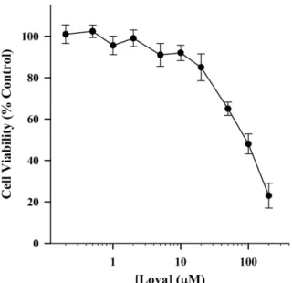
로바스타틴이 세포 독성에 미치는 농도를 조사하여 향후 실험 에 사용될 농도 범위를 결정하기 위해서 MTT 분석을 시행하였 다. B16 흑색종 세포에 대한 로바스타틴의 세포독성을 측정한 결과, 10 µM 이하의 농도로 처리했을 때 세포생존율은 90% 이 상으로 나타났으며, 50 µM을 처리했을 때 생존율이 유의성 있게 저하되었다(Fig. 1). 따라서 향 후 실험에서 로바스타틴은 10 µM 이하의 농도로 사용하였다. 로바스타틴은 다른 세포에서도 본 실 험에서 사용한 B16 세포에서의 결과와 비슷한 양상을 보이고 있 으며11)다른 스타틴 약물인 심바스타틴도 심장 섬유아세포에서 비슷한 농도에서 세포독성을 나타낸다.14)
로바스타틴의 멜라닌 생성 촉진 효과
B16 세포를 이용하여 로바스타틴이 멜라닌 합성에 미치는 영 향을 확인하였다. 로바스타틴을 처리하고 48시간 동안 배양 후
Fig. 1− Effects of lovastatin on cell viability in B16 melanoma cells.
Cells were incubated with or without each concentration of lovastatin (Lova) for 48 hr. Cell viability assay was done by the MTT staining method. Results are expressed as percent change of control condition in which cells were grown in medium without drugs. Data points represent the mean values of four replications with bars indicating SEM.
*p<0.05 compared to control in which the cells were incubated with lovastatin-free medium.
Fig. 2− Effect of lovastatin on the production of melanin in B16 melanoma cells. Melanin content was measured by the method described in the Method section. Briefly, the cells were initially grown for 12 hr in order to attach them to the bottom of the culture flasks. Then, the cells were incubated with each concentration of lovastatin (Lova) for 48 hr. Each column represents the mean value of four replications with bars indicating SEM. *p<0.05 compared to control in which the cells were incubated with lovastatin-free medium.
그 효과를 관찰하였는데 그 이유는 이 시간대에서 대조군, 즉 약 물을 가하지 않은 상태에서 분석이 가능한 멜라닌 합성이 이루 어지기 때문이다. 배양 후 B16 세포를 수집하여 멜라닌 양을 측 정한 결과, 로바스타틴을 처리한 군에서 농도 의존적으로 멜라 닌 합성이 증가되었으며 20 µM 농도 처리군에서 대조군에 비해 약 2배가 증가하여 통계적으로 유의한 촉진 효과를 나타내었다 (Fig. 2).
화장품 산업이 발달함에 따라 미백 기능성을 가진 물질의 개 발이 활발한 가운데 식물유래 폴리페놀 화합물이 멜라닌 합성을 저해한다는 연구결과가 발표되었다.15)그러나 이와는 반대로 어 떤 플라보노이드 화합물은 오히려 멜라닌 합성을 증가한다는 연 구결과도 발표되었다.16)본 연구에서 사용한 스타틴계 약물이 멜 라닌 합성에 미치는 영향에 관한 연구는 플루바스타틴의 효과에 대한 보고뿐이며13)여기에서 플루바스타틴은 멜라닌 합성을 촉 진하였는데 로바스타틴에 대한 본 연구결과와 일치한다.
로바스타틴에 의한 멜라닌 생성 촉진 효과에 미치는 콜레스테롤 대 사의 역할
로바스타틴은 HMG-CoA 환원효소를 저해하여 콜레스테롤의 합성을 억제하는 효과를 가지고 있으므로 그 작용기전에 콜레스 테롤 대사의 변화가 연관되어 있을 것으로 생각된다. 이것을 증 명하기 위하여 HMG-CoA 환원 효소의 대사물질인 mevalonate,
FPP, 또는 GGPP를 첨가하여 로바스타틴에 의한 멜라닌 합성 촉 진작용이 상쇄되는지를 확인하였다. 하지만 mevalonate(100 µM), FPP(10µM), 또는 GGPP(10 µM)의 첨가는 로바스타틴(5 µM)의 효과에 유의한 영향을 끼치지 못했으며, 이 물질 자체도 멜라닌 합성에 유의한 변화를 초래하지 않았다(Fig. 3). 이 결과로 미루 어 보아 로바스타틴에 의한 멜라닌 합성 촉진 효과와 HMG-CoA 환원효소의 억제를 통한 콜레스테롤의 합성 저해와는 서로 상관 관계가 없음을 시사한다.
최근 Schallreuter 등은 콜레스테롤이 cAMP 경로를 직접적으 로 자극하여 멜라닌 합성을 촉진한다는 결과를 발표하였다.12) 또한 Jin 등은 콜레스테롤 특유의 결합 물질인 methyl-β- cyclodextrin을 사용하여 콜레스테롤의 감소가 멜라닌 합성을 억 제한다는 사실을 밝혔다.17)하지만 본 연구의 결과로는 콜레스 테롤의 대사과정과 멜라닌 합성과는 연관관계가 없는 것으로 판 단된다. 서로 상반된 결과에 대한 이유는 현재로선 정확히 알 수 없으나 각 연구에서 사용한 세포의 종류가 다르기 때문일 가능 성이 있으며 이에 대해선 향후 좀 더 체계적인 연구가 필요할 것 으로 사료된다. 실제로 이전의 많은 연구에서도 본 연구의 결과 와 마찬가지로 스타틴 약물의 작용이 HMG-CoA 환원효소를 억 제하는 능력과 관계없이 일어나는 경우가 보고된 바 있다.18)
로바스타틴에 의한 멜라닌 생성 촉진 효과에 미치는 cAMP의 역할 멜라닌 합성은 세포내 여러 신호 전달 경로를 통하여 일어나는 데 멜라닌 합성관련 실험에서 흔히 사용되는 양성 대조군 화합물 인 α-melanoyte stimulating factor(α-MSH)에 의한 멜라닌 합성 촉진작용은 cAMP 경로를 통하여 일어난다고 알려져 있다.19) cAMP는 cAMP-dependent protein kinase A(PKA) 및 cAMP- response element binding protein(CREB) 전사인자의 활성화를 통하여 microphthalmia-associated transcription factor(MITF)의 발현을 증가함으로써 tyrosinase 유전자 발현을 자극하여 멜라닌 의 합성을 촉진한다.20)
따라서 다음은 로바스타틴에 의한 멜라닌 합성 촉진 작용에 미치는 기전에 관여하는 cAMP 경로의 역할을 조사하였다. 로바 스타틴은 멜라닌을 촉진하는 농도(1~10 µM)에서 cAMP 농도의 변화를 초래하지 않았으나 cAMP 경로를 상향조절하는 양성 대 조 물질인 FK(10 µM)에 의해서는 현저한 cAMP 농도의 증가를 보였다(Fig. 4A). 또한 FK(10 µM)는 약 3배의 멜라닌 합성을 증 가시켰으며 이 효과는 강력한 PKA 억제제인 H89(10 µM)의 전 처리에 의해 유의성 있게 감소되었다. 하지만 로바스타틴(5 µM) 에 의한 멜라닌 합성 증가는 H89에 의해 유의성 있는 변화를 보 이지 않았다(Fig. 4B). 이 결과는 로바스타틴에 의한 멜라닌 합 성 촉진 작용의 기전에 cAMP 경로는 중요한 역할을 하지 않음 을 시사한다.
많은 연구에서 멜라닌 합성을 촉진하거나 억제하는 물질들의 Fig. 3− Role of cholesterol metabolism in the lovastatin-induced
production of melanin in B16 melanoma cells. Melanin content was measured by the method described in Fig. 2.
In these experiments mevalonate (100µM), FPP (10 µM), and GGPP (10µM) were used as a cholesterol metabolite.
These drugs were added 30 min before lovastatin application.
Each column represents the mean value of four replications with bars indicating SEM. *p<0.05 compared to control in which the cells were incubated with lovastatin-free medium.
작용기전에 cAMP 경로가 중요하게 연루되어 있음이 보고되었 다.21) 흥미롭게도 사람 멜라닌 세포에서도 HMG-CoA 환원효소 를 통하여 콜레스테롤이 합성될 수 있고 이 과정에 cAMP 경로 가 중요한 매개 역할을 하고 있다는 사실이 밝혀졌다.12)하지만 콜레스테롤 대사와 멜라닌 합성과는 상관관계가 없어 보이는 본 연구 결과의 이유는 현재로는 정확히 알 수 없으며 이를 위해선 향후 체계적이고 다양한 접근을 통한 연구가 필요하다고 하겠다.
Fig. 4− Role of cAMP pathway in the lovastatin-induced production of melanin in B16 melanoma cells. In the data (A) cAMP was measured with each designated concentration of lovastatin (Lova) or 10µM of forskolin (FK), used as a positive control. Each column represents the mean value of four replications with bars indicating SEM. *p<0.05 compared to control in which the cells were incubated with drug-free medium. The data (B) represent the effects of H89, an inhibitor of protein kinase A, on the production of melanin induced by either Lova (5µM) or FK (10 µM).
H89 (10µM) was added 30 min before treatment with Lova or FK. Each column represents the mean value of four replications with bars indicating SEM. *p<0.05 compared to control in which the cells were incubated with drug-free medium. #p<0.05 compared to FK alone.
Fig. 5− Role of intracellular Ca2+ in the lovastatin-induced production of melanin in B16 melanoma cells. Intracellular Ca2+ concentration was assessed by Fura-2 fluorescence technique, and the data (A) represent intracellular Ca2+
changes with time. The arrow shows the time point for addition of lovastatin (5µM). In the data (B) intracellular Ca2+ increases are expressed as a percent change of control condition in which the cells were incubated with lovastatin-free medium. Each column represents the mean value of four replications with bars indicating SEM.
*p<0.05 compared to control. The data (C) represent the effect of BAPTA/AM, an intracellular Ca2+ chelator, on the production of melanin induced by lovastatin (Lova).
BAPTA/AM (1µM) was added 30 min before treatment with Lova (5µM). Each column represents the mean value of four replications with bars indicating SEM. *p<0.05 compared to control. #p<0.05 compared to Lova alone.
로바스타틴에 의한 멜라닌 생성 촉진 효과에 미치는 Ca2+신호의 역할
멜라닌 합성에 관련된 세포내 신호로는 cAMP 뿐만 아니라 칼 슘신호 또한 중요한 역할을 담당하고 있다는 사실이 알려지고 있 다.22)따라서 본 연구에서는 로바스타틴에 의한 멜라닌 합성 촉
진 작용에 미치는 칼슘신호의 역할을 알아보고자 하였다.
Fura-2 형광법을 통한 세포내 칼슘 측정에서 로바스타틴은 멜 라닌 합성을 촉진하는 농도(5 µM)에서 빠르게 세포내 칼슘 농도 를 증가시켰으며(Fig. 5A), 이 작용은 농도 의존적으로 나타났다 (Fig. 5B). 세포내 칼슘 킬레이트인 BAPTA/AM(1 µM)의 전처리 는 로바스타틴에 의한 멜라닌 합성을 유의성 있게 억제하였다 (Fig. 5C). 이 결과는 로바스타틴의 멜라닌 합성 촉진 작용에 세 포내 칼슘이 중요한 매개 신호로 역할을 하고 있음을 시사한다.
다음은 로바스타틴에 의한 세포내 칼슘 증가가 어떤 경로를 통해 일어나는 지를 추적하기 위해 1 mM EGTA를 함유하는 칼 슘-비포함 용액을 이용하여 세포내 칼슘변화를 관찰하였다. 이 실험 조건하에서는 세포외 칼슘 농도를 효과적으로 감소할 수 있 기 때문에 칼슘유입을 차단할 수가 있게 된다. 여기에서 로바스 타틴(5 µM)은 여전히 세포내 칼슘을 증가시켰으며(Fig. 6A) 멜 라닌 합성 능력에도 유의성 있는 변화를 보이지 않았다(Fig. 6B).
하지만 흥미롭게도 소포체막에 존재하는 ryanodine 수용체와 IP3 수용체를 각각 억제함으로써 소포체로부터 세포내 칼슘 유리를 방해하는 약물인 dantrolene(50 µM) 및 TMB-8(20 µM)은 로바 스타틴에 의한 칼슘증가와(Fig. 6A) 멜라닌 합성을(Fig. 6B) 유 의성 있게 감소시켰다. 이 결과는 칼슘 유리가 로바스타틴에 의 한 멜라닌 합성 촉진 작용을 매개한다는 중요한 신호라는 사실 을 입증한다.
세포내 칼슘신호는 세포의 분열, 분화 및 사멸은 물론 근육 수 축과 호르몬이나 신경전달물질의 외포작용 등 세포 기능에 중요 한 역할을 담당하고 있다고 알려져 있지만 멜라닌 합성에 미치 는 영향에 대해서는 많은 연구가 되어 있지 않다. Carsberg 등 의 연구에서 보면 자외선의 조사에 의해 세포내 칼슘 농도 및 멜 라닌 합성이 증가되고 인위적으로 세포외 칼슘농도를 높였을 때 도 멜라닌 합성이 증가되었다.23)이와는 반대로 칼슘 통로 억제 제인 verapamil이 멜라닌 합성을 촉진시킨다는 결과도 발표되었 다.24) 하지만 이 연구에서는 verapamil의 멜라닌 합성 촉진 작용 과 세포내 칼슘 동태와의 상관성에 대해선 연구하지 않았다. 본 연구 결과에서도 보듯이 세포내 칼슘은 멜라닌 합성을 촉진시키 는 과정에 관여할 것으로 생각되지만 좀 더 확실한 결론을 도출 하기 위해서는 이를 입증하는 많은 연구가 필요할 것으로 사료된다.
그럼에도 불구하고 본 연구에서 밝힌 로바스타틴이 멜라닌 합 성을 촉진한다는 사실은 이 약물이 백반증과 같은 멜라닌 합성 조절 이상에 의한 저색소질환의 치료용도로 활용될 가치가 충분 하다고 판단된다.
결 론
본 연구에서는 HMG-CoA 환원 효소 억제제인 로바스타틴이 멜라닌 합성에 미치는 영향을 알아보고 그 기전을 규명하고자 하 Fig. 6− Role of intracellular Ca2+ release in the lovastatin-induced
production of melanin in B16 melanoma cells. In the data (A) intracellular Ca2+ concentration was assessed by Fura- 2 fluorescence technique. In these experiment dantrolene (50µM) and TMB-8 (20 µM), intracellular Ca2+ release blockers, were added 5 min before lovastatin (Lova, 5µM) application. In the EGTA group, a nominal Ca2+-free medium containing 100µM EGTA was used. The data are expressed as percent changes of intracellular Ca2+ levels compared to the control condition in which the cells were incubated with Lova-free medium. Each column represents the mean values of four replications with bars indicating SEM. *p<0.05 compared to control. #p<0.05 compared to Lova alone. In the data (B) the effects of these inhibitors of Ca2+ signal on the lovastatin-induced production of melanin was observed. In these experiments EGTA (100µM), dantrolene (50µM) and TMB-8 (20 µM) were added 30 min before treatment with Lova (5µM). Each column represents the mean values of four replications with bars indicating SEM. *p<0.05 compared to control. #p<0.05 compared to Lova alone.
였다. 실험결과에서 로바스타틴은 B16 세포에서 농도의존적으로 멜라닌 합성을 증가시켰다. 로바스타틴에 의한 멜라닌 합성 촉 진작용은 mevalonate, FPP 및 GGPP 등 HMG-CoA 환원효소 의 억제를 상쇄시킬 수 있는 대사체의 처리에 의해 유의성 있는 변화를 보이지 않았으므로 로바스타틴의 작용은 콜레스테롤 대 사과정과는 무관한 것으로 생각된다. 또한 로바스타틴은 세포내 cAMP 농도를 변화시키지 않았고 PKA 억제제인 H89를 처리해 도 로바스타틴에 의한 멜라닌 합성은 영향을 받지 않은 결과는 로바스타틴의 작용에 cAMP 경로가 매개 신호로 작용하고 있지 않음을 시사한다. 하지만 로바스타틴은 세포내 칼슘 농도를 농 도의존적으로 증가시켰고 세포내 칼슘 킬레이트인 BAPTA/AM 의 처리에 의해 로바스타틴의 멜라닌 합성촉진 작용이 유의성있 게 감소되었다. 로바스타틴에 의한 칼슘 증가 및 멜라닌 합성 작 용은 세포외 칼슘 킬레이트인 EGTA에 의해서는 변화가 엇었으 나 세포내 칼슘 유리 억제 약물인 dantrolene 과 TMM-8에 의 해서는 완전히 차단되는 것으로 보아 칼슘신호는 로바스타틴의 이런 작용에 깊이 연루되어 있다고 판단된다. 본 연구에서 밝힌 로바스타틴의 멜라닌 합성 촉진 작용은 멜라닌 합성장애에 의한 저색소질환의 치료제 개발연구에 활용될 충분한 가치가 있다고 판단된다.
감사의 말씀
본 연구는 덕성여자대학교 2012년도 교내연구비 지원에 의해 수행되었음.
참고문헌
1) Boissy, R. E. : Melanosome transfer to and translocation in the keratinocyte. Exp. Dermatol. 12 Suppl 2, 5 (2003).
2) Gillbro, J. M. and Olsson, M. J. : The melanogenesis and mechanisms of skin-lightening agents-existing and new approaches. Int. J. Cosmet. Sci.
33, 210 (2011).
3) Schallreuter, K. U., Kothari, S., Chavan, B. and Spencer, J. D. : Regulation of melanogenesis-controversies and new concepts.
Exp. Dermatol. 17, 395 (2008).
4) Chang, T. S. : An updated review of tyrosinase inhibitors. Int.
J. Mol. Sci. 10, 2440 (2009).
5) Sposito, A. C. and Chapman, M. J. : Statin therapy in acute coronary syndromes: mechanistic insight into clinical benefit.
Arterioscler. Thromb. Vasc. Biol. 22, 1524 (2002).
6) Muck, A. O., Seeger, H. and Wallwiener, D. : Inhibitory effect of statins on the proliferation of human breast cancer cells. Int.
J. Clin. Pharmacol. Ther. 42, 695 (2004).
7) Hatzigeorgiou, C. and Jackson, J. L. : Hydroxymethylglutaryl- coenzyme A reductase inhibitors and osteoporosis: a meta-
analysis. Osteoporos. Int. 16, 990 (2005).
8) Szczepanska-Szerej, A., Kurzepa, J., Wojczal, J. and Stelmasiak, Z. : Simvastatin-induced prevention of the increase in TNF- level in the acute phase of ischemic stroke. Pharmacol. Rep.
59, 94 (2007).
9) Saito, A., Saito, N., Mol, W., Furukawa, H., Tsutsumida, A., Oyama, A., Sekido, M., Sasaki, S. and Yamamoto, Y. : Simvastatin inhibits growth via apoptosis and the induction of cell cycle arrest in human melanoma cells. Melanoma Res. 18, 85 (2008).
10) Glynn, S. A., O'Sullivan, D., Eustace, A. J., Clynes, M. and O'Donovan, N. : The 3-hydroxy-3-methylglutaryl-coenzyme A reductase inhibitors, simvastatin, lovastatin and mevastatin inhibit proliferation and invasion of melanoma cells. BMC Cancer 8, 9 (2008).
11) Lee, Y. S. : Lovastatin induces apoptotic cell death by activation of intracellular Ca2+ signal in HepG2 human hepatoma cells. J. Appl. Pharmacol. 15, 137 (2007).
12) Schallreuter, K. U., Hasse, S., Rokos, H., Chavan, B., Shalbaf, M., Spencer, J. D. and Wood, J. M. : Cholesterol regulates melanogenesis in human epidermal melanocytes and melanoma cells. Exp. Dermatol. 18, 680 (2009).
13) Galus, R., Niderla, J., Sladowski, D., Sajjad, E., Wlodarski, K.
and Jozwiak, J. : Fluvastatin increases tyrosinase synthesis induced by a-melanocyte-stimulating hormone in B16F10 melanoma cells. Pharmacol. Rep. 62, 164 (2010).
14) Copaja, M., Venegas, D., Aranguiz, P., Canales, J., Vivar, R., Avalos, Y., Garcia, L., Chiong, M., Olmedo, I., Catalan, M., Leyton, L., Lavandero, S. and Diaz-Araya, G. : Simvastatin disrupts cytoskeleton and decreases cardiac fibroblast adhesion, migration and viability. Toxicology 294, 42 (2012).
15) Yoshioka, S., Terashita, T., Yoshizumi, H. and Shirasaka, N. : Inhibitory effects of whisky polyphenols on melanogenesis in mouse B16 melanoma cells. Biosci. Biotechnol. Biochem. 75, 2278 (2011).
16) Huang, Y. C., Liu, K. C. and Chiou, Y. L. : Melanogenesis of murine melanoma cells induced by hesperetin, a Citrus hydrolysate-derived flavonoid. Food Chem. Toxicol. 50, 653 (2012).
17) Jin, S. H., Lee, Y. Y. and Kang, H. Y. : Methyl-β-cyclodextrin, a specific cholesterol-binding agent, inhibits melanogenesis in human melanocytes through activation of ERK. Arch. Dermatol.
Res. 300, 451 (2008).
18) Zhou, Q. and Liao, J. K. : Pleiotropic effects of statins. Basic research and clinical perspectives. Circ. J. 74, 818 (2010).
19) Busca, R. and Ballotti, R. : Cyclic AMP a key messenger in the regulation of skin pigmentation. Pigment Cell Res. 13, 60 (2000).
20) Bentley, N. J., Eisen, T. and Goding, C.R. : Melanocyte-specific
expression of the human tyrosinase promoter: activation by the microphthalmia gene product and role of the initiator. Mol.
Cell. Biol. 14, 7996 (1994).
21) Liang, C. H., Chou, T. H., Tseng, Y. P. and Ding, H. Y. : trans- Caffeic acid stearyl ester from Paeonia suffruticosa inhibits melanin synthesis by cAMP-mediating down-regulation of α- melanocyte-stimulating hormone-stimulated melanogenesis signaling pathway in B16 cells. Biol. Pharm. Bull. 35, 2198 (2012).
22) deOliveira, A. R., Castrucci, A. M., Visconti, M. A. : Cellular
signalling in vertebrate pigment cells. Braz. J. Med. Biol. Res.
29, 1743 (1996).
23) Carsberg, C. J., Jones, K. T., Sharpe, G. R. and Friedmann, P. S. : Intracellular calcium modulates the responses of human melanocytes to melanogenic stimuli. J. Dermatol. Sci. 9, 157 (1995).
24) Parker, C. and Sherbet, G. V. : The Ca2+ channel blocker verapamil enhances melanogenesis without altering metastatic potential in the B16 murine melanoma. Melanoma Res. 3, 347 (1993).