230
Original Article
Korean Circulation J 2007;37:230-233
ISSN1738-5520
ⓒ 2007, The Korean Society of Circulation CASE REPORT
Very Late Stent Thrombosis in Coronary Bare-Metal Stent Implantation:
A Case Report
Yoon-Sung Cho, MD, Hyun-Gwang Jung, MD, Tae-Woo Kim, MD, Kang-Min Kim, MD, Jae-Hoon Chang, MD, Dong-Hyun Rho, MD, Chang-Hun Lee, MD,
Joon-Sang Lee, MD, Byung-Jae Ahn, MD and Joon-Hoon Jeong, MD Department of Internal Medicine, Wallace Memorial Baptist Hospital, Busan, Korea ABSTRACT
Stent thrombosis is generally a fatal complication after percutaneous coronary intervention. Despite the incidence of stent thrombosis has reduced with improved techniques and drugs, stent thrombosis persists at a rate of 0.5-2%
in elective cases, and up to 6% in patients with acute coronary syndromes. It almost always causes acute myocardial infarction or sudden cardiac death. While very late stent thrombosis, occurring beyond 1 year, is not uncommon with the use of drug-eluting stents, it is distinctly unusual with the use of bare-metal stents. We report a case of very late thrombosis of a bare-metal stent occurring 880 days after coronary stent implantation. (Korean Circulation J 2007;37:230-233)
KEY WORDS:Coronary thrombosis;Stents.
Introduction
Stent thrombosis remains the primary cause of death after percutaneous coronary intervention(PCI). Despite improvement of PCI, stent thrombosis persists at a rate of 0.5-2% in elective cases, and up to 6% in patients with acute coronary syndromes.1)2) Stent thrombosis most often develops within the first 48 hours after the PCI, and rarely after a week of stent implantation.2) It almost always causes acute myocardial infarction(AMI) or sud- den cardiac death.3-5) While very late stent thrombosis (VLST), occurring beyond 1 year, is not uncommon with the use of drug-eluting stents(DES), it is distinctly unusual with the use of bare-metal stents(BMS).3)4)6) We report a case of very late thrombosis of a bare-metal stent occurring 880 days after implantation.
Case
A 48-year-old male presented to our hospital with increasing exertional chest pain for a month on Nove-
mber 7, 2003. His chest pain was characterized by squee- zing pattern and this was located in the substernal area and it radiated to his both shoulder. He had a managed for diabetes mellitus and hyperlipidemia. His initial blood pressure was 120/80 mmHg and pulse rate 88 beats per minute. The electrogram on admission showed T wave inversion in lead V1-4. Echocardiography reve- aled normal systolic function without any regional wall motion abnormality.
Cononary angiography revealed a subtotal occlusion of the proximal left anterior descending coronary artery (LAD)(Fig. 1), and an Arthos®(AMG, Faesfeld-Erle, Ger- many) stent(3.0×38 mm at 14 atm) was implanted with an excellent angiographic result(Fig. 2). He was disch- arged in stable condition and medicated with aspirin, clopidogrel, statin and oral hypoglycemic agents. Six months later, the patient was visit to our hospital for re- curring exertional chest pain in May 2004. He had been taking clopidogrel(75 mg/day) for the initial 1 month post-stent implantation, but aspirin was continued. His coronary angiography revealed about 90% in-stent re- stenosis(ISR) of the previously stented segment of the proximal LAD(Fig. 3A), and the cutting balloon angio- plasty(CBA) was performed(Fig. 3B). Clopidogrel was given for 6 months, and then it was discontinued there- after, while aspirin was continued. After the procedure, the patient underwent follow-up coronary angiography with an excellent angiographic result in December 2004
Received:February 19, 2007 Revision Received:March 21, 2007 Accepted:May 2, 2007
Correspondence:Joon-Hoon Jeong, MD,Department of Internal Medicine, Wallace Memorial Baptist Hospital, 374-75 Namsan-dong, Geumjeong- gu, Busan 609-728, Korea
Tel: 82-51-580-1202, Fax: 82-51-583-6200 E-mail: [email protected]
Yoon-Sung Cho, et al:Very Late Stent Thrombosis in Bare-Metal Stent Implantation·231
(Fig. 4A, B). The patient was completely asymptomatic until April 2006. On April 5, 2006, the patient visited to other hospital with sudden severe chest pain. His electrocardiogram(ECG) revealed ST-segment elevation and Q-wave in precordial leads V1-4(Fig. 5). The patient was diagnosed as ST-elevation acute myocardial infar- ction in the anteroseptal localization. So, fibrinolytic agent was administrated in other hospital. And then, he was transferred to our hospital one day later. His ECG still revealed ST-segment elevation and Q-wave in precordial leads V1-4, and his troponin I increased up to 34.5 ng/mL on April 6, 2006. The antithrombin III, protein C and protein S levels were 25 mg/dL, 77.51% and 91.86%, respectively, in within normal limits.
The patient was given aspirin and clopidogrel and was also started on heparin and nitroglycerin infusions in the emergency room. On April 7, 2006, his ECG reve- aled normalization of ST-segment elevation with Q- wave. He was transferred to the cardiac catheterization laboratory on April 8, 2006. Coronary angiography revealed a thrombotic occlusion of the previously stented segment of the proximal LAD, with 30% ISR(Fig. 6).
But, the patient did not undergo any further coronary procedure because of restoration of perfusion to distal TIMI 3 flow. The patient was discharged with aspirin, clopidogrel and warfarin. After the six-month, follow- up coronary angiography revealed no thrombus with good patency of previuos stented site and IVUS(Fig. 7) showed good expansion of the stent with mild intimal hyperplasia.
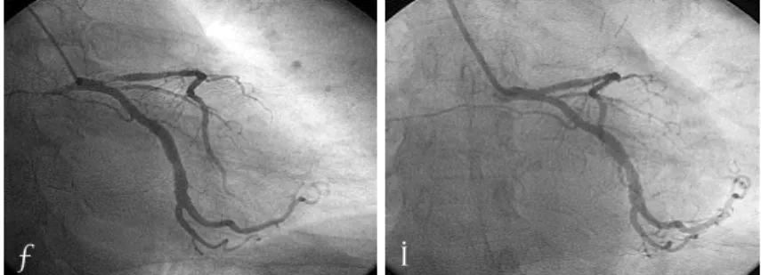
Fig. 1. AP-caudal view. The coronary angiograms on initial admission demonstrated near total occlusion of the proximal LAD. AP: anterio- posterior, LAD: left anterior descending coronary artery.
Fig. 2. AP-caudal view. The post-stenting angiography showed a good appearance with TIMI 3 flow. The arrow indicated the stent deployed in the proximal LAD. AP: anterioposterior, LAD: left anterior des- cending coronary artery.
Fig. 4. The follow-up coronary angiograms, six months after CBA, showed 30% ISR of the previously CBA segment of the proximal LAD. A: AP- caudal view. B: spider view. CBA: cutting balloon angioplasty, ISR: in-stent restenosis, LAD: left anterior descending coronary artery, AP: anterio- posterior.
A B
Fig. 3. AP-caudal views. A: the angiography showed 90% in-stent restenosis in the proximal LAD at 6 months after stent deployment. B: the post- procedural angiography after performing CBA showed good patency of the stent without restenosis. AP: anterioposterior, LAD: left anterior desc- ending coronary artery, CBA: cutting balloon angioplasty.
A B
232·Korean Circulation J 2007;37:230-233
Discussion
Stent thrombosis is generally a fatal complication after PCI. Coronary stent has dramatically improved upon the acute procedural success and also reduced the res- tenosis rates observed with balloon angioplasty alone.
Significant improvements in the prevention of stent thrombosis have been achieved through the technical development of stent implantations and the use of po- tent antiplatelet agents.3)7) Stent thrombosis is classified as either subacute stent thrombosis(SAT), occurring wi- thin 30 days, or as late stent thrombosis(LST), occurring beyond 30 days.4)8-10) While VLST is uncommon with the use of DESs, it is distinctly unusual with the use of BMSs.4) Since BMSs are known to be re-endothelialized
within a few weeks after the procedure, this rapid endo- thelialization of BMSs makes LST exceedingly rare.3)10)11) However, delayed endothelialization associated with the implantation of a DES may increase the risk of LST.10)
In the recently published TAXUS V trial, the inci- dence of stent thrombosis in the bare-metal arm of the study group was 0.5% at 30 days, 0.2% at 6 months and 0% at 9 months.6) There have been no studies looking specifically at the determinants and incidence of VLST with the use of BMS. Presumed causes of BMS throm- bosis, both early and late, include noncompliance with antiplatelet agents, exercise-induced procoagulant state, brachytherapy, small stent size and underdeployment of the stent.3)9)12)13) Furthermore, longer stent length, num- ber of implanted stents, stent malapposition, residual dissections, reduced TIMI flow, gene polymorphisms, and resistance to the antiplatelet effects of acetylsalicylic acid(ASA) and potentially thienopyridines have been reported to increase the risk for stent thrombosis.1)3)7)9)10)
Premature discontinuation of antiplatelet therapy is the most common precipitation factor of stent thrombosis10) and coronary brachytherapy is also the most important risk factor of late stent thrombosis in BMSs.3) We could not identify any potential explanations for this throm- botic event in our patient except for the possibility of relatively small stent size(3.0 mm), longer stent length (38 mm) and resistance to antiplatelet agents, but IVUS in follow-up period, showed good expansion and appo- sition and our patient was persistently compliant with his drugs. Stent thrombosis of our patient may be new plague rupture or erosion in previuos stented segment.
The management of VLST is similar to that of SAT
A B
C D
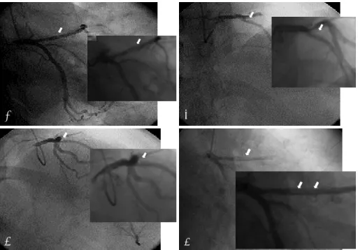
Fig. 6. The coronary angiograms at 880 days after stent deployment. The coronary angiograms demonstrated a huge thrombus (arrows) in the previously stented segment of the proximal LAD. A: AP-caudal view. B: AP-cranial view. C: RAO-cranial view. D: RAO-caudal view. LAD: left anterior descending coronary artery, AP: anterioposterior, RAO: right anterior oblique.
Fig. 5. The electrocardiogram showed regular sinus rhythm with Q- wave and ST segment elevation in precordial leads V1-4 at 880 days after stent deployment.
Yoon-Sung Cho, et al:Very Late Stent Thrombosis in Bare-Metal Stent Implantation·233
and LST, and consists of the restoration of perfusion to distal TIMI 3 flow - most commonly by a variety of percutaneous techniques such as balloon angioplasty, AngioJet®(Possis Medical, Inc., Minneapolis, Minnesota) thrombectomy, or re-stenting with subsequent long-term dual antiplatelet therapy.4) Our patient did not undergo any other coronary procedure because of restoration of perfusion to distal TIMI 3 flow. In other words, throm- bolytic therapy was successful in restoration of perfusion.
The optimum duration of antiplatelet therapy for patients with coronary artery stents still remains to be determined. In the era of BMSs, it has been customary to prescribe dual antiplatelet therapy for the duration of 1 month followed by the indefinite treatment with ASA alone.3) More recently, randomized clinical trials tested the benefit of extended(9-12 months) dual anti- platelet therapy compared with treatment with ASA alone. Our report shows that stent thrombosis may arise as late as 880 days after performing successful PCI, even when treating this patient with antiplatelet agents.
Therefore, clinicians should be concerned about the possibility of VLST in those patients who have under- gone BMS implantation. Most importantly, education regarding the importance of compliance with combi- nation anti-platelet therapy needs to be emphasized.
In addition, careful attention should be paid to assure adequate high-pressure inflation during deployment.13)
Further large-scaled studies are needed to determine the optimal combination and duration for antiplatelet ther- apy that should be used to prevent these serious throm- botic events.
REFERENCES
1) Wenaweser P, Rey C, Eberli FR, et al. Stent thrombosis following bare-metal stent implantation: success of emergency percutan- eous coronary intervention and predictors of adverse outcome.
Eur Heart J 2005;26:1180-7.
2) Apostolovic S, Perisic Z, Tomasevic M, Stankovic G, Pavlovic M, Salinger-Martinovic S. Late thrombosis of coronary bare-metal stent-case report. Srp Arh Celok Lek 2006;134(3-4). Abstract.
3) Park DW, Park SW. Stent thrombosis in the era of the drug- eluting stent. Korean Circ J 2005;35:791-4.
4) Manjappa N, Agarwal A, Cavusoglu E. Very late bare-metal stent thrombosis. A case report and review of the literature. J Invasive Cardiol 2006;18:E203-6.
5) Moussa I, Di Mario C, Reimers B, et al. Subacute stent throm- bosis in the era of intravascular ultrasound-guided coronary stenting without anticoagulation: frequency, predictors and cli- nical outcome. J Am Coll Cardiol 1997;29:6-12.
6) Stone GW, Ellis SG, Cannon L, et al. Comparison of a polymer- based paclitaxel-eluting stent with a bare metal stent in patients with complex coronary artery disease: a randomized controlled trial. JAMA 2005;294:1215-23.
7) Park SH, Hong GR, Seo HS, Tahk SJ. Stent thrombosis after successful drug-eluting stent implantation. Korean Circ J 2005;
35:163-71.
8) Katayama T, Nakashima H, Takagi C, et al. Predictors of sub- acute stent thrombosis in acute myocardial infarction patients following primary coronary stenting with bare-metal stent. Circ J 2006;70:151-5.
9) Shin DH, Kwon DA, Chung JW, et al. Late stent thrombosis after intracoronary brachytherapy: learning from brachytherapy experiences in the drug-eluting stent era. Korean Circ J 2006;
36:324-7.
10) Choi BR, Lee CW, Park SW. Late stent thrombosis associated with late stent malapposition after drug-eluting stenting: a case report. Korean Circ J 2006;36:472-5.
11) Leon MB, Teirstein PS, Moses JW, et al. Localized intracoronary gamma-radiation therapy to inhibit the recurrence of restenosis after stenting. N Engl J Med 2001;344:250-6.
12) Parodi G, Antoniucci D. Late coronary stent thrombosis associ- ated with exercise testing. Cathether Cardiovasc Interv 2004;61:
515-7.
13) Lee EM, Oh DJ, Kim HC, et al. Optimal balloon inflation press- ures for stent deployment-high pressure is always good? Korean Circ J 1998;28:1272-9.
Fig. 7. Intravascular ultrasound (IVUS) showing good expansion and apposition with mild intimal hyperplasia.