• 검색 결과가 없습니다.

C-Type Natriuretic Peptide as a Surrogate Marker in Variant Angina Pectoris

N/A
N/A
Protected

Academic year: 2022

Share "C-Type Natriuretic Peptide as a Surrogate Marker in Variant Angina Pectoris"

Copied!
6
0
0

로드 중.... (전체 텍스트 보기)

전체 글

(1)

Korean Circulation Journal

Introduction

Variant angina pectoris (VAP) was first described by Prinzmetal et al.,

1)

and is a syndrome of ischemic pain accompanied by ST-segment

http://dx.doi.org/10.4070/kcj.2013.43.3.168 Print ISSN 1738-5520 • On-line ISSN 1738-5555

C-Type Natriuretic Peptide as a Surrogate Marker in Variant Angina Pectoris

Dong-Hyeon Lee, MD, Ho-Joong Youn, MD, Yun-Seok Choi, MD, Jong-Min Lee, MD, Chul-Soo Park, MD, Hae-Ok Jung, MD, Hui-Kyung Jeon, MD, and Man-Young Lee, MD

Division of Cardiology, Department of Internal Medicine, Seoul St. Mary’s Hospital, The Catholic University of Korea College of Medicine, Seoul, Korea

Background and Objectives: The purpose of this study was to assess the value of C-type natriuretic peptide (CNP) as a surrogate marker for detection of coronary artery spasm in variant angina pectoris (VAP).

Subjects and Methods: Sixty-six patients (mean age: 51±11 years, M : F=40 : 26) who underwent coronary angiography on suspicion of angina and who were diagnosed with VAP by the acetylcholine-induced spasm provocation test (SPT) were enrolled and divided into a SPT (-) group (n=23) and a SPT (+) group (n=43). Concentrations of CNP and other markers were determined by immunoassay in both groups.

Results: Plasma CNP and creatine kinase myoglobin band (CK-MB) concentrations were significantly increased in the SPT (+) group relative to the SPT (-) group (CNP, 5.268±1.800 pg/mL vs. 3.342±1.150 pg/mL, p=0.002; CK-MB, 2.54±1.03 ng/dL vs. 1.86±0.96 ng/dL, p=0.019, respectively) while plasma high sensitivity C-reactive protein (hs-CRP) and N-terminal pro-brain natriuretic peptide (NT pro-BNP) con- centrations were not significantly different between the SPT (-) group and SPT (+) group (hs-CRP, 2.76±4.99 mg/L vs. 3.13±4.88 mg/L, p=0.789; NT pro-BNP, 49±47 pg/mL vs. 57±63 pg/mL, p=0.818, respectively). Plasma CNP concentration was independently associated with the VAP via SPT {odds ratio: 2.014 (95% confidence interval: 1.016-3.992), p=0.045}. A CNP cut-off value of 4.096 pg/mL was found to have a sensitivity of 68.2% and a specificity of 40.0% for predicting the probability of VAP via SPT.

Conclusion: Increased plasma CNP concentration in patients with VAP may have an impact on the regulation of endothelial function in accordance with the progression of atherosclerosis. Further analysis is warranted to develop clinical applications of this finding. (Korean Circ J 2013;43:168-173)

KEY WORDS: Natriuretic peptide, C-type; Angina pectoris, variant; Endothelium; Acetylcholine.

Received: October 30, 2012

Revision Received: November 15, 2012 Accepted: December 6, 2012

Correspondence: Ho-Joong Youn, MD, Division of Cardiology, Depart- ment of Internal Medicine, Seoul St. Mary’s Hospital, The Catholic Universi- ty of Korea College of Medicine, 222 Banpo-daero, Seocho-gu, Seoul 137- 701, Korea

Tel: 82-2-2258-6029, Fax: 82-2-591-1506 E-mail: [email protected]

• The authors have no financial conflicts of interest.

This is an Open Access article distributed under the terms of the Creative Commons Attribution Non-Commercial License (http://creativecommons.

org/licenses/by-nc/3.0) which permits unrestricted non-commercial use, distribution, and reproduction in any medium, provided the original work is properly cited.

elevation that occurs at rest and without exertion. Transient increas- es in coronary vasomotor tone or vasospasm are the pathogenic mechanism underlying Prinzmetal or VAP.

2)

Coronary artery spasm in VAP has been found to be associated with systemic alteration in nitric oxide (NO) production or an imbalance between endothelium- derived relaxing and contracting factors that results in complica- tions, most often myocardial infarction, life-threatening arrhyth- mias, or sudden cardiac death.

3-5)

Natriuretic peptides, comprising atrial natriuretic peptide (ANP),

brain natriuretic peptide (BNP) and C-type natriuretic peptide (CNP),

are a family of polypeptide mediators that play an important role in

the regulation of cardiovascular homeostasis by maintaining blood

pressure and extracellular fluid volume.

6-8)

ANP and BNP are media-

tors that are mainly secreted by the myocardium in response to many

pathophysiological conditions,

6-8)

while CNP is mainly produced by

vascular endothelium and contributes to smooth muscle relaxation,

where it may exert its autocrine/paracrine roles in vascular tone and

(2)

muscle cell growth.

6-8)

The contribution of endothelial mediators to the effects of CNP on the coronary circulation is unclear and poorly characterized. Therefore, we investigated the value of CNP as a sur- rogate marker for detection of coronary artery spasm in VAP, and estimated a threshold value for this purpose.

Subjects and Methods

Patients

Between July 2005 and May 2008, a total of sixty-six patients (mean age: 51±11 years, M : F=40 : 26) who underwent coronary an- giography on suspicion of angina and who were diagnosed with VAP by acetylcholine-induced spasm provocation test (SPT) were enroll- ed. Patients were divided into two groups: the SPT (-) group (n=23) and the SPT (+) group (n=43), members of which were diagnosed with VAP based on the definition of VAP applicable for acetylcho- line-induced SPT, that is, focal occlusion greater than 70% of the luminal diameter with clinical symptoms or ST-segment change on electrocardiography (ECG).

9)

This study was approved by the Ethics Committee of St. Mary’s Hos- pital. Patients were informed of the investigative nature of the study and written informed consent was obtained before enrollment.

Coronary angiography

Coronary angiography was performed in all patients using a fe- moral approach with standard catheters placed in conventional po- sitions. Nitroglycerin was not administered before angiography. For SPT of the coronary artery, intracoronary injection of acetylcholine was administered according to the method of Okumura et al.

10)

In- cremental doses of 20 and 50 μg acetylcholine were injected into the right coronary artery, and 20, 50, and 100 μg doses were inject- ed into the left coronary artery. ECG and coronary angiography were repeated to confirm the presence of coronary artery spasm. The angiograms were analyzed by two experienced angiographers.

Biochemical assays

Concurrently, blood samples for the analysis of plasma CNP and N-terminal pro-brain natriuretic peptide (NT pro-BNP) concentration were drawn into ethylenediaminetetraacetic acid-plasma vacuum containers. Plasma CNP (catalog No. E0721h) concentration was as- sayed with an immunoassay (USCNLife Science & Technology co., Ltd., Wuhan, China) in which there is no cross-reactivity with plasma NT pro-BNP. The detectable range for assay was CNP 0-500 pg/mL (sensitivity 3.9 pg/mL).

Statistical analysis

All analyses were performed using the SAS statistical software

version 9.1 (SAS Institute Inc., Cary, NC, USA). For comparisons be- tween the SPT (-) and (+) groups, mean±standard deviations and proportions were compared using the unpaired t-test and chi-sq- uare test, respectively. The relationships between multiple parame- ters and plasma CNP or NT pro-BNP concentration were assessed using Spearman’s correlation coefficient by rank. After adjustment for age and gender, multivariate logistic regression analysis was performed for detection of the independent factors associated with Table 1. Baseline characteristics

Variables SPT (-) group

(n=23)

SPT (+) group

(n=43) p

Age (year) 49±12 51±10 0.500

Gender, male (%) 10 (43.5) 30 (70.0) 0.037

Hypertension, n (%) 13 (56.5) 18 (41.9) 0.255

Diabetes mellitus, n (%) 1 (4.3) 5 (11.6) 0.327

BMI (kg/cm

2

) 24.1±2.5 25.4±2.6 0.059

Smoking, n (%) 6 (26.1) 17 (39.5) 0.275

SBP (mm Hg) 122±24 119±15 0.619

DBP (mm Hg) 71±12 73±13 0.502

HR (bpm) 66±14 67±10 0.744

WBC, counts/L (%) 6529±2058 7460±2283 0.098

Neutrophil 61.4±10.0 56.2±13.2 0.097

Lymphocyte 30.2±7.8 33.8±12.0 0.164

Monocyte 6.1±2.3 6.7±2.2 0.394

Eosinophil 1.9±1.6 2.8±2.4 0.122

Basophil 0.4±0.2 0.6±0.5 0.039

Hb (g/dL) 13.6±1.3 14.2±1.5 0.112

Platelet (counts×10

3

/L) 258±79 247±53 0.540

Fasting plasma glucose

(mg/dL) 108±26 112±28 0.575

HbA1c (%) 5.2±0.6 5.6±0.7 0.068

Total cholesterol (mg/dL) 200±40 182±36 0.090

Triglyceride (mg/dL) 148±94 148±88 0.995

HDL-C (mg/dL) 46±10 49±40 0.666

LDL-C (mg/dL) 124±36 115±29 0.331

Blood urea nitrogen

(mg/dL) 13.2±2.8 15.8±4.9 0.014

Creatinine (mg/dL) 0.91±0.15 0.15±1.07 0.170

hs-CRP (mg/L) 2.76±4.99 3.13±4.88 0.789

CK-MB (ng/dL) 1.86±0.96 2.54±1.03 0.019

NT pro-BNP (pg/mL) 49±47 57±63 0.818

CNP (pg/mL) 3.342±1.150 5.268±1.800 0.002

SPT: spasm provocation test, BMI: body mass index, SBP: systolic blood pres- sure, DBP: diastolic blood pressure, HR: heart rate, WBC: white blood cell, Hb:

hemoglobin, HDL-C: high density lipoprotein-cholesterol, LDL-C: low density lipoprotein-cholesterol, hs-CRP: high sensitivity C-reactive protein, CK-MB:

creatine kinase myoglobin band, NT pro-BNP: N-terminal pro-brain natriuretic

peptide, CNP: C-type natriuretic peptide

(3)

VAP via acetylcholine-induced SPT. A receiver operating curve was used to determine the cut-off value for plasma CNP concentration as a predictor of the probability of the presence of VAP via acetyl- choline-induced SPT. Statistical significance was considered at p of less than 0.05.

Results

Clinical characteristics

Among the sixty-six patients subjected to the intracoronary ac- etylcholine-induced SPT, 43 were SPT positive {SPT (+) group; mean age: 51±10 years, M : F=30 : 13} and 23 were SPT negative {SPT (-) group; mean age: 49±12 years, M : F=10 : 13} (Table 1).

Results for comparison according to acetylcholine-induced spasm provocation test

Basophil percentage and blood urea nitrogen concentration were significantly increased in the SPT (+) group relative to the SPT (-) gr- oup (basophil percentage, 0.6±0.5% vs. 0.4±0.2%, p=0.039; blood urea nitrogen, 15.8±4.9 mg/dL vs. 13.2±2.8 mg/dL, p=0.014, respec- tively) (Table 1).

Plasma CNP and creatine kinase myoglobin band (CK-MB) con- centrations were significantly increased in the SPT (+) group relative to the SPT (-) group {CNP, 5.268±1.800 pg/mL vs. 3.342±1.150 pg/

mL, p=0.002 (Fig. 1); CK-MB, 2.54±1.03 ng/dL vs. 1.86±0.96 ng/dL, p=0.019, respectively} while plasma high sensitivity C-reactive pro- tein (hs-CRP) and NT pro-BNP concentrations were not significantly different between the SPT (-) group and SPT (+) groups (hs-CRP, 2.76±4.99 mg/L vs. 3.13±4.88 mg/L, p=0.789; NT pro-BNP, 49±47 pg/mL vs. 57±63 pg/mL, p=0.818, respectively) (Table 1).

Associations among biomarkers

Plasma NT pro-BNP concentration was positively correlated with that of CK-MB (r=0.543, p<0.0001) and negatively correlated with heart rate (r=-0.339, p=0.026, respectively) (Table 2). Plasma CNP concentration was negatively correlated with heart rate (r=-0.403, p=0.030) and total cholesterol level (r=-0.369, p=0.041, respecti- vely) (Table 2).

Predictability of coronary artery spasm

Plasma CNP concentration was independently associated with coronary artery spasm in patients with VAP via acetylcholine-in- duced SPT after adjustment for age and gender {odds ratio: 2.014 (95% confidence interval: 1.016-3.992), p=0.045} (Table 3). A CNP cut-off value of 4.096 pg/mL was found to have a sensitivity of

Fig. 1. Difference in CNP levels between the SPT (-) group and the SPT (+) group. CNP: C-type natriuretic peptide, SPT: spasm provocation test.

12.000

9.000

6.000

3.000

0.000

C- ty pe n at riu re tic p ep tid e (p g/ m L)

SPT (-) SPT (+) 3.342±1.150

5.268±1.800 p=0.002

Table 2. The relationships between levels of either NT pro-BNP or CNP and other factors

Variables NT pro-BNP CNP

Coefficient p Coefficient p

BMI -0.018 0.791 0.093 0.619

SBP -0.041 0.907 0.229 0.215

DBP -0.189 0.219 0.303 0.098

HR -0.339 0.026 -0.403 0.030

WBC -0.024 0.876 -0.176 0.345

Neutrophil -0.076 0.621 -0.347 0.056

Lymphocyte 0.113 0.458 0.315 0.084

Monocyte -0.056 0.716 0.195 0.294

Eosinophil -0.087 0.570 0.128 0.493

Basophil 0.133 0.383 0.266 0.147

Hb 0.000 0.999 0.180 0.334

Platelet -0.191 0.209 -0.236 0.200

Fasting plasma glucose -0.049 0.751 -0.156 0.410

HbA1c -0.028 0.861 -0.028 0.887

Total cholesterol -0.011 0.945 -0.369 0.041

Triglyceride -0.194 0.213 -0.132 0.479

HDL-C 0.042 0.791 0.233 0.207

LDL-C -0.053 0.736 -0.323 0.076

Blood urea nitrogen -0.093 0.545 0.102 0.584

Creatinine -0.098 0.524 0.252 0.172

hs-CRP -0.087 0.574 -0.264 0.151

CK-MB 0.543 <0.0001 0.001 0.995

NT pro-BNP - - 0.190 0.333

CNP 0.190 0.333 - -

BMI: body mass index, SBP: systolic blood pressure, DBP: diastolic blood pressure, HR: heart rate, WBC: white blood cell, Hb: hemoglobin, HDL-C:

high density lipoprotein-cholesterol, LDL-C: low density lipoprotein-choles- terol, hs-CRP: high sensitivity C-reactive protein, CK-MB: creatine kinase myoglobulin, NT pro-BNP: N-terminal pro-brain natriuretic peptide, CNP:

C-type natriuretic peptide

(4)

68.2% and a specificity of 40.0% for predicting the probability of VAP presence via SPT (Fig. 2).

Discussion

The natriuretic peptide system includes three known peptides:

ANP, BNP, and CNP. Although ANP and BNP rise excessively in pati- ents with cardiovascular diseases and are powerful neurohormonal indicators of left ventricular function and prognosis in congestive heart failure (CHF), the role of plasma CNP in human circulation has appeared to be minor and has been poorly characterized.

6-8)

C-type natriuretic peptide, a new member of the natriuretic pep- tide family identified in both the animal and human brain, termi- nates in the second cysteine residue, thereby lacking a further C-ter- minal extension, and its level in humans is 10-fold higher than those of ANP and BNP.

11)12)

Recently, some studies have reported the pathophysiological role of CNP as a predictor of endothelial vasomotor response in patients with CHF.

13)14)

Although it is well known that CNP is a peptide pro- duced by the vascular endothelium with vasodilative properties,

15-17)

our study is the first to demonstrate the pathophysiological role of CNP in patients with VAP.

The most important finding in the present data was that plasma CNP concentration was higher in the SPT (+) rather than SPT (-) gr- oup. Until recently, most research has reported that CNP level was not only inversely related to a number of cardiovascular risk factors but also that CNP exhibited significant negative associations with pulse wave velocity for arterial stiffness and intima-media thickness for early atherosclerosis and positive association with flow mediat- ed dilation for endothelial function.

18-21)

This has been well demon- strated by previous reports into the association with endothelial dysfunction in patients with VAP

22)

as well as those with microvas- cular angina.

23-26)

However, our present result showed that plasma CNP concentr- ation was higher in the SPT (+) group, which is known to be a type of endothelial dysfunction. Our investigators could carefully elicit a new understanding as follows. Coronary atherosclerosis is initiat- ed by endothelial dysfunction due to inflammation and increased low density lipoprotein-cholesterol (LDL-C) levels, followed by the deposition of lipids and macrophages from the lumen into the in- ner layer of the coronary artery, resulting in plaque formation.

27)

In short, endothelial dysfunction is likely to be the earliest event in the process of atherosclerotic lesion formation. Therefore, plasma CNP concentrations in present study may have an impact on the regul- ation of endothelial function in accordance with atherosclerotic progression in patients with VAP. Further analysis is warranted to develop clinical applications of this concept.

Additionally, in terms of the vascular endothelium, Palmer et al.

21)

demonstrated that circulating CNP concentration was related to cardiac hemodynamic load and ischemic burden. In contrast, Naruko et al.

19)

suggested that a paracrine and autocrine system of CNP may be important in controlling the neointimal growth of vascular smooth muscle cells after percutaneous coronary intervention in hu- mans. Moreover, in animal studies addressing the role of the car- diovascular NO system in the actions of CNP, Costa et al.

20)

dem- onstrated that CNP increased NO excretion, which would induce a reduction in cardiac myocyte contractility and relaxation of the vas- cular smooth muscle, thereby suggesting an association with endo- thelial function. Although our results are in agreement with previ- ous reports, further research using a larger population is warranted.

Kuo et al.

28)

reported that the myocardium regularly produces or releases CNP in patients with symptomatic paroxysmal supraven- tricular tachycardia (PSVT) and normal left ventricular systolic func- Table 3. Multivariate logistic regression analysis

Variables Odds ratio 95% confidence

interval p

Basophil percentage 114.561 0.165–79479.400 0.155

Blood urea nitrogen 1.232 0.821–1.848 0.313

CK-MB 1.404 0.417–4.722 0.584

NT pro-BNP 0.996 0.987–1.006 0.463

CNP 2.014 1.016–3.992 0.045

Plasma CNP concentration was independently associated with coronary artery spasm in patients with VAP diagnosed by acetylcholine-induced SPT after adjustment for age and gender. CNP: C-type natriuretic peptide, VAP:

variant angina pectoris, SPT: spasm provocation test, CK-MB: creatine kinase myoglobin band, NT pro-BNP: N-terminal pro-brain natriuretic peptide

Fig. 2. ROC curve. A cut-off value of CNP of 4.096 pg/mL shows a sensitivity of 86.4% and a specificity of 50.0% for predicting the probability of the pres- ence of VAP via acetylcholine-induced SPT. ROC: receiver operating charac- teristics, AUC: area under curve, CI: confidence interval, CNP: C-type natri- uretic peptide, VAP: variant angina pectoris, SPT: spasm provocation test.

1.0

0.8

0.6

0.4

0.2

0.0

Se ns iti vi ty

1-specificity

0.0 0.2 0.4 0.6 0.8 1.0 AUC=0.770 95% CI (0.605-0.936) Sensitivity 68.2%

Specificity 40.0%

(5)

tion and brief periods of rapid atrial pacing, and that PSVT or elec- trophysiological studies did not change the production and/or re- lease of CNP. Lupattelli et al.

29)

suggested that CNP was not altered in patients affected by hypercholesterolemia. Nevertheless, the levels of high density lipoprotein-cholesterol were closely related to those of CNP. In the present study, heart rate and total cholesterol level were significantly correlated with the CNP level and thus further analysis is warranted to address this discrepancy.

This study has some limitations. First, blood samples were not col- lected from the coronary sinus but from the femoral artery. Second, associations with various vasoactive agents, such as endothelin, angiotensin II, and serotonin, were not assessed. Lastly, there was a selection bias in this study’s patients. To overcome this methodolog- ical limitation, more research in patients with significant coronary artery disease (>50% stenosis in ≥1 coronary artery) is warranted.

In conclusion, coronary atherosclerosis is initiated by endothelial dysfunction due to inflammation and increased LDL-C levels, which is followed by the deposition of lipids and macrophages from the lumen into the inner layer of the coronary artery, resulting in plaque formation. Therefore, increased plasma CNP concentration in pa- tients with VAP may have an impact on the regulation of endothelial function in accordance with the progression of atherosclerosis. Fur- ther analysis is warranted to develop clinical applications of this finding.

Acknowledgments

This study was supported by a grant from the Seoul R&BD Program in the Republic of Korea (10526).

References

1. Prinzmetal M, Kennamer R, Merliss R, Wada T, Bor N. Angina pectoris. I.

A variant form of angina pectoris; preliminary report. Am J Med 1959;

27:375-88.

2. Mayer S, Hillis LD. Prinzmetal’s variant angina. Clin Cardiol 1998;21:

243-6.

3. Hillis LD, Braunwald E. Coronary-artery spasm. N Engl J Med 1978;299:

695-702.

4. Marx JL. Coronary artery spasms and heart disease. Science 1980;208:

1127-30.

5. Braunwald E. Coronary artery spasm. Mechanisms and clinical relev- ance. JAMA 1981;246:1957-9.

6. Yandle TG. Biochemistry of natriuretic peptides. J Intern Med 1994;235:

561-76.

7. Komatsu Y, Nakao K, Itoh H, Suga S, Ogawa Y, Imura H. Vascular natri- uretic peptide. Lancet 1992;340:622.

8. Suzuki T, Yamazaki T, Yazaki Y. The role of the natriuretic peptides in the cardiovascular system. Cardiovasc Res 2001;51:489-94.

9. Baim DS, Grossman W. Grossman’s Cardiac Catheterization, Angiog-

raphy, and Intervention. 6th ed. Philadelphia: Lippincott Williams &

Wilkins;2000. p.247-51.

10. Okumura K, Yasue H, Horio Y, et al. Multivessel coronary spasm in pa- tients with variant angina: a study with intracoronary injection of ace- tylcholine. Circulation 1988;77:535-42.

11. Sudoh T, Minamino N, Kangawa K, Matsuo H. C-type natriuretic pep- tide (CNP): a new member of natriuretic peptide family identified in porcine brain. Biochem Biophys Res Commun 1990;168:863-70.

12. Ogawa Y, Nakao K, Nakagawa O, et al. Human C-type natriuretic pep- tide. Characterization of the gene and peptide. Hypertension 1992;19 (6 Pt 2):809-13.

13. Kalra PR, Clague JR, Bolger AP, et al. Myocardial production of C-type natriuretic peptide in chronic heart failure. Circulation 2003;107:571-3.

14. Del Ry S, Maltinti M, Cabiati M, Emdin M, Giannessi D, Morales MA. C- type natriuretic peptide and its relation to non-invasive indices of left ventricular function in patients with chronic heart failure. Peptides 2008;29:79-82.

15. Komatsu Y, Itoh H, Suga S, et al. Regulation of endothelial production of C-type natriuretic peptide in coculture with vascular smooth muscle cells. Role of the vascular natriuretic peptide system in vascular growth inhibition. Circ Res 1996;78:606-14.

16. Chun TH, Itoh H, Ogawa Y, et al. Shear stress augments expression of C-type natriuretic peptide and adrenomedullin. Hypertension 1997;29:

1296-302.

17. Kelsall CJ, Chester AH, Sarathchandra P, Singer DR. Expression and lo- calization of C-type natriuretic peptide in human vascular smooth mu- scle cells. Vascul Pharmacol 2006;45:368-73.

18. Vlachopoulos C, Ioakeimidis N, Terentes-Printzios D, et al. Amino-ter- minal pro-C-type natriuretic peptide is associated with arterial stiff- ness, endothelial function and early atherosclerosis. Atherosclerosis 2010;211:649-55.

19. Naruko T, Itoh A, Haze K, et al. C-Type natriuretic peptide and natri- uretic peptide receptors are expressed by smooth muscle cells in the neointima after percutaneous coronary intervention. Atherosclerosis 2005;181:241-50.

20. Costa MA, Elesgaray R, Caniffi C, Fellet A, Arranz C. Role of cardiovas- cular nitric oxide system in C-type natriuretic peptide effects. Biochem Biophys Res Commun 2007;359:180-6.

21. Palmer SC, Prickett TC, Espiner EA, Yandle TG, Richards AM. Regional release and clearance of C-type natriuretic peptides in the human cir- culation and relation to cardiac function. Hypertension 2009;54:612-8.

22. Hwang HJ, Chung WB, Park JH, et al. Estimation of coronary flow ve- locity reserve using transthoracic Doppler echocardiography and cold pressor test might be useful for detecting of patients with variant an- gina. Echocardiography 2010;27:435-41.

23. Park CS, Youn HJ, Ihm SH, et al. Relation between peripheral vascular endothelial function and coronary flow reserve in patients with chest pain and normal coronary angiogram. Korean Circ J 2004;34:485-91.

24. Park CS, Youn HJ, Kim JH, et al. Relation between peripheral vascular endothelial function and coronary flow reserve in patients with chest pain and normal coronary angiogram. Int J Cardiol 2006;113:118-20.

25. Egashira K, Inou T, Hirooka Y, Yamada A, Urabe Y, Takeshita A. Evidence

of impaired endothelium-dependent coronary vasodilatation in pa-

(6)

tients with angina pectoris and normal coronary angiograms. N Engl J Med 1993;328:1659-64.

26. Camici PG, Crea F. Coronary microvascular dysfunction. N Engl J Med 2007;356:830-40.

27. Libby P, Ridker PM, Hansson GK. Progress and challenges in translat- ing the biology of atherosclerosis. Nature 2011;473:317-25.

28. Kuo JY, Yeh HI, Chang SH, et al. C-type natriuretic peptide in individu- als with normal left ventricular systolic function. Scand Cardiovasc J 2007;41:155-9.

29. Lupattelli G, Marchesi S, Siepi D, et al. Natriuretic peptides levels are

related to HDL-cholesterol with no influence on endothelium depen-

dent vasodilatation. Vasa 2006;35:215-20.

수치

Fig. 1. Difference in CNP levels between the SPT (-) group and the SPT (+)  group. CNP: C-type natriuretic peptide, SPT: spasm provocation test.
Fig. 2. ROC curve. A cut-off value of CNP of 4.096 pg/mL shows a sensitivity  of 86.4% and a specificity of 50.0% for predicting the probability of the  pres-ence of VAP via acetylcholine-induced SPT

참조

관련 문서

Brain natriuretic peptide predicts right heart failure in patients with acute pulmonary embolism. Kucher N, Printzen

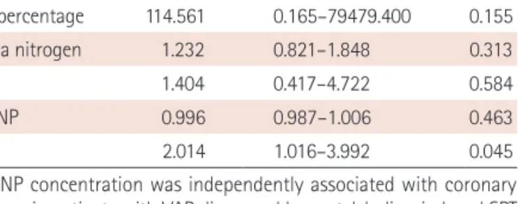
MACE, major adverse cardiac events; Tn-I, troponin-I; NT-proBNP, N- terminal proB-type natriuretic peptide; hs-CRP, high-sensitivity C-reactive protein; IHD, ischemic

The present study was designed to examine whether the bas- al C-peptide level correlates with cardiovascular risk in T2DM patients via comparison with IMT, the 10-year coronary heart

The multiple regression analysis also showed that basal C-peptide significantly correlated with IMT (P=0.043) but did not correlate with the ten-year coronary heart disease (CHD)