Agric. Chem. Biotechnol. 48(3), 148-150 (2005)
Short Communication Solution Structure of Human Atrial
Natriuretic Peptides (7-28)
Yukyung Lee and Yoongho Lim*
Bio/Molecular Informatics Center, Konkuk University, Seoul 143-701, Korea
Received July 22, 2005; Accepted August 25, 2005 Key words: atrial natriuretic peptides, nuclear magnetic
resonance, molecular modeling
The natriuretic peptides consist of three families, atrial natriuretic peptide (ANP), brain natriuretic peptide (BNP), and C-type natriuretic peptide (CNP). CNP is observed in the central and peripheral tissues, functioning as an autocrine or paracrine regulator, whereas ANP and BNP are produced by the atrium and ventricle and function as cardiac hormones.1) ANP was first isolated from human atrial extracts, and BNP and CNP from porcine brain.2) Natriuretic peptides have a ring structure by a disulfide bond. Seventeen residues, which show a high sequence homology, are included in the ring structure.
Therefore, exocyclic N-terminal and C-terminal residues make differences in natriuretic peptides, which cause different binding affinities for natriuretic peptide receptors.2)
Up to now, three dimensional (3D) structures of two different ANP fragments, human ANP(1-28) and rat ANP(7- 23), have been determined.3,4) A disulfide bond is formed between Cys7 and Cys23. Human ANP(1-28) and rat ANP(7- 23) differ in their 12th residues (human, Met; rat, Ile). Authors determined 3D structure of the other human ANP fragment, which consists of Cys7-Tyr28, using nuclear magnetic resonance (NMR) spectroscopy. A structural comparison of these three fragments may give information about different binding affinities for natriuretic peptide receptors. In addition, to obtain the first 3D solution structure of ANP fragment under aqueous condition, we performed this study.
Human ANP(7-28), Cys-Phe-Gly-Gly-Arg-Met-Asp-Arg- Ile-Gly-Ala-Gln-Ser-Gly-Leu-Gly-Cys-Asn-Ser-Phe-Arg- Tyr, (approximately 1 mM) American Peptide Co. Inc., Sunnyvale, CA, USA) was dissolved in a mixture 650 ul H2O and D2O (9 : 1) and transferred into a 5-mm NMR tube. NMR experiments were carried out on a Bruker Avance 600 (14.1 Tesla, Bruker, Karlsruhe, Germany) with a cryoprobe at 298 K. For 1H-NMR, 128 transients were acquired with a 1-sec
relaxation delay using 32 K data points. The 90o pulse was 9.9 msec with a spectral width of 6,009 Hz. Two-dimensional spectra were acquired with 2,048 and 256 data points for t2 and t1, respectively, using time proportional phase increments, except that magnitude mode was applied in COSY experiments.
The nuclear Overhauser exchanged and spectroscopy (NOESY) experiment was performed at a mixing time of 500 msec. The mixing time for the total correlated spectroscopy (TOCSY) experiment with MLEV17 spin-lock pulse program was 80 msec.5) In all experiments water peaks were suppressed by presaturation. Prior to fourier transformation, zero filling of 2 K and sine squared bell window function were applied using XWIN-NMR (Bruker).6) The 1H-NMR data are listed in Table 1.
The dihedral angles were obtained based on the Karplus equation as follows:
J= 6.51cos2(θ-60) – 1.76cos(θ-60) + 0.9
where J and θ denote vicinal coupling constant and dihedral angle, respectively.7) Distance constraints were calculated using the relationship ηij/ηkl= (rkl/rij)6, where η and r are the nOe intensity and distance for the hydrogen atom pairs i-j and k-l, respectively.8)
To obtain refined structures from 62 nOe cross peaks and 13 dihedral angles, the molecular modeling calculations were carried out on an O2 R12,000 Silicon Graphics workstation.
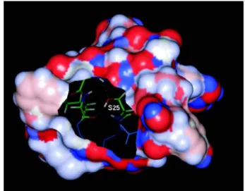
The simulated annealing followed the previously published method.6) The energy profile collected from molecular dynamics was analyzed using the Analysis module included in InsightII. Among 200 conformers, 20 showing low total energy were selected for superimposition. Subsequently, the conformer with the lowest energy was subjected to PROCHECK for statistical evaluation.5) Results of the Ramachandran plot obtained from PROCHECK showing 46.7% of the most favored region and 53.3% of the additional allowed region validated the reliability of the three- dimensional structure of human ANP(7-28). The root mean squared deviation (RMSD) value of the 20 conformers superimposed was 0.57Å. When the ring part without C- terminal region was superimposed, the RMSD value was 0.39Å, lower than the 0.57Å obtained as expected due to the flexibility of C-terminal. The superimposed structures of the peptide are shown in Fig. 1. The connolly surface of the peptide was built using InsightII program. The surface of Ser19 is hydrophilic, whereas that of Ser25 is hydrophobic.
Because the hydroxyl group of Ser25 forms an H-bond with the amide group of Gly20 and ketone group of Leu21, Ser25 OH and CH2β faced to the core and the surface, respectively (Fig. 2), the side chain of Ser25 thereby contributing to the hydrophobicity.
ANP binds to the ANP receptor (ANPR), whose crystallographic structure was deposited in Protein Data Bank (1T34.PDB).9) 1T34.PDB includes a complex of two ANPRs and a rat
*Corresponding author
Phone: 82-2-450-3760; Fax: 82-2-453-3761 E-mail: [email protected]
Abbreviations: ANP, atrial natriuretic peptide; 3D, three dimensional;
COSY, correlated spectroscopy
Solution Structure of Human Atrial Natriuretic Peptides 149
ANP(7-27) fragment, which is placed between the two ANPRs. A residue difference existed between human Met12 and rat Ile12. The crystallographic structure of rat ANP(7-27) extracted from 1T34.PDB was compared with the solution structure of human ANP(7-28) determined in this work. The RMSD value for the backbone structures was 8.03Å. This large RMSD value may have been caused by the solution structure and the crystallographic structure. While rat ANP(7- 27) had a flat form, human ANP(7-28) had a twisted form due
to the difference in their numbers of hydrogen bonds, 21 for human ANP, whereas only 2 for rat ANP. Met12 of human ANP faces to the surface and Ile12 of rat ANP to the core. On the other hand, the reverse was observed in Leu21. Unlike Ser25 of human ANP, the hydroxyl group of Ser25 of rat ANP faces to the surface, such that the side chain of Ser25 causes the hydrophilicity as with Ser19.
As mentioned above, solution structural studies on human ANP(1-28) and rat ANP(7-23) were carried out by Kobayashi
et al.3) and Olejniczak et al. using dimethylsulfoxide and Table 1. Assignments of the 1H-NMR data of human ANP(7-28)
Amino acid NH Hα Hβ Hγ and Others
Cys7 - 4.21 3.12, 2.94 -
Phe8 8.89 4.58 2.98 -a
Gly9 8.41 3.76 - -
Gly10 7.81 3.81 - -
Arg11 8.13 4.19 1.67, 1.74 1.49 Hδ 3.07 NH 7.24
Met12 8.39 4.38 1.89, 2.45 CH3 1.94
Asp13 8.21 4.51 2.64 -
Arg14 8.07 4.23 1.65, 1.72 1.46 Hδ 3.05 NH 7.08
Ile15 8.01 4.02 1.76 CH2γ 1.36, 1.06 CH3γ 0.79 CH3δ 0.74
Gly16 8.39 3.92 - -
Ala17 8.01 4.20 1.30 -
Gln18 8.32 4.25 1.89 2.27 NH2 7.41, 6.76
Ser19 8.17 4.31 3.77 -
Gly20 8.32 3.87 - -
Leu21 8.03 4.25 1.59, 1.55 1.49 CH3δ 0.82, 0.74
Gly22 8.44 3.86 - -
Cys23 8.28 4.57 3.10, 2.88 -
Asn24 8.51 4.62 2.71, 2.63 NH2 7.49, 6.84
Ser25 8.69 4.27 3.65 -
Phe26 8.02 4.45 2.92, 2.85 Hδ 7.04 Hε 7.15 Hζ 7.21
Arg27 7.91 4.14 1.48, 1.56 1.36 Hδ 3.01 NH 7.01
Tyr28 7.68 4.30 2.99, 2.76 Hδ 6.71 Hε 7.01
a: not observed.
Fig. 1. Superimposition of 20 refined structures of human ANP(7-28).
Fig. 2. Connolly surface of human ANP(7-28) built using InsightII program.
150 Yukyung Lee and Yoongho Lim sodium dodecyl sulfate as solvents, respectively, whereas our
experiments were performed in aqueous solvent (H2O : D2O = 9 : 1). This study thus shows the first three-dimensional solution structure of ANP fragment under aqueous condition.
Acknowledgment
This work was supported by Konkuk University.
References
1. Chusho, H., Tamura, N., Ogawa, Y., Yasoda, A., Suda, M., Miyazawa, T., Nakamura, K., Nakao, K., Kurihara, T., Komatsu, Y., Itoh, H., Tanaka, K., Saito, Y., Katsuki, M.
and Nakao, K. (2001) Dwarfism and early death in mice lacking C-type natriuretic peptide. PNAS.98, 4016-4021.
2. Mimeault, M., Lean A., Lafleur, M., Bonenfant, D. and Fournier, A. (1995) Evaluation of conformational and bind- ing characteristics of various natriuretic peptides and related analogs. Biochemistry34, 955-964.
3. Kobayashi, Y., Ohkubo, T., Kyogoku, Y., Koyama, S., Kobayashi, M. and Go, N. (1988) The conformation of a- human atrial natriuretic polypeptide in solution. J. Bio-
chem. 104, 322-325.
4. Olejniczak, E., Gampe, R., Rockway, T. and Fesik S. (1988) NMR study of the solution conformation of rat atrial natri- uretic factor 7-23 in sodium dodecyl sulfate micelles. Bio- chemistry27, 7124-7131.
5. Koh, D., Park, K., Jung, J., Yang, H., Mok, K. and Lim, Y.
(2001) Complete assignment of the 1H and 13C NMR spec- tra of resveratrol derivatives. Magn. Reson. Chem. 39, 768- 770.
6. Yang, H. and Lim, Y. (2005) Solution Structure Determina- tion of Four Diploptera punctata Allatostatins by NMR Spectroscopy and molecular modeling. Bull. Korean Chem.
Soc. 26, 845-848.
7. Karplus, M. (1963) Vicinal proton coupling in nuclear mag- netic resonance. J. Am. Chem. Soc. 85, 2870-2871.
8. Wüthrich, K. (1986) In NMR of Proteins and Nucleic Acids (1st ed.) John Wiley & Sons, New York, USA.
9. Ogawa, H., Qiu, Y., Ogata, C. M., Misono, K. S. (2004) Crystal structure of hormone-bound atrial natriuretic pep- tide receptor extracellular domain: rotation mechanism for transmembrane signal transduction. J. Biol. Chem. 279, 28625-28631.