Respiratory Distress Syndrome
1Divison of Respiratory & Critical Care Medicine, 2Divison of Cardiology Medicine, Department of Internal Medicine, Kangnam St. Mary's Hospital, Seoul, Korea
Chin Kook Rhee, M.D.1, Young Bin Joo, M.D.1, Seok Chan Kim, M.D.1, Sung Hak Park, M.D.1, Sook Young Lee, M.D.1, Yoon Seok Koh, M.D.2, Young Kyoon Kim,, M.D.1
급성호흡곤란증후군 환자에 있어서 B-type Natriuretic Peptide의 유용성
이진국1, 주영빈1, 김석찬1, 박성학1, 이숙영1, 고윤석2, 김영균1
1가톨릭대학교 의과대학 내과, 2순환기 내과, 강남성모병원
연구배경: B-type natriuretic peptide(BNP)는 심인성 및 다른 쇼크 상태를 포함하는 심혈관 질환에서 사망을 예측하는 좋은 인자 중의 하나로 알려져 있다. 그러나, 급성호흡곤란증후군환자에서 이런 관계가 잘 알려져 있지 않는 바, 저자들은 BNP가 급성호흡곤란증후군 환자에서 사망을 예측할 수 있는 지를 연구하였다.
방 법: 본 연구는 전향적 관찰로 시행되었다. 급성호흡곤란증후군으로 진단된 환자들에게 심초음파 검사를 시행한 후, 좌심실 구획률이 50% 미만이거나 확장성 심부전 양상을 보인 환자들을 제외하였다. 2003년 12월부터 2006년 2월까지 총 47명의 환자가 채택되었다. 등록된 환자들은 24시간 내로 BNP를 포함한 여러 검사실 수치를 얻었으며, APACHE (Acute Physiology and Chronic Health Evaluation) II 점수를 구하였다.
결 과: BNP와 APACHE II 점수 평균값은 생존자군과 사망자군에서 유의한 차의를 보였다(BNP: 219.5±57.7 pg/mL vs 492.3±88.8 pg/mL; p=0.013, APACHE II 점수: 17.4±1.6 vs 23.1±1.3, p=0.009). BNP 는 혈중 크레아티닌 수치와 양의 상관 관계를 보였으나(r=0.374, p=0.01), 좌심실 구획률과는 유의한 관계가 없었다. Receiver operating characteristic 곡선상, BNP 수치를 585 pg/mL로 잡았을 때 사망을 예측하는데 있어서 94%의 특이도를 보였으며, APACHE II 점수의 경우에는 15.5를 기준으로 하였을 때 87%의 민감도를 보였다. 이 두 요소를 결합하여 ‘아파치II 점수+11×logBNP’ 수치를 계산하여 기준점을 46.14로 했을 경우, 사망 예측에 있어서 민감도 63%, 특이도 82%의 결과를 얻을 수 있었다.
결 론: 좌심실 기능 부전을 보이지 않는 급성호흡곤란증후군 환자에서 BNP 수치는 생존자군과 사망자군에서 유의한 차이를 보였으며 사망을 예측할 수 있었다. 향후 급성호흡곤란증후군 환자에 있어서 BNP와 관련된 연구가 더 필요하다고 생각된다. (Tuberc Respir Dis 2007; 62: 389-397)
Key Words: Acute Respiratory Distress Syndrome (ARDS), B-type natriuretic peptide (BNP), Mortality, Prognosis.
Address for correspondence: Young Kyoon Kim, M.D.
Divison of Respiratory & Critical Care Medicine, Department of Internal Medicine, Kangnam St. Mary's Hospital, The Catholic University of Korea, 505 Banpo-dong, Seocho-gu, Seoul, 137-701, Korea Phone: 82-2-590-2756, Fax: 82-2-599-3589 E-mail: [email protected]
Received: Mar. 9. 2007 Accepted: Apr. 27. 2007
INTRODUCTION
Among patients with congestive heart failure (CHF), B-type natriuretic peptide (BNP) con- centrations reflect left ventricular end-diastolic pressure, left ventricular ejection fraction, and New York Heart Association heart failure class
1-5. Assay for BNP has shown comparable accuracy in
diagnosing, monitoring and establishing prognosis
in patients with heart failure
6-9. Although there
were some reports that BNP can be elevated in
patients with ARDS
10,11, little is known about the
correlation between BNP and mortality in patients
with ARDS. Some articles about BNP in patients
with shock or respiratory failure were published,
but the enrolled patients were not 100% ARDS
patients
12-14. Moreover, many of enrolled patients
showed low ejection fraction, and this definitely
affected the BNP concentration. We performed this
study to see if BNP concentrations were elevated in
patients with ARDS who had normal ejection
fraction and evaluate the relationship between BNP
and other variables. We also investigated if BNP
and other variables could show difference between
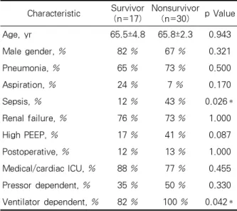
Characteristic Survivor (n=17)
Nonsurvivor
(n=30) p Value
Age, yr 65.5±4.8 65.8±2.3 0.943
Male gender, % 82 % 67 % 0.321
Pneumonia, % 65 % 73 % 0.500
Aspiration, % 24 % 7 % 0.170
Sepsis, % 12 % 43 % 0.026*
Renal failure, % 76 % 73 % 1.000
High PEEP, % 17 % 41 % 0.087
Postoperative, % 12 % 13 % 1.000
Medical/cardiac ICU, % 88 % 77 % 0.455 Pressor dependent, % 35 % 50 % 0.330 Ventilator dependent, % 82 % 100 % 0.042*
PEEP: Positive End-Expiratory Pressures
*: p<0.05
Table 1. Baseline characteristics of the study patients
survivors and nonsurvivors and could predict mortality.
METHODS Study Design
This was a prospective observational study.
Consecutive patients admitted to medical, cardiac, and surgical intensive care units of The Kangnam St. Mary’s Hospital who met inclusion criteria were enrolled. The study period was from December, 2003 to February, 2006. Written informed consent was obtained from either patient or next of kin. The Kangnam St. Mary’s Hospital Institutional Review Board approved the study.
Patients
Patients were included who had acute respiratory failure (partial pressure of oxygen in arterial blood (PaO2) / fractional concentration of inspired oxygen (FiO2) ≤ 200 mmHg), bilateral infiltration on chest radiography, and no clinical evidence of increased left arterial pressure. Echocardiographic study was done and patients with ejection fraction (EF) lower than 50% or showing any features of diastolic dysfunction were ruled out. We also excluded patients younger than 18 years.
Measurements
BNP, PaO2, FiO2, C-reactive protein (CRP), Acute Physiology and Chronic Health Evaluation II (APACHE II) score, and EF were obtained within 24h hours at the time of enrollment. Patients were followed up, and 30-day survival was documented.
Blood samples were obtained from peripheral blood.
BNP was measured using the quantitative immuno-
fluorescence assay Triage BNP (Biosite, San Diego, USA) on 5 ml of whole blood drawn in an ethylenediaminetetraacetic acid (EDTA) tube. This assay can detect BNP levels in the 5-5000 pg/mL range. APACHE II scores were calculated using data available from the 24-hr period at the time of enrollment. Transthoracic echocardiography is performed routinely in patients with ARDS. Systolic left ventricle (LV) dysfunction was defined as an EF below 50%. In patients without systolic LV dysfunction, diastolic dysfunction was defined as transmitral flow parameters consistent with one of the three patterns of diastolic dysfunction: impaired relaxation (LV peak E-wave velocity / LV peak A-wave velocity (E/A) < 1 and E-wave decelera- tion time (EDT) > 240 ms), pseudonormalization (1
< E/A < 2 and 150 < EDT < 200 ms) or restricted
filling (E/A > 2 and EDT < 150 ms)
15. Right
ventricle (RV) dysfunction was defined as an
RV-end diastolic area (EDA)/LV-EDA ratio grater
than 0.6
16. Pulmonary hypertension was defined
when pulmonary regurgitation velocity > 2m/sec
17.
Figure 1. Box and whiskers plots of median B-type natriuretic peptide (BNP) concentrations among surviving patients vs. those dying during the study.
The median BNP concentrations among those dying were significantly higher than those surviving (p = 0.013).
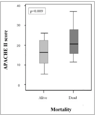
Figure 2. Box and whiskers plots of median acute physiology and chronic health evaluation (APACHE) II scores among surviving patients vs. those dying during the study. The median APACHE II scores among those dying were significantly higher than those surviving (p = 0.009).
High Positive End-Expiratory Pressures (PEEP) was defined as more than 15 cmH
2O. The patients, researchers, physicians, and echocardiographers were blinded to the BNP concentrations.
Statistical Analysis
Independent samples T test was done to compare means of continuous variables. Chi-square test was used for analysis of categorical variables.
Correlations between each variable were evaluated by bivariate analysis with Pearson correlation.
Receiver operating characteristic (ROC) curves for BNP and APACHE II score were constructed to predict mortality. All statistical analyses were performed with SPSS software (Chicago, IL, USA).
RESULTS
Clinical characteristics and mortality
From 2003 December to 2006 February, 103 patients were diagnosed as ARDS. 17 patients refused to be enrolled. 14 patients were excluded because of failure of BNP sampling or echocar- diographic examination within 24h hours of diagnosis. 25 patients showed LV dysfunction. So among 103 patients, 47 patients met inclusion criteria. 33 patients were on the intubated state during BNP sampling, 36 were during echocar- diographic examination.
Seventeen patients were alive and thirty patients
were dead at day 30. The baseline characteristics of
the study group are shown in Table 1. Percentage
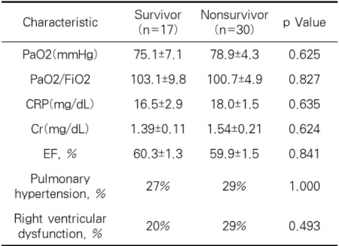
Characteristic Survivor (n=17)
Nonsurvivor
(n=30) p Value PaO2(mmHg) 75.1±7.1 78.9±4.3 0.625
PaO2/FiO2 103.1±9.8 100.7±4.9 0.827 CRP(mg/dL) 16.5±2.9 18.0±1.5 0.635 Cr(mg/dL) 1.39±0.11 1.54±0.21 0.624 EF, % 60.3±1.3 59.9±1.5 0.841 Pulmonary
hypertension, % 27% 29% 1.000
Right ventricular
dysfunction, % 20% 29% 0.493
PaO2: partial pressure of oxygen in arterial blood; FiO2:
fractional concentration of inspired oxygen; CRP:
C-reactive protein; Cr: creatinine; EF: ejection fraction Table 2. Comparison of parameters except APACHE II score and BNP
Variables Age BNP PaO2/FiO2 CRP Cr APACHE Ⅱ EF
BNP r 0.086 1.000 -0.014 0.101 0.374 0.087 0.138
p value 0.567 0.000 0.925 0.548 0.010† 0.562 0.355
APACHE Ⅱ r -0.033 0.087 -0.292 0.212 0.375 1.000 0.079
p value 0.824 0.562 0.047* 0.201 0.009† 0.000 0.597
BNP: B-type natriuretic peptide; PaO2: partial pressure of oxygen in arterial blood; FiO2: fractional concentration of inspired oxygen; CRP: C-reactive protein; Cr: creatinine; APACHE: Acute Physiology and Chronic Health Evaluation; EF:
ejection fraction
* p < 0.05, †p < 0.01
Table 3. Correlation between variables
of sepsis and ventilator dependent showed signi- ficant differences (Sepsis, 12% vs 43%; p=0.026, Ventilator dependent, 82% vs 100%; p=0.042, respectively). Others did not differ significantly.
BNP concentration and APACHE II score differ significantly between those who survived and those who died (BNP, 219.5 ± 57.7 pg/mL vs 492.3± 88.8 pg/mL; p=0.013, APACHE II score, 17.4 ± 1.6 vs 23.1 ± 1.3: p=0.009, respectively). Figure 1 and 2 show these differences. Parameters other than APACHE II score and BNP failed to show difference (Table 2). BNP concentration and EF were not different significantly between non-intubated
and intubated group (BNP ; non-intubated : 331.7 ± 109.8 pg/mL vs intubated : 419.8 ± 77.3 pg/mL, p=0.517, EF ; non-intubated : 59.3 ± 2.1 % vs intubated : 60.3 ± 1.2 %, p=0.686).
Correlation
BNP concentration was correlated with serum creatinine level (r = 0.374, p = 0.010), but not with EF, age, PaO2, FiO2, PaO2/FiO2, CRP, and APACHE II score. APACHE II score was correlated with PaO2/FiO2 (r = -0.292, p = 0.047), Cr (r = 0.375, p = 0.009) (Table 3).
BNP concentration
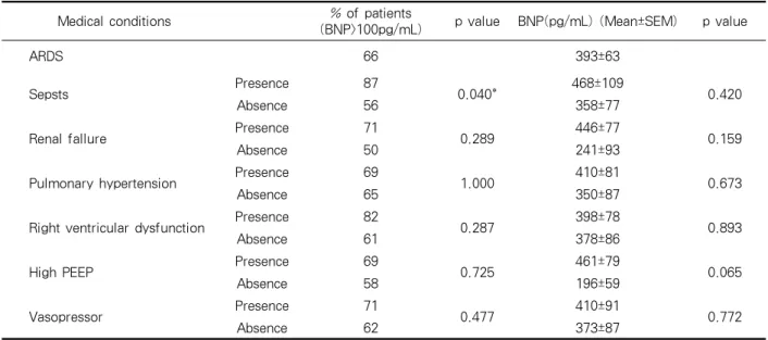
In total enrolled patients, mean BNP concen- tration was 393 ± 63 pg/mL, and 66% of patients showed elevated BNP level (>100 pg/mL). We examined percentage of patients who are BNP >
100 pg/mL and mean BNP concentration between
variable medical conditions(Table 4). These conditions
can be causes of raised plasma BNP level
18. Sepsis
patients showed significantly increased percentage
of BNP elevation, but mean BNP didn’t differ
significantly. In other conditions, although higher
percentages were seen, p values were more than
0.05.
Area Under Curve = 0.678 Area Under Curve = 0.710
Figure 3. Receiver operating characteristic curve for BNP (left) APACHE II score (right) for the prediction of mortality.
BNP: B-type natriuretic peptide; APACHE: Acute Physiology and Chronic Health Evaluation
Medical conditions % of patients
(BNP>100pg/mL) p value BNP(pg/mL) (Mean±SEM) p value
ARDS 66 393±63
Sepsts Presence 87
0.040* 468±109
0.420
Absence 56 358±77
Renal fallure Presence 71
0.289 446±77
0.159
Absence 50 241±93
Pulmonary hypertension Presence 69
1.000 410±81
0.673
Absence 65 350±87
Right ventricular dysfunction Presence 82
0.287 398±78
0.893
Absence 61 378±86
High PEEP Presence 69
0.725 461±79
0.065
Absence 58 196±59
Vasopressor Presence 71
0.477 410±91
0.772
Absence 62 373±87
BNP: B-type natriuretic peptide; PEEP: Positive End-Expiratory Pressures
* p < 0.05
Table 4. Comparison of B-type natriuretic peptide (BNP) concentration, as a function of medical conditions
Receiver operating characteristic (ROC) curve for the prediction of mortality
With the use of the threshold value for BNP of 585 pg/mL, the sensitivity for the prediction of mortality was 43%, with specificity of 94%, and area under the curve (AUC) of 0.678. The threshold
value for APACHE II of 15.5 showed sensitivity of 87%, specificity of 47%, and AUC of 0.710 (Figure 3). In order to combine these two factors, we used log-transformed BNP values. ‘APACHE II + 11×logBNP’ showed the best result of AUC (0.747).
Using the threshold value for 46.14, sensitivity was
63%, and specificity was 82% (Figure 4).
Sensitivity Specificity PPV NPV AUC p value
BNP≥585pg/dL 0.433 0.941 0.929 0.485 0.678 0.044*
APACHE Ⅱ≥15.5 0.867 0.471 0.743 0.667 0.710 0.018*
APACHE Ⅱ+11×logBNP ≥46.16 0.633 0.824 0.864 0.560 0.747 0.005†
PPV: positive predictive value; NPV: negative predictive value; AUC: area under the curve; BNP: B-type natriuretic peptide; APACHE: Acute Physiology and Chronic Health Evaluation
Table 5. Sensitivity, specificity, PPV, NPV, AUC for BNP, APACHE II, and APACHE II + 11×logBNP.
Area Under Curve = 0.747
Figure 4. Receiver operating characteristic curve for
‘APACHE II score + 11×log BNP’ for the prediction of mortality.
BNP: B-type natriuretic peptide; APACHE: Acute Physiology and Chronic Health Evaluation
Sensitivity, specificity, PPV, NPV, AUC for BNP, APACHE II, and APACHE II + 11×logBNP are given in Table 5.
DISCUSSION
BNP can be elevated not only in CHF patients but also other critically ill patients
12,19. Little is known in ARDS patients. Maeder and colleagues
10reported one case of ARDS patient who showed marked elevation of BNP in spite of normal ejection fraction. Although there are some articles about
BNP in respiratory failure patients
13,14, none of them were about only ARDS patients. We wanted to evaluate the relationship between BNP and mortality in ARDS patients, and also to exclude the effect of LV dysfunction on BNP. So, we enrolled patients who met the diagnostic criteria of ARDS, and ruled out any patients who had less than 50%
of ejection fraction and/or any features of diastolic dysfunction. This can explain the relatively small number of patients in this study in spite of long study period.
BNP concentrations among those who died were higher than those who survived in shock patients
12. Although APACHE II score is well known predictor of mortality in critically ill patients, there was no significant difference in APACHE II score between two groups in that study. In a Jefic D et al’s study, mean BNP concentrations did not differ between the survivors and nonsurvivors in respiratory failure patients, and mean APACHE II scores were higher in nonsurvivors than in survivors
13. In our study, not only BNP concentrations but also APACHE II scores are elevated significantly in patients who died. Our study is unique in that we enrolled only ARDS patients and ruled out LV dysfunction patients. This may be the reason of the different result compared to previous two similar studies.
What we tried to show was BNP could be elevated
in ARDS patients without the effect of LV
dysfunction, and this elevation could be related to
the mortality. We clearly showed that BNP
concentrations and APACHE II scores were elevated significantly in nonsurvivors. BNP is useful biomarkers for the diagnosis of HF.
However, BNP might be raised to different degrees in critical illness and various pulmonary diseases. In these situations, BNP may also serve as markers of severity and prognosis
18. ARDS is one of these diseases, and we showed BNP > 585 pg/mL has a high specificity for the prediction of mortality.
APACHE II score is well known predictor of mortality in critical illness patients. BNP also seems to be the predictor of mortality. APACHE II score and BNP were not correlated significantly in critically ill patients
19. In our study, the result was also same. We hypothesized that BNP could reflect certain conditions which APACHE II score couldn’t cover. So, we combined there two markers hoping they could produce synergistic result in predicting mortality. BNP concentrations showed wide variation compared to APCHE II score, so log BNP was used to correct this problem. The concept of log BNP was adapted from previous published study
12. Although there was no reference in combination of these two scores, we could prove
‘APACHE II + 11×logBNP’ was better than BNP or APACHE II in predicting mortality.
It is well known that the BNP is elevated in patients with low ejection fraction. However, in our study, there was no correlation between BNP concentration and EF. This may be due to the exclusion of patients who showed low ejection fraction. This result can tell we effectively eliminated the effect of LV dysfunction on BNP in our study, and we could examine more pure effect of ARDS on BNP concentration.
Sepsis, renal failure, pulmonary hypertension, right ventricular dysfunction, high PEEP, and shock state can be the cause of raised plasma BNP levels
18. In ARDS patients, these conditions may
coexist commonly. This may explain the reason of elevated level of BNP in ARDS. The high mortality rate in patients with high BNP levels may reflect these conditions. However, in our study, only sepsis patients showed just increased percentage of BNP elevation. We are not sure the elevation of BNP is due to sepsis in our study. No other medical conditions affected significantly the level of BNP.
BNP concentration was also seemed not to be affected by the intubated state. Maybe this is because the number of enrolled patients was small, and/or there were another factors that we don’t know which can elevate BNP levels. Recently, Karmpaliotis et al published similar result as ours
20. BNP appears useful in excluding cardiogenic pulmonary edema, but some ARDS patients showed elevation of BNP in the absence of CHF. Among those patients, high BNP concentration was associated with mortality. Unfortunately, they also could not explain the pathogenesis of this result.
The limitation of our study is the small number of patients. A second limitation is that we only measured a single BNP concentration at the time of enrollment. As BNP is known to fluctuate, serial measurement of BNP may be more useful than single. Another limitation is that we did not measure N-terminal pro BNP (NT-pro BNP) which is also well known useful marker for the diagnosis of heart failure.
Our results are useful as they reflect information of only ARDS patients. All enrolled patients were compatible with the diagnostic criteria of ARDS.
They are also unique in that they had normal
ejection fraction. We showed that BNP and
APACHE II score could predict mortality. When we
combined these two factors, we could get better
results. Further study about BNP in ARDS patients
should be done.
SUMMARY
Background
B-type natriuretic peptide (BNP) has been shown to be strong mortality predictors in a wide variety of cardiovascular syndromes. Little is known about BNP in patients with acute respiratory distress syndrome (ARDS). We studied whether BNP can predict mortality in patients with ARDS.
Method
Echocardiographic study was done to all patients with ARDS, and we excluded patient with low ejection fraction (less than 50%) or showing any features of diastolic dysfunction. 47 patients were enrolled between December, 2003 and February, 2006. Parameters including BNP were obtained within 24h hours at the time of enrollment.
Result
Mean BNP concentrations and APACHE II scores differed between the survivors and nonsurvivors (BNP, 219.5 ± 57.7 pg/mL vs 492.3± 88.8 pg/mL;
p=0.013, APACHE II score, 17.4 ± 1.6 vs 23.1 ± 1.3, p=0.009, respectively). With the use of the threshold value for BNP of 585 pg/mL, the specificity for the prediction of mortality was 94%. The threshold value for APACHE II of 15.5 showed sensitivity of 87%. ‘APACHE II + 11×logBNP’ showed sensitivity 63%, and specificity 82%, using threshold value for 46.14.
Conclusion
BNP concentrations and APCHE II scores were more elevated in nonsurvivors than survivors in patients with ARDS who have normal ejection fraction. BNP can predict mortality. Further study should be done.
REFERENCES
1. Maisel AS, Krishnaswamy P, Nowak RM, McCord J, Hollander JE, Duc P, et al. Rapid measurement of B-type natriuretic peptide in the emergency diagnosis of heart failure. N Engl J Med 2002;347:161-7.
2. Vasan RS, Benjamin EJ, Larson MG, Leip EP, Wang TJ, Wilson PW, et al. Plasma natriuretic peptides for community screening for left ventricular hypertrophy and systolic dysfunction: the Framingham heart study. JAMA 2002;288:1252-9.
3. Maisel AS, Koon J, Krishnaswamy P, Kazenegra R, Clopton P, Gardetto N, et al. Utility of B-natriuretic peptide as a rapid, point-of-care test for screening patients undergoing echocardiography to determine left ventricular dysfunction. Am Heart J 2001;141:
367-74.
4. Omland T, Aakvaag A, Bonarjee VV, Caidahl K, Lie RT, Nilsen DW, et al. Plasma brain natriuretic peptide as an indicator of left ventricular systolic function and long-term survival after acute myocar- dial infarction. Comparison with plasma atrial natriu- retic peptide and N-terminal proatrial natriuretic peptide. Circulation 1996;93:1963-9.
5. Krishnaswamy P, Lubien E, Clopton P, Koon J, Kaza- negra R, Wanner E, et al. Utility of B-natriuretic peptide levels in identifying patients with left ventricular systolic or diastolic dysfunction. Am J Med 2001;111:274-9.
6. Hammerer-Lercher A, Neubauer E, Muller S, Pac- hinger O, Puschendorf B, Mair J. Head-to-head com- parison of N-terminal pro-brain natriuretic peptide, brain natriuretic peptide and N-terminal pro-atrial natriuretic peptide in diagnosing left ventricular dysfunction. Clin Chim Acta 2001;310:193-7.
7. Mair J, Hammerer-Lercher A, Puschendorf B. The impact of cardiac natriuretic peptide determination on the diagnosis and management of heart failure. Clin Chem Lab Med 2001;39:571-88.
8. Bettencourt P. NT-proBNP and BNP: biomarkers for heart failure management. Eur J Heart Fail 2004;6:
359-63.
9. Yeo KT, Wu AH, Apple FS, Kroll MH, Christenson RH, Lewandrowski KB, et al. Multicenter evaluation of the Roche NT-proBNP assay and comparison to the Biosite Triage BNP assay. Clin Chim Acta 2003;338:107-15.
10. Maeder M, Ammann P, Rickli H, Diethelm M.
Elevation of B-type natriuretic peptide levels in acute respiratory distress syndrome. Swiss Med Wkly 2003;133:515-8.
11. Mitaka C, Hirata Y, Nagura T, Tsunoda Y, Itoh M, Amaha K. Increased plasma concentrations of brain natriuretic peptide in patients with acute lung injury.
J Crit Care 1997;12:66-71.
12. Tung RH, Garcia C, Morss AM, Pino RM, Fifer MA, Thompson BT, et al. Utility of B-type natriuretic peptide for the evaluation of intensive care unit shock.
Crit Care Med 2004;32:1643-7.
13. Jefic D, Lee JW, Jefic D, Savoy-Moore RT, Rosman HS. Utility of B-type natriuretic peptide and N-terminal pro B-type natriuretic peptide in evaluation of respira- tory failure in critically ill patients. Chest 2005;
128:288-95.
14. Bal L, Thierry S, Brocas E, Van de Louw A, Pottecher J, Hours S, et al. B-type natriuretic peptide (BNP) and N-terminal-proBNP for heart failure diagnosis in shock or acute respiratory distress. Acta Anaesthesiol Scand 2006;50:340-7.
15. Garcia MJ, Thomas JD, Klein AL. New Doppler echocardiographic applications for the study of dias- tolic function. J Am Coll Cardiol 1998;32:865-75.
16. Vieillard-Baron A, Prin S, Chergui K, Dubourg O, Jardin F. Echo-Doppler demonstration of acute cor pulmonale at the bedside in the medical intensive care unit. Am J Respir Crit Care Med 2002;166:
1310-9.
17. Feigenbaum H, Armstrong WF, Ryan T. Feigenbaum’s Echocardiography. 6th ed. Philadelphia: Lippincott Williams & Wilkins; 2005. p. 209.
18. Phua J, Lim TK, Lee KH. B-type natriuretic peptide:
issues for the intensivist and pulmonologist. Crit Care Med 2005;33:2094-13.
19. Berendes E, Van Aken H, Raufhake C, Schmidt C, Assmann G, Walter M. Differential secretion of atrial and brain natriuretic peptide in critically ill patients.
Anesth Analg 2001;93:676-82.
20. Karmpaliotis D, Kirtane AJ, Ruisi CP, Polonsky T, Malhotra A, Talmor D, et al. Diagnostic and prog- nostic utility of brain natriuretic Peptide in subjects admitted to the ICU with hypoxic respiratory failure due to noncardiogenic and cardiogenic pulmonary edema. Chest 2007;131:964-71.