Solitary Paraaortic Lymph Node Metastasis of Primary Hepatocellular Carcinoma Misdiagnosed as Primary Retroperitoneal Tumor
6
0
0
전체 글
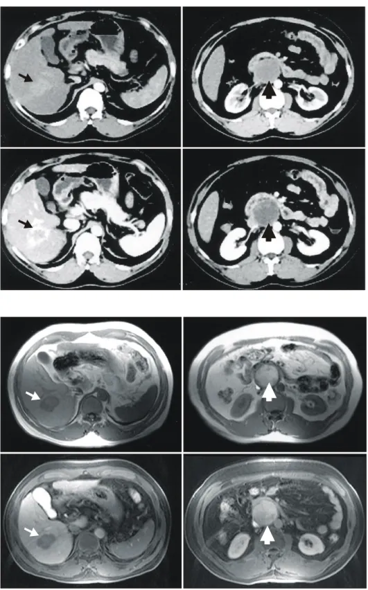
(2) 78. 대한외과학회지:제 70 권 제 1 호 2006. ꠏꠏꠏꠏꠏꠏꠏꠏꠏꠏꠏꠏꠏꠏꠏꠏꠏꠏꠏꠏꠏꠏꠏꠏꠏꠏꠏꠏꠏꠏꠏꠏꠏꠏꠏꠏꠏꠏꠏꠏꠏꠏꠏꠏꠏꠏꠏꠏꠏꠏꠏꠏꠏꠏꠏꠏꠏꠏꠏꠏꠏꠏꠏꠏꠏꠏꠏꠏꠏꠏꠏꠏꠏꠏꠏꠏꠏꠏꠏꠏꠏꠏꠏꠏꠏꠏꠏꠏꠏꠏꠏꠏꠏꠏꠏꠏꠏꠏꠏꠏꠏꠏꠏꠏꠏꠏꠏꠏꠏꠏꠏꠏꠏꠏꠏ. Fig. 1. Preoperative CT scan showed 3.4 cm sized peripheral enhanced mass on S6 of liver (small arrow), and 4.7 cm sized well encapsulated low attenuated mass in posterior aspect of third portion of duodenum (large arrow).. Fig. 2. Preoperative MRI scan showed 3.9×2.7 cm sized mass in right liver (small arrow) and 5.0 cm sized well-encapsulated mass in posterior aspect of third portion of duodenum (large arrow). The signal intensites of both masses are different on T1 and T2 weighted image.. 강이 되지 않는 4.7 cm 크기의 피낭성 종괴가 관찰되었다 (Fig. 1). 복부 자기공명영상에서도 T1과 T2 증강 영상에서 서로 다른 신호강도를 보이는 종괴가 간우엽과 십이지장 3번째 부위 뒤에서 관찰되었다(Fig. 2). 간동맥조영술에서 간우엽에 과혈관성의 단일종양이 관찰되었으며, 후복막종. 괴는 조영되지 않았다(Fig. 3). 양성자방출스캔에서 간우엽 의 종괴에 FDG 흡수가 중등도로 나타났으며, 우측 신문부 에 높은 FDG 흡수를 보이고 중앙부 괴사를 동반한 후복막 종괴가 관찰되었다(Fig. 4). 이상의 방사선 소견으로 원발성 간암 및 원발성 후복막종양의 진단하에 후복막종양의 정확.
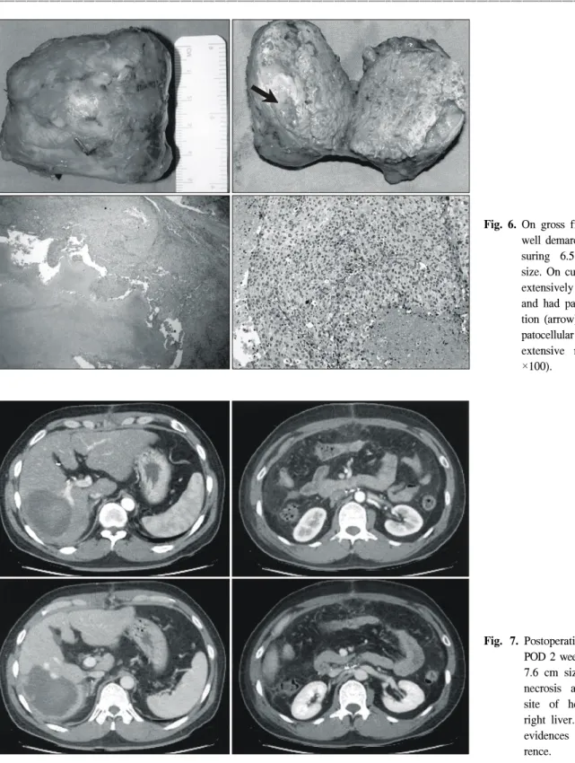
(3) 이재길 외:후복막종양으로 오인된 원발성간암의 단독 대동맥주위림프절 전이. 79. ꠏꠏꠏꠏꠏꠏꠏꠏꠏꠏꠏꠏꠏꠏꠏꠏꠏꠏꠏꠏꠏꠏꠏꠏꠏꠏꠏꠏꠏꠏꠏꠏꠏꠏꠏꠏꠏꠏꠏꠏꠏꠏꠏꠏꠏꠏꠏꠏꠏꠏꠏꠏꠏꠏꠏꠏꠏꠏꠏꠏꠏꠏꠏꠏꠏꠏꠏꠏꠏꠏꠏꠏꠏꠏꠏꠏꠏꠏꠏꠏꠏꠏꠏꠏꠏꠏꠏꠏꠏꠏꠏꠏꠏꠏꠏꠏꠏꠏꠏꠏꠏꠏꠏꠏꠏꠏꠏꠏꠏꠏꠏꠏꠏꠏꠏ. Fig. 3. Preoperative HAA showed single hypervascular mass in right liver. There was no definite vascularity of extrahepatic mass.. Fig 4. Preoperative PET scan showed moderate uptake of FDG on mass in right liver (small arrow). Another high uptake of FDG was seen on retroperitoneal mass at renal hilum level with extensive central necrosis (large arrow).. Fig. 5. NAB for retroperitoneal mass showed degenerated atypical cells with plump eosinophilic cytoplasm in a blackground of extensive coagulative necrosis. The pathologic diagnosis was possibly metastatic carcinoma, but it's origin was unknown (H&E, ×200).. 한 진단을 위해 시행한 세침흡인검사에서 조직의 대부분이 괴사되어 있었고 전이성 악성종양이 의심되었으나 그 원발 부위는 알 수 없었다(Fig. 5). 수술 소견상 간표면은 심한 대 결절성 간경변증 소견을 보였으며 수술 중 시행한 초음파 검사에서 4.0×2.5 cm 크기의 종괴가 5번과 6번 분절에 걸 쳐 1개가 있었으며 맥관계로의 침윤소견은 보이지 않았다. 복강 내에도 전이를 의심할 수 있는 소견은 보이지 않았다. 췌두부 뒤 후복막의 하대정맥과 대동맥 사이의 좌신정맥 바로 아래에 약 6.0×5.0 cm 크기의 종괴가 있었다. 후복막 종괴절제술을 시행하였고 육안소견에서 중앙부 괴사를 동 반한 타원형의 종괴로 동결절편검사에서 전이성 간세포암 으로 진단되었다(Fig. 6). 원발성 간암에 대해서는 3.5 cm Ⓡ TM Laveen needle과 RF 3000 (Boston Scientific, CA, USA)을 이용하여 460 kHz의 고주파열치료(RFA)를 5회 시행하였다. 수술 후 2주째 시행한 복부전산화단층촬영에서 잔존간에 잔존암이나 재발을 의심할만한 소견은 없었으며, RFA 부위 에 광범위한 괴사 소견이 보였다(Fig. 7). 환자는 수술 후 16 일째 퇴원하였고, 현재 재발 소견없이 전신항암화학요법을 시행하는 중이다..
(4) 80. 대한외과학회지:제 70 권 제 1 호 2006. ꠏꠏꠏꠏꠏꠏꠏꠏꠏꠏꠏꠏꠏꠏꠏꠏꠏꠏꠏꠏꠏꠏꠏꠏꠏꠏꠏꠏꠏꠏꠏꠏꠏꠏꠏꠏꠏꠏꠏꠏꠏꠏꠏꠏꠏꠏꠏꠏꠏꠏꠏꠏꠏꠏꠏꠏꠏꠏꠏꠏꠏꠏꠏꠏꠏꠏꠏꠏꠏꠏꠏꠏꠏꠏꠏꠏꠏꠏꠏꠏꠏꠏꠏꠏꠏꠏꠏꠏꠏꠏꠏꠏꠏꠏꠏꠏꠏꠏꠏꠏꠏꠏꠏꠏꠏꠏꠏꠏꠏꠏꠏꠏꠏꠏꠏ. Fig. 6. On gross finding, it was a well demarcated mass measuring 6.5×5×3.5 cm in size. On cut section, it was extensively central necrotic and had partial viable portion (arrow). It showed hepatocellular carcinoma, with extensive necrosis (H&E, ×100).. Fig. 7. Postoperative CT scan at POD 2 weeks showed about 7.6 cm sized hemorrhagic necrosis at the previous site of hepatic mass in right liver. There were no evidences of tumor recurrence.. 고. 찰. 간세포암의 간외전이는 비교적 흔하게 나타나며 약 15∼ 35%로 보고하고 있으며,(4-8) 전이 부위는 폐가 가장 흔하 며, 림프절, 근골격계 및 부신 등으로 잘 전이된다.(5) 드물. 게 뇌, 소화기계, 비장 등으로 전이가 있을 수 있으며, 유방 과 치은으로의 전이도 보고되었다.(11-13) Katyal 등(5)에 의 하면 간세포암의 림프절 전이는 간외전이의 53%로 폐전이 에 이어 두 번째 호발 부위로, 대부분은 복강림프절과 간십 이지장림프절로 전이되며 대동맥주위림프절 전이는 약 3∼6% 정도이며 대부분 타부위 림프절 전이를 동반하였다..
(5) 이재길 외:후복막종양으로 오인된 원발성간암의 단독 대동맥주위림프절 전이. 81. ꠏꠏꠏꠏꠏꠏꠏꠏꠏꠏꠏꠏꠏꠏꠏꠏꠏꠏꠏꠏꠏꠏꠏꠏꠏꠏꠏꠏꠏꠏꠏꠏꠏꠏꠏꠏꠏꠏꠏꠏꠏꠏꠏꠏꠏꠏꠏꠏꠏꠏꠏꠏꠏꠏꠏꠏꠏꠏꠏꠏꠏꠏꠏꠏꠏꠏꠏꠏꠏꠏꠏꠏꠏꠏꠏꠏꠏꠏꠏꠏꠏꠏꠏꠏꠏꠏꠏꠏꠏꠏꠏꠏꠏꠏꠏꠏꠏꠏꠏꠏꠏꠏꠏꠏꠏꠏꠏꠏꠏꠏꠏꠏꠏꠏꠏ. 그러나 본 증례처럼 대동맥주위림프절의 단독전이만을 보 인 증례는 없었다. 간세포암의 간외 전이의 치료에 대한 적응증과 그 장기 적 결과에 대해서는 명확한 보고는 없지만, 최근 들어 수술 적 절제로 좋은 결과를 보이는 보고들도 있다. Lo 등(6)에 의하면 간세포암의 단독 간외전이 환자는 가능하다면 수술 적 절제가 장기생존의 유일한 기회라고 하였고, Aramaki 등 (7)은 단독 간외전이에 대한 수술적 절제나 국소치료를 한 경우에 예후가 좋다고 하였다. Poon 등(8)은 간외전이는 절 제를 시행하고 간내재발에 대해서는 국소치료를 시행한 경 우가 간외전이에 대해 전신항암화학요법을 시행한 경우에 비해 생존율이 유의하게 높음을 보고하였다. 간암의 간외 전이 중 폐전이의 치료에 대해서는 많은 보고들이 있었으 며, 적응이 되는 경우 수술적 절제를 시행한 예에서 생존율 이 높았다.(14,15) Uenishi 등(16)은 본 예와 다르지만 광범위 하게 간문부, 간십이지장인대, 총간동맥, 대동맥 주위의 림 프절에 전이를 동반한 간세포암 환자에서 간엽절제술과 림 프절곽청술 후 15개월동안 생존하였음을 보고하였다. 이상 에서 살펴보면 간세포암의 간외전이의 경우 전이병소가 단 독전이이거나 절제가 가능하다면 수술적 절제를 하는 것이 환자의 생존에 도움이 된다. 간세포암의 간외전이에 대한 전신항암화학요법의 효과 에 대해서는 논란의 여지가 많다. 일부에서는 간세포암의 절제 불가능한 폐전이의 예에서 항암화학요법이 효과가 있 다는 보고도 있으나 이들은 증례보고이거나 증례의 수가 적다.(17-19) Okusaka 등(9)은 간세포암의 간외전이 환자에 서 전신항암화학요법이 효과가 없음을 보고하였고, Aramaki 등(7)도 간내재발 및 간외전이 환자에서 전신항암화학 요법 단독치료는 생존율을 증가시키지 못하였으며, 간내재 발에 대한 국소치료와 더불어 전신항암화학요법을 병합하 여 시행하였을 때 더 좋은 예후를 보인다고 보고하였다. Poon 등(8)은 간세포암의 간외전이 환자에서 절제 후 시행 한 보조항암화학요법이 생존율에 영향을 미치지 못함을 보 고하였다. 이상으로 간세포암의 간외전이 환자에서 생존율을 높이 기 위한 최선의 치료는 가능한 한 절제를 하거나 국소치료 를 시행하는 것이다. 그러기 위해서는 간세포암의 간내병 변이 치료되었거나 치료가 가능한 상태여야 한다. 그리고 보조항암화학요법은 좀더 연구가 필요할 것으로 사료되지만 좋은 결과를 보인 보고들이 있어서 시도해볼 만할 것이다.. REFERENCES 1) Nagasue N, Uchida M, Makino Y, Takemoto Y, Yamanoi A, Hayashi T, et al. Incidence and factors associated with intrahepatic recurrence following resection of hepatocellular carcinoma, Gastroenterology 1993;105:488-94.. 2) Chen MF, Hwang TL, Jeng LB, Wang CS, Jan YY, Chen SC. Postoperative recurrence of hepatocellular carcinoma. Two hundred five consecutive patients who underwent hepatic resection in 15 years. Arch Surg 1994;129:738-42. 3) Hanazaki K, Kajikawa S, Shimozawa N, Mihara M, Shimada K, Hiraguri M, et al. Survival and recurrence after hepatic resecton of 386 consecutive patients with hepatocellular carcinoma. J Am Coll Surg 2000;191:381-8. 4) Shuto T, Hirohashi K, Kubo S, Tanaka H, Yamamoto T, Kakemura S, et al. Distant metastatic recurrence after hepatic resection for hepatocellular carcinoma. Osaka City Med J 2002;48: 17-21. 5) Katyal S, Oliver III JH, Peterson MS, Ferris JV, Carr BS, Baron RL. Extrahepatic metastases of hepatocellular carcinoma. Radiology 2000;216:698-703. 6) Lo CM, Lai CS, Fan ST, Choi TK, Wong J. Resection for extrahepatic recurrence of hepatocellular carcinoma. Br J Surg 1994;81:1019-21. 7) Aramaki M, Kawano K, Kai T, Yokoyama H, Morii Y, Sasaki A, et al. Treatment for extrahepatic metastasis of hepatocellular carcinoma following successful hepatic resection. HepatoGastroenterology 1999;46:2931-4. 8) Poon R, Fan ST, O'Suilleebhain CB, Wong J. Aggressive management of patients with extrahepatic and intrahepatic recurrences of hepatocellular carcinoma by combined resection and locoregional therapy. J Am Coll Surg 2002;195:311-8. 9) Okusaka T, Okada S, Ishii H, Nose H, Nagahama H, Nakasuka H, et al. Prognosis of hepatocellular carcinoma patients with extrahepatic metastases. Hepato-gastroenterology 1997;44:251-7. 10) Imamura I. Prognostic efficacy of treatment for extrahepatic metastasis after surgical treatment of hepatocellular carcinoma. Kureme Med J 2003;50:41-8. 11) Chang L, Chen YL, Kao MC. Intracranial metastasis of hepatocellular carcinoma: review of 45 cases. Surg Neurol 2004; 62:172-7. 12) Lo HC, Lee KF, Yeh CN, Chen MF. Breast metastasis from hepatocellular carcinoma. Hepatogastroenterology 2004;51:38790. 13) Choi SJ, Kim YS, Kim NR, Jeong SW, Lee SH, Jeong JS, et al. A case of hepatocellular carcinoma with metastasis to gingival mucosa. Korean J Hepatol 2002;8:495-9. 14) Lam CM, Lo CM, Yuen WK, Liu CL, Fan ST. Prolonged survival in selected patients following surgical resection for pulmonary metastasis from hepatocellular carcinoma. Br J Surg 1998;85:1198-200. 15) Gwak GY, Jung JO, Sung SW, Lee HS. Long-term survival after pulmonary metastatectomy of hepatocellular carcinoma; treatment outcome or natural history? Hepato-gastroenterology 2004;51:1428-33. 16) Uenishi T, Hirohashi K, Tanaka H, Yamamoto T, Kubo S, Kinoshita H. A surgically treated case of hepatocellular carcinoma with extensive lymph node metastases. Hepato-Gast-.
(6) 82. 대한외과학회지:제 70 권 제 1 호 2006. ꠏꠏꠏꠏꠏꠏꠏꠏꠏꠏꠏꠏꠏꠏꠏꠏꠏꠏꠏꠏꠏꠏꠏꠏꠏꠏꠏꠏꠏꠏꠏꠏꠏꠏꠏꠏꠏꠏꠏꠏꠏꠏꠏꠏꠏꠏꠏꠏꠏꠏꠏꠏꠏꠏꠏꠏꠏꠏꠏꠏꠏꠏꠏꠏꠏꠏꠏꠏꠏꠏꠏꠏꠏꠏꠏꠏꠏꠏꠏꠏꠏꠏꠏꠏꠏꠏꠏꠏꠏꠏꠏꠏꠏꠏꠏꠏꠏꠏꠏꠏꠏꠏꠏꠏꠏꠏꠏꠏꠏꠏꠏꠏꠏꠏꠏ roenterology 2000;47:1714-6. 17) Nakao A, Sato S, Nakajima A, Nabeyama A, Okada Y, Sakagami K. Efficacy of treatment with frequent and low-dose epirubicin in two cases of pulmonary metastases after surgery of liver cancer. Gan to Kagaku Ryoho 1995;22:1831-4. 18) Chung YH, Song IH, Song BC, Lee GC, Koh MS, Yoon HK, et al. Combined therapy consisting of intraarterial cisplatin. infusion and systemic interferon-alpha for hepatocellular carcinoma patients with major portal vein thrombosis or distant metastasis. Cancer 2000;88:1986-91. 19) Seki S, Yamada T, Kawakita N, Masuichi H, Kitada T, Sakaguchi H. A new chemotherapeutic regimen for advanced unresectable hepatocellular carcinoma. Hepato-Gastroenterology 2003;50:1598-602..
(7)
수치

관련 문서
So we investigated a clinico-statistical evaluation on cervical lymph node metastasis in 183 patients who were diagnosed with oral squamous cell carcinoma at the Department of
Key Words: Single node metastasis, Gastric carcinoma, Sentinel lymph node 중심 단어: 단일 림프절 전이, 위암, 감시 림프절.. Purpose: In order to examine the significance of
Conclusion: These results suggest that the prognosis for mediastinal lymph node metastasis in differentiated thyroid carcinoma can be improved by an aggressive mediastinal
유두상 갑상선암(papillary thyroid carcinoma)은 림프절 전 이(lymph node metastasis)를 잘 하는 특성이 있으며 임상적 으로 20∼50%에서 경부림프절 전이(cervical
We aimed to develop a radiomics signature using US images of the primary tumor to preopera- tively predict lateral lymph node metastasis (LNM) in patients with conventional
EXPRESSIONS OF METASTASIS-RELATED FACTORS IN ORTHOTOPIC TUMOR MODELS OF ORAL SQUAMOUS CELL CARCINOMA
Key words: Oral squamous cell carcinoma, Lymph node metastasis, Metastasis-related factors, Mouth
Pathologic features by clinical node stage at pre-operative imaging for 440 renal cell carcinoma patients without distant metastasis (cM0) treated with nephrectomy and lymph
Tumor size(p=0.000), ETE(p=0.001), multifocality(p=0.014), and bilaterality(p=0.001) were significantly related factors for cervical lymph node metastasis clinically
